

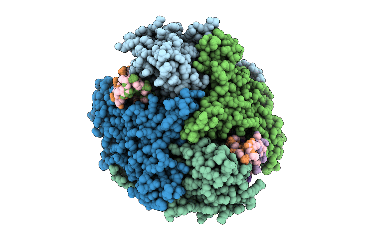
1FIU image
Deposition Date 2000-08-07
Release Date 2001-02-07
Last Version Date 2024-02-07
Entry Detail
PDB ID:
1FIU
Keywords:
Title:
TETRAMERIC RESTRICTION ENDONUCLEASE NGOMIV IN COMPLEX WITH CLEAVED DNA
Biological Source:
Source Organism(s):
Expression System(s):
Method Details:
Experimental Method:
Resolution:
1.60 Å
R-Value Free:
0.20
R-Value Work:
0.17
Space Group:
P 21 21 21
Macromolecular Entities
Structures with similar UniProt ID
Protein Blast
Polymer Type:polypeptide(L)
Molecule:TYPE II RESTRICTION ENZYME NG
Gene (Uniprot):ngoMIVR
Chain IDs:I (auth: A), J (auth: B), K (auth: C), L (auth: D)
Chain Length:286
Number of Molecules:4
Biological Source:Neisseria gonorrhoeae
Primary Citation
Structure of the tetrameric restriction endonuclease NgoMIV in complex with cleaved DNA.
Nat. Struct. Biol. 7 792 799 (2000)
PMID: 10966652 DOI: 10.1038/79032

Abstact

The crystal structure of the NgoMIV restriction endonuclease in complex with cleaved DNA has been determined at 1.6 A resolution. The crystallographic asymmetric unit contains a protein tetramer and two DNA molecules cleaved at their recognition sites. This is the first structure of a tetrameric restriction enzyme-DNA complex. In the tetramer, two primary dimers are arranged back to back with two oligonucleotides bound in clefts on opposite sides of the tetramer. The DNA molecules retain a B-type conformation and have an enclosed angle between their helical axes of 60 degrees. Sequence-specific interactions occur in both the major and minor grooves. Two Mg2+ ions are located close to the cleaved phosphate at the active site of NgoMIV. Biochemical experiments show that interactions between the recognition sites within the tetramer greatly increase DNA cleavage efficiency.

Legend

Protein

Chemical

Disease

Primary Citation of related structures
Feedback Form
Name
Email
Institute
Feedback