1COT image
Deposition Date 1994-07-06
Release Date 1994-09-30
Last Version Date 2024-11-20
Entry Detail
PDB ID:
1COT
Title:
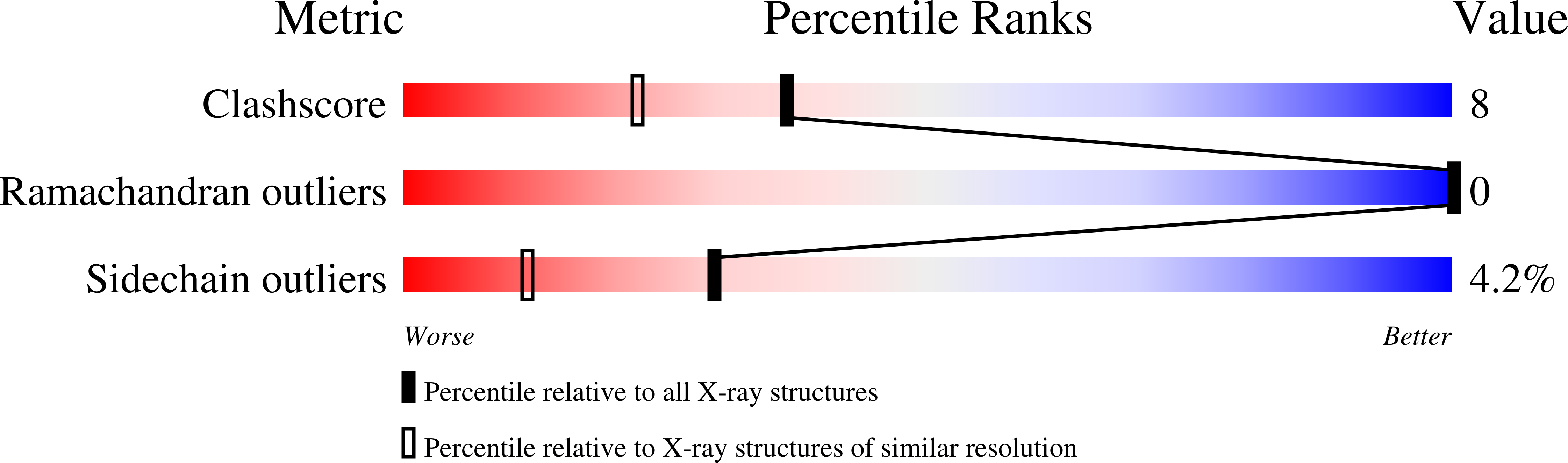
X-RAY STRUCTURE OF THE CYTOCHROME C2 ISOLATED FROM PARACOCCUS DENITRIFICANS REFINED TO 1.7 ANGSTROMS RESOLUTION
Biological Source:
Source Organism(s):
Method Details:
Experimental Method:
Resolution:
1.70 Å
R-Value Observed:
0.17
Space Group:
P 21 21 21
Macromolecular Entities
Structures with similar UniProt ID
Protein Blast
Polymer Type:polypeptide(L)
Molecule:CYTOCHROME C2
Gene (Uniprot):cycA
Chain IDs:A
Chain Length:129
Number of Molecules:1
Biological Source:Paracoccus denitrificans
Ligand Molecules
Primary Citation
X-Ray structure of the cytochrome c2 isolated from Paracoccus denitrificans refined to 1.7-A resolution.
Arch. Biochem. Biophys. 310 460 466 (1994)
PMID: 8179333 DOI: 10.1006/abbi.1994.1193

Abstact

The cytochrome c2 (formerly c550) isolated from Paracoccus denitrificans is one of the larger bacterial c-type proteins examined thus far. The molecular structure of this cytochrome has been redetermined and refined to 1.7-A resolution with a crystallographic R-factor of 17.5% for all measured X-ray data. Like other, smaller c-type cytochromes, the molecule consists of five alpha-helices that wrap around the heme group. In addition, this bacterial cytochrome contains two strands of anti-parallel beta-sheet, five Type I turns, and three Type II turns. The present model differs from the originally determined structure in several regions including the N-terminus, the loop delineated by Asp 25 to Lys 31, the region defined by Trp 86 to Val 88, and the C-terminus. A total of 103 water molecules has been positioned into the electron density map. Six of these waters are directly involved in heme binding.

Legend

Protein

Chemical

Disease

Primary Citation of related structures
Feedback Form
Name
Email
Institute
Feedback