1BCF image
Deposition Date 1993-12-06
Release Date 1994-12-20
Last Version Date 2024-02-07
Entry Detail
PDB ID:
1BCF
Title:
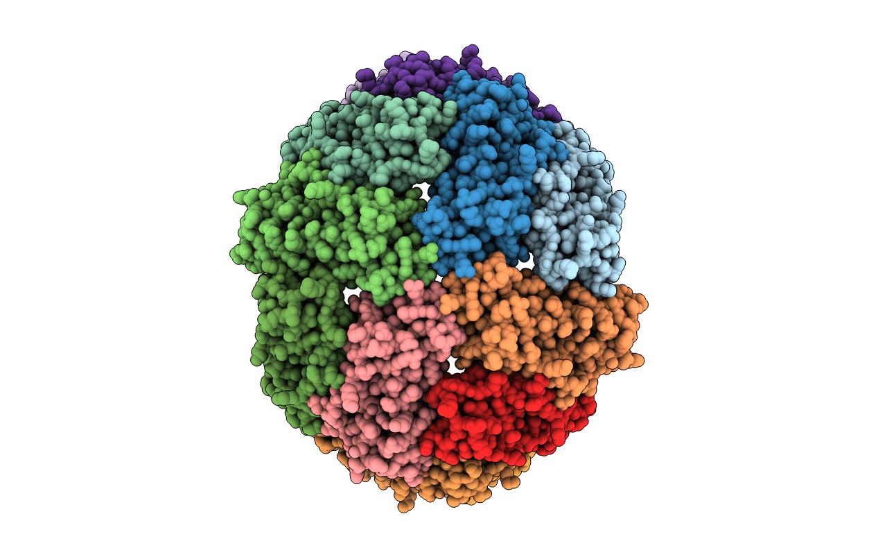
THE STRUCTURE OF A UNIQUE, TWO-FOLD SYMMETRIC, HAEM-BINDING SITE
Biological Source:
Source Organism(s):
Escherichia coli (Taxon ID: 562)
Method Details:
Experimental Method:
Resolution:
2.90 Å
R-Value Work:
0.20
R-Value Observed:
0.20
Space Group:
P 42 21 2
Macromolecular Entities
Structures with similar UniProt ID
Protein Blast
Polymer Type:polypeptide(L)
Molecule:BACTERIOFERRITIN
Gene (Uniprot):bfr
Chain IDs:A, B, C, D, E, F, G, H, I, J, K, L
Chain Length:158
Number of Molecules:12
Biological Source:Escherichia coli
Primary Citation
Structure of a unique twofold symmetric haem-binding site.
Nat. Struct. Biol. 1 453 460 (1994)
PMID: 7664064 DOI: 10.1038/nsb0794-453

Abstact

Bacterioferritin of Escherichia coli, also known as cytochrome b1, is a hollow, nearly spherical shell made up of 24 identical protein subunits and 12 haems. We have solved this structure in a tetragonal crystal form at 2.9 A resolution. We find that each haem is bound in a pocket formed by the interface between a pair of symmetry-related subunits. The quasi-twofold axis of the haem is closely aligned with the local twofold axis relating these subunits. The axial ligands of the haem are sulphurs of two equivalent methionyl residues (Met 52) from the symmetry-related subunits. A cluster of four water molecules is trapped in the gap between the upper edge of the haem and two extended protein loops which close off the haem from the outer aqueous environment. This is the first structure of a bis-methionine ligated haem-binding site and the first case of a twofold symmetric haem-binding site.

Legend

Protein

Chemical

Disease

Primary Citation of related structures
Feedback Form
Name
Email
Institute
Feedback