Deposition Date
1997-11-27
Release Date
1998-03-18
Last Version Date
2024-02-07
Entry Detail
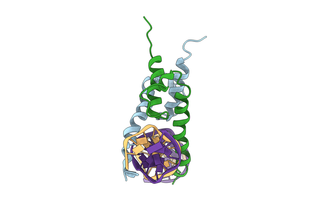
PDB ID:
1A0A
Keywords:
Title:
PHOSPHATE SYSTEM POSITIVE REGULATORY PROTEIN PHO4/DNA COMPLEX
Biological Source:
Source Organism(s):
Saccharomyces cerevisiae (Taxon ID: 4932)
Expression System(s):
Method Details:
Experimental Method:
Resolution:
2.80 Å
R-Value Free:
0.28
R-Value Work:
0.23
Space Group:
P 21 21 21