

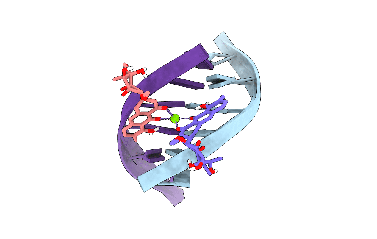
146D image
Deposition Date 1993-11-09
Release Date 1995-03-31
Last Version Date 2024-05-22
Entry Detail
PDB ID:
146D
Keywords:
Title:
SOLUTION STRUCTURE OF THE MITHRAMYCIN DIMER-DNA COMPLEX
Method Details:
Experimental Method:
Conformers Calculated:
2
Conformers Submitted:
2
Selection Criteria:
all calculated structures submitted
Macromolecular Entities
Polymer Type:polydeoxyribonucleotide
Molecule:DNA (5'-D(*TP*CP*GP*CP*GP*A)-
Chain IDs:A, B
Chain Length:6
Number of Molecules:2
Biological Source:
Primary Citation
Solution structure of the mithramycin dimer-DNA complex.
Biochemistry 32 6588 6604 (1993)
PMID: 8329387 DOI: 10.1021/bi00077a012

Abstact

We have characterized the NMR parameters for the complexes formed by the Mg(2+)-coordinated mithramycin dimer with self-complementary d(T-G-G-C-C-A) and d(T-C-G-C-G-A) duplexes. The solution structure of the latter complex has been determined using a combined NMR-molecular dynamics study including relaxation matrix refinement. The Mg(2+)-coordinated mithramycin dimer-d(T-C-G-C-G-A) complex exhibits a 2-fold center of symmetry with the divalent cation coordinated aglycons positioned opposite the central (G3-C4).(G3-C4) segment such that the aglycon C8 hydroxyl oxygens form symmetrical sequence-specific hydrogen bonds to guanine amino protons in the complex. The C-D-E trisaccharide segments of each monomer in the mithramycin dimer adopt extended conformations, are positioned inside the minor groove, and are directed toward either end of the duplex. The C-D saccharide component of one monomer and the aglycon of the other monomer in the mithramycin dimer share a widened minor groove with the hydrophobic edges of the C and D sugars interacting with individual strands of the duplex. The E-sugar ring is positioned in the floor of the minor groove, and its hydroxyl-bearing face interacts with both strands of the duplex through hydrogen-bonding and hydrophobic intermolecular interactions. The A-B disaccharide and the hydrophilic side chain form intermolecular contacts with the sugar-phosphate backbone in the complex. The antiparallel alignment of divalent cation coordinated monomers in the mithramycin dimer results in the two outwardly directed C-D-E trisaccharide segments generating a right-handed continuous hexasaccharide domain that spans six base pairs in the minor groove of the duplex. The solution structure of the mithramycin dimer-DNA complex reported in this study and the solution structure of the chromomycin dimer-DNA complex reported previously [Gao, X., Mirau, P., & Patel, D. J. (1992) J. Mol. Biol. 223, 259-279] show global similarities, as well as local differences that are of interest. All four nucleotides in the tetranucleotide segment of the duplex centered about the sequence-specific (G-C).(G-C) step adopt A-DNA sugar puckers and glycosidic torsion angles in the chromomycin dimer-DNA complex, while only the central cytidine adopts an A-DNA sugar pucker and glycosidic torsion angle in the mithramycin dimer-DNA complex.(ABSTRACT TRUNCATED AT 400 WORDS)

Legend

Protein

Chemical

Disease

Primary Citation of related structures
Feedback Form
Name
Email
Institute
Feedback