10LX image
Deposition Date 2026-01-27
Release Date 2026-05-20
Last Version Date 2026-05-20
Entry Detail
PDB ID:
10LX
Keywords:
Title:
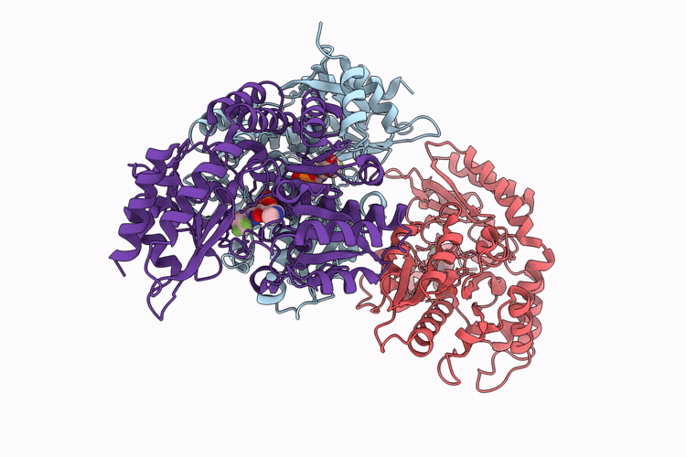
High Stable Quinonoid Intermediate of Human Ornithine Aminotransferase Complexed with (1R,4S)-4-Amino-3-(trifluoromethyl)cyclopent-2-ene-1-carboxylic Acid
Biological Source:
Source Organism(s):
Homo sapiens (Taxon ID: 9606)
Expression System(s):
Method Details:
Experimental Method:
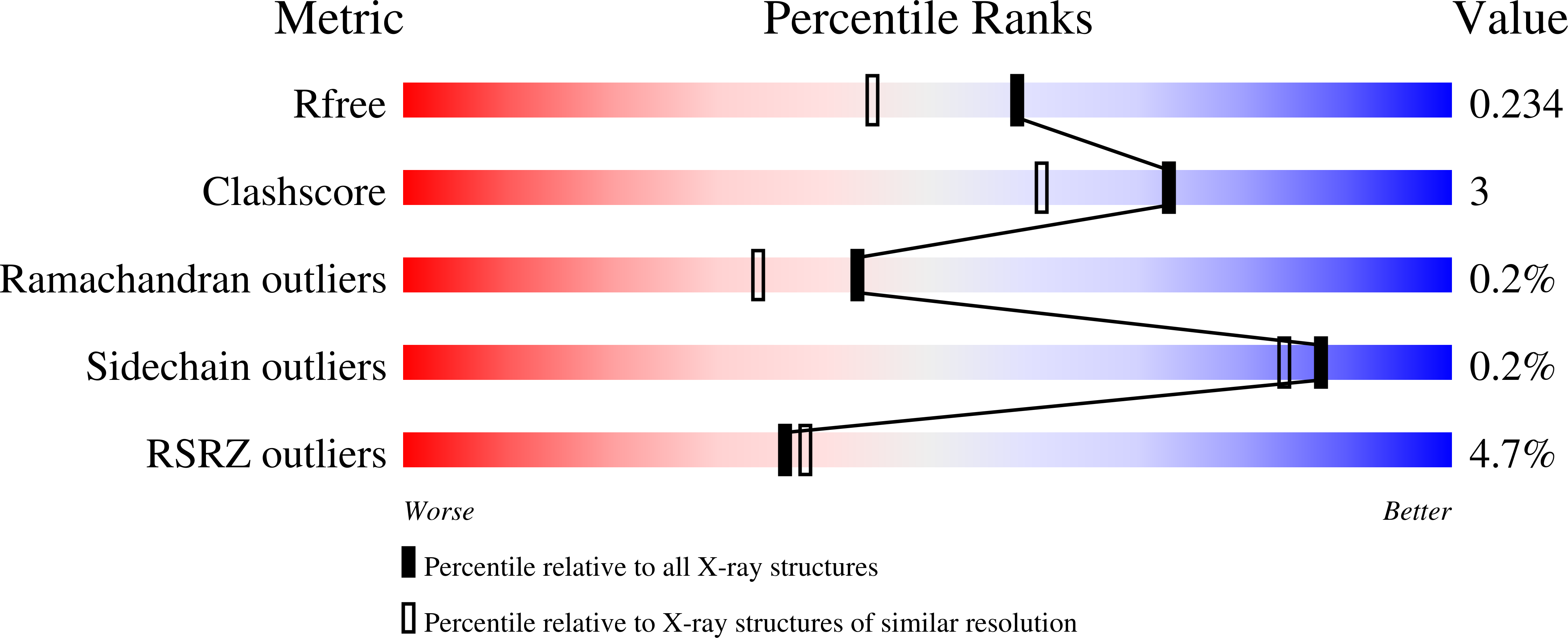
Resolution:
1.83 Å
R-Value Free:
0.23
R-Value Work:
0.19
R-Value Observed:
0.20
Space Group:
P 32 2 1
Macromolecular Entities
Structures with similar UniProt ID
Protein Blast
Polymer Type:polypeptide(L)
Molecule:Ornithine aminotransferase, m
Gene (Uniprot):OAT
Chain IDs:A, B, C
Chain Length:439
Number of Molecules:3
Biological Source:Homo sapiens
Ligand Molecules
Primary Citation
Inactivation of ornithine aminotransferase by (1 R ,4 S )-4-Amino-3-(trifluoromethyl)cyclopent-2-ene-1-carboxylic acid via a stable quinonoid intermediate.
Med.Chem.Res. 35 792 803 (2026)
PMID: 42078119 DOI: 10.1007/s00044-026-03538-1

Abstact

Ornithine aminotransferase (OAT), a pyridoxal 5'-phosphate (PLP)-dependent enzyme, is a key contributor to glutamine supply in cancer cells, suggesting its therapeutic potential for hepatocellular carcinoma (HCC), the most common form of liver cancer. To identify an initial set of OAT inactivators, we have tested inactivators of gamma-aminobutyric acid aminotransferase (GABA-AT), a homologous PLP-dependent enzyme, with human OAT (hOAT) and identified several co-inactivators. Among the active molecules, (1R,4S)-4-amino-3-(trifluoromethyl)cyclopent-2-ene-1-carboxylic acid (2) has not been thoroughly investigated for its time-dependent kinetics and mechanistic pathways with OAT. In this study, we evaluated the time-dependent inactivation of hOAT by 2 and investigated the underlying mechanism, primarily based on X-ray crystallography. The results demonstrated that 2 acts as a time-dependent OAT inactivator with an inactivation efficiency (k (inact)/K (I) = 5.1 min(-1)mM(-1)) approximately 30-fold higher than that for GABA-AT (k (inact)/K (I) = 0.17 min(-1)mM(-1)) and, notably, revealed an inactivation pathway that proceeds via a stable quinonoid intermediate, as evidenced by the UV-Vis spectroscopy.

Legend

Protein

Chemical

Disease

Primary Citation of related structures
Feedback Form
Name
Email
Institute
Feedback