

Search Count: 21
 |
Cryo-Em Structure Of Atubab Heterodimer From Heimdallarchaeia
Organism: Candidatus heimdallarchaeota archaeon
Method: ELECTRON MICROSCOPY Resolution:3.50 Å Release Date: 2026-06-10 Classification: UNKNOWN FUNCTION Ligands: GDP |
Organism: Candidatus heimdallarchaeota archaeon
Method: ELECTRON MICROSCOPY
Release Date: 2026-06-10
Ligands: GDP
 |
Cryo-Em Structure Of Atubab Mini Microtubules From Heimdallarchaeia
Organism: Candidatus heimdallarchaeota archaeon
Method: ELECTRON MICROSCOPY Release Date: 2026-06-10 Classification: UNKNOWN FUNCTION Ligands: GDP |
Organism: Candidatus heimdallarchaeota archaeon
Method: ELECTRON MICROSCOPY
Release Date: 2026-06-10
Ligands: GDP
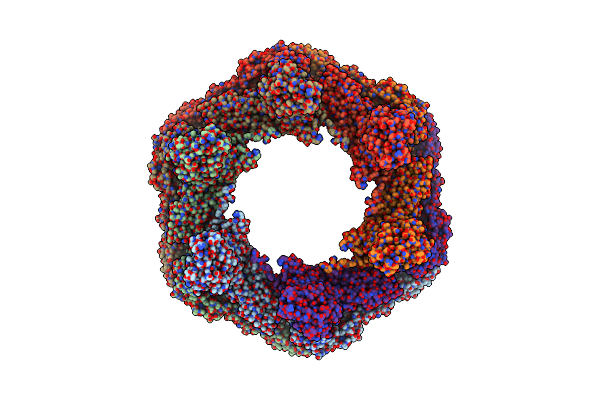 |
In Situ Structure Of The Nitrosopumilus Maritimus S-Layer - Six-Fold Symmetry (C6)
Organism: Nitrosopumilus maritimus scm1
Method: ELECTRON MICROSCOPY Release Date: 2024-04-17 Classification: STRUCTURAL PROTEIN |
Organism: Nitrosopumilus maritimus scm1
Method: ELECTRON MICROSCOPY
Release Date: 2024-04-17
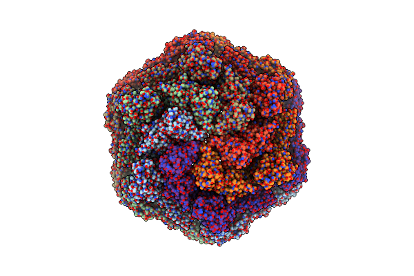 |
In Situ Structure Of The Nitrosopumilus Maritimus S-Layer - Composite Map Between C2 And C6
Organism: Nitrosopumilus maritimus scm1
Method: ELECTRON MICROSCOPY Release Date: 2024-04-17 Classification: STRUCTURAL PROTEIN |
Organism: Nitrosopumilus maritimus scm1
Method: ELECTRON MICROSCOPY
Release Date: 2024-04-17
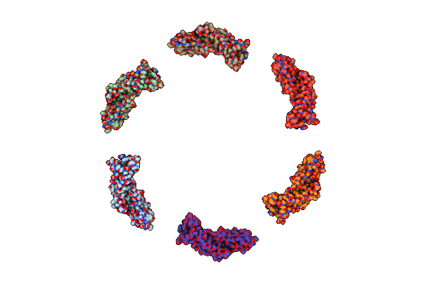 |
In Vitro Structure Of The Nitrosopumilus Maritimus S-Layer - Six-Fold Symmetry (C6)
Organism: Nitrosopumilus maritimus scm1
Method: ELECTRON MICROSCOPY Resolution:2.87 Å Release Date: 2024-04-10 Classification: STRUCTURAL PROTEIN |
Organism: Nitrosopumilus maritimus scm1
Method: ELECTRON MICROSCOPY
Release Date: 2024-04-10
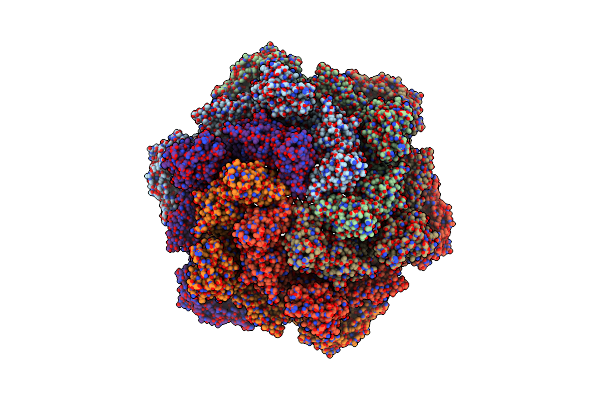 |
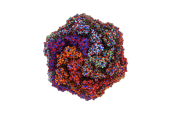
In Vitro Structure Of The Nitrosopumilus Maritimus S-Layer - Two-Fold Symmetry (C2)
Organism: Nitrosopumilus maritimus scm1
Method: ELECTRON MICROSCOPY Resolution:2.71 Å Release Date: 2024-04-10 Classification: STRUCTURAL PROTEIN |
Organism: Nitrosopumilus maritimus scm1
Method: ELECTRON MICROSCOPY
Release Date: 2024-04-10
 |
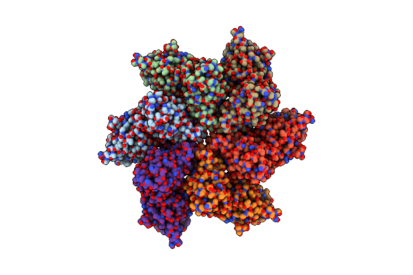
In Vitro Structure Of The Nitrosopumilus Maritimus S-Layer - Composite Map Between Two And Six-Fold Symmetrised
Organism: Nitrosopumilus maritimus scm1
Method: ELECTRON MICROSCOPY Resolution:2.87 Å Release Date: 2024-04-10 Classification: STRUCTURAL PROTEIN |
Organism: Nitrosopumilus maritimus scm1
Method: ELECTRON MICROSCOPY
Release Date: 2024-04-10
 |
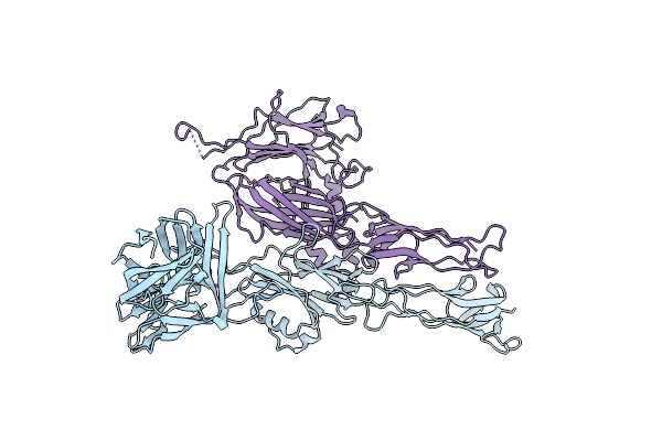
In Situ Structure Of The Nitrosopumilus Maritimus S-Layer - Two-Fold Symmetry (C2)
Organism: Nitrosopumilus maritimus scm1
Method: ELECTRON MICROSCOPY Release Date: 2024-04-10 Classification: STRUCTURAL PROTEIN |
Organism: Nitrosopumilus maritimus scm1
Method: ELECTRON MICROSCOPY
Release Date: 2024-04-10
 |
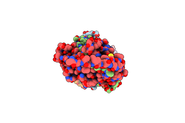
Deinococcus Radidurans Hpi S-Layer
Organism: Deinococcus radiodurans (strain atcc 13939 / dsm 20539 / jcm 16871 / lmg 4051 / nbrc 15346 / ncimb 9279 / r1 / vkm b-1422)
Method: ELECTRON MICROSCOPY Resolution:2.52 Å Release Date: 2023-04-19 Classification: STRUCTURAL PROTEIN |
 |
Cryo-Em Structure Of The Cupe Pilus From Pseudomonas Aeruginosa
Organism: Pseudomonas aeruginosa pao1
Method: ELECTRON MICROSCOPY Release Date: 2023-03-22 Classification: PROTEIN FIBRIL |
Organism: Pseudomonas aeruginosa pao1
Method: ELECTRON MICROSCOPY
Release Date: 2023-03-22
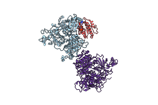 |
Copii Inner Coat
Organism: Saccharomyces cerevisiae
Method: ELECTRON MICROSCOPY Release Date: 2023-01-18 Classification: PROTEIN TRANSPORT Ligands: MG, GNP |
Organism: Saccharomyces cerevisiae
Method: ELECTRON MICROSCOPY
Release Date: 2023-01-18
Ligands: MG, GNP
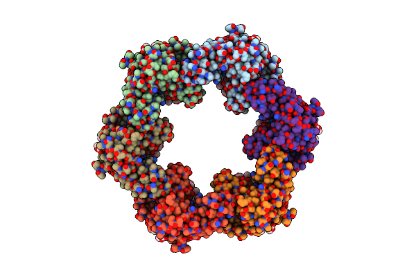 |
In Situ Structure Of The Caulobacter Crescentus S-Layer
Organism: Caulobacter vibrioides na1000
Method: ELECTRON MICROSCOPY Release Date: 2022-12-28 Classification: STRUCTURAL PROTEIN Ligands: CA |
Organism: Caulobacter vibrioides na1000
Method: ELECTRON MICROSCOPY
Release Date: 2022-12-28
Ligands: CA
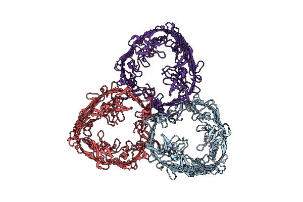 |
Structure Of Trimeric Slpa Outer Membrane Protein
Organism: Deinococcus radiodurans
Method: ELECTRON MICROSCOPY Release Date: 2022-11-30 Classification: STRUCTURAL PROTEIN Ligands: CA |
Organism: Deinococcus radiodurans
Method: ELECTRON MICROSCOPY
Release Date: 2022-11-30
Ligands: CA
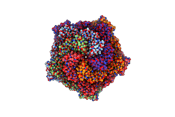 |
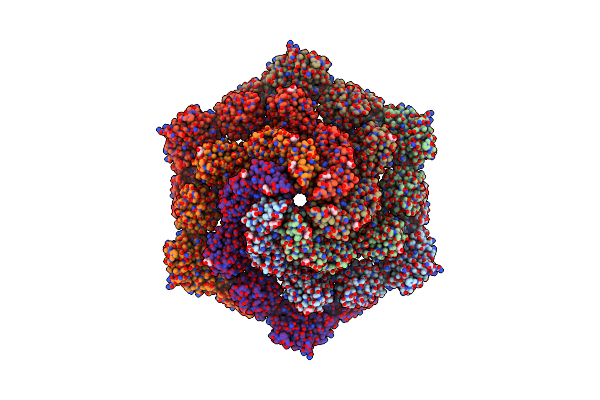
In-Situ Structure Of Pentameric S-Layer Protein
|
 |
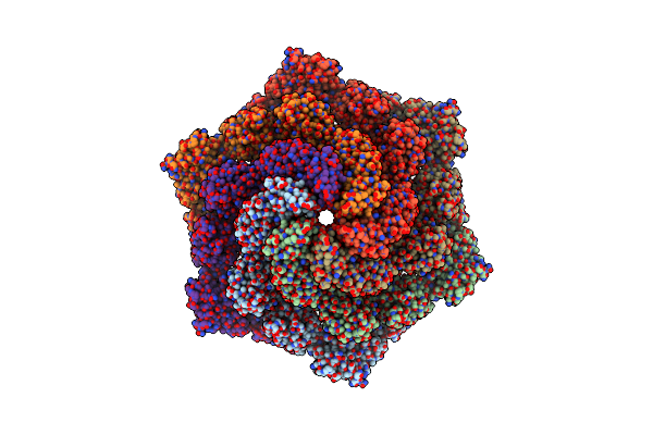
Structure Of Hexameric S-Layer Protein From Haloferax Volcanii Archaea
Organism: Haloferax volcanii ds2
Method: ELECTRON MICROSCOPY Release Date: 2021-12-15 Classification: STRUCTURAL PROTEIN Ligands: CA, BGC |
Organism: Haloferax volcanii ds2
Method: ELECTRON MICROSCOPY
Release Date: 2021-12-15
Ligands: CA, BGC
 |
In-Situ Structure Of Hexameric S-Layer Protein
Organism: Haloferax volcanii ds2
Method: ELECTRON MICROSCOPY Release Date: 2021-12-15 Classification: STRUCTURAL PROTEIN |
Organism: Haloferax volcanii ds2
Method: ELECTRON MICROSCOPY
Release Date: 2021-12-15
 |
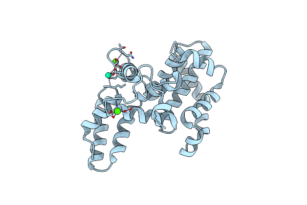
Structure Of Pentameric S-Layer Protein From Halofaerax Volcanii
Organism: Haloferax volcanii ds2
Method: ELECTRON MICROSCOPY Release Date: 2021-12-15 Classification: STRUCTURAL PROTEIN Ligands: BGC |
Organism: Haloferax volcanii ds2
Method: ELECTRON MICROSCOPY
Release Date: 2021-12-15
Ligands: BGC
 |
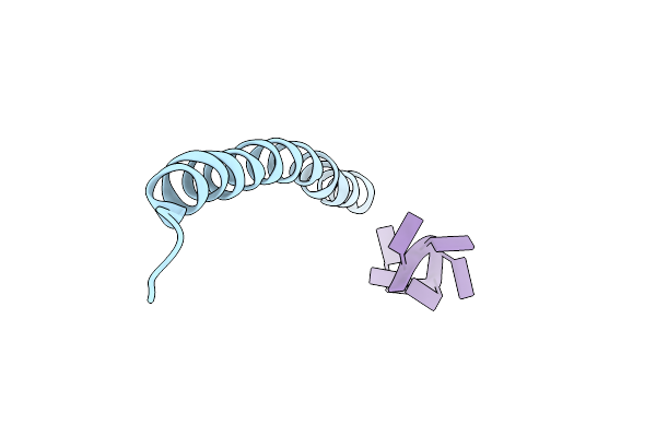
Structure Of The Caulobacter Crescentus S-Layer Protein Rsaa N-Terminal Domain Bound To Lps And Soaked With Holmium
Organism: Caulobacter vibrioides
Method: ELECTRON MICROSCOPY Resolution:4.37 Å Release Date: 2021-12-01 Classification: STRUCTURAL PROTEIN Ligands: CA, HO |
Organism: Caulobacter vibrioides
Method: ELECTRON MICROSCOPY
Release Date: 2021-12-01
Ligands: CA, HO
 |
Cryo-Em Structure Of Pf4 Bacteriophage Coat Protein With Single-Stranded Dna
Organism: Pseudomonas virus pf1, Pseudomonas aeruginosa pao1
Method: ELECTRON MICROSCOPY Release Date: 2020-02-26 Classification: VIRUS |
Organism: Pseudomonas virus pf1, Pseudomonas aeruginosa pao1
Method: ELECTRON MICROSCOPY
Release Date: 2020-02-26
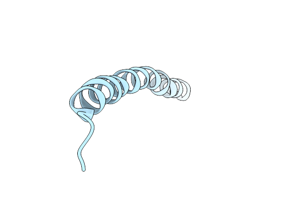 |
Cryo-Em Structure Of Pf4 Bacteriophage Coat Protein Without Ssdna
Organism: Pseudomonas virus pf1
Method: ELECTRON MICROSCOPY Release Date: 2020-02-26 Classification: VIRUS |
Organism: Pseudomonas virus pf1
Method: ELECTRON MICROSCOPY
Release Date: 2020-02-26