

Search Count: 40
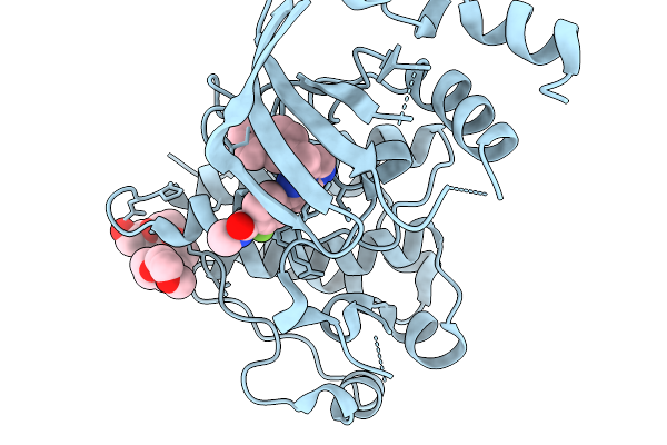 |
Crystal Structure Of Wild-Type C-Met Bound By Capmatinib.
Organism: Homo sapiens
Method: X-RAY DIFFRACTION Resolution:1.32 Å Release Date: 2026-02-18 Classification: TRANSFERASE Ligands: A1JRF, 12P |
Organism: Homo sapiens
Method: X-RAY DIFFRACTION
Release Date: 2026-02-18
Ligands: A1JRF, 12P
 |
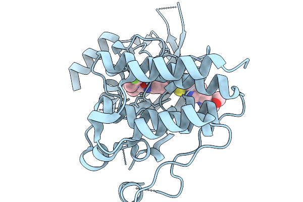
Crystal Structure Of Y1230H C-Met Bound By Capmatinib.
Organism: Homo sapiens
Method: X-RAY DIFFRACTION Resolution:2.29 Å Release Date: 2026-02-18 Classification: TRANSFERASE Ligands: A1JRF |
Organism: Homo sapiens
Method: X-RAY DIFFRACTION
Release Date: 2026-02-18
Ligands: A1JRF
 |
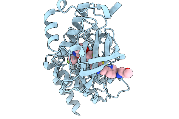
Crystal Structure Of Wild-Type C-Met Bound By Sitravatinib.
Organism: Homo sapiens
Method: X-RAY DIFFRACTION Resolution:1.46 Å Release Date: 2026-02-18 Classification: TRANSFERASE Ligands: A1JSO |
Organism: Homo sapiens
Method: X-RAY DIFFRACTION
Release Date: 2026-02-18
Ligands: A1JSO
 |
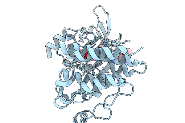
Crystal Structure Of D1228V C-Met Bound By Sitravatinib.
Organism: Homo sapiens
Method: X-RAY DIFFRACTION Resolution:1.54 Å Release Date: 2026-02-18 Classification: TRANSFERASE Ligands: A1JSO |
Organism: Homo sapiens
Method: X-RAY DIFFRACTION
Release Date: 2026-02-18
Ligands: A1JSO
 |
Crystal Structure Of Wild-Type C-Met Bound By Glesatinib
Organism: Homo sapiens
Method: X-RAY DIFFRACTION Resolution:1.20 Å Release Date: 2026-02-18 Classification: TRANSFERASE Ligands: A1JSR |
Organism: Homo sapiens
Method: X-RAY DIFFRACTION
Release Date: 2026-02-18
Ligands: A1JSR
 |
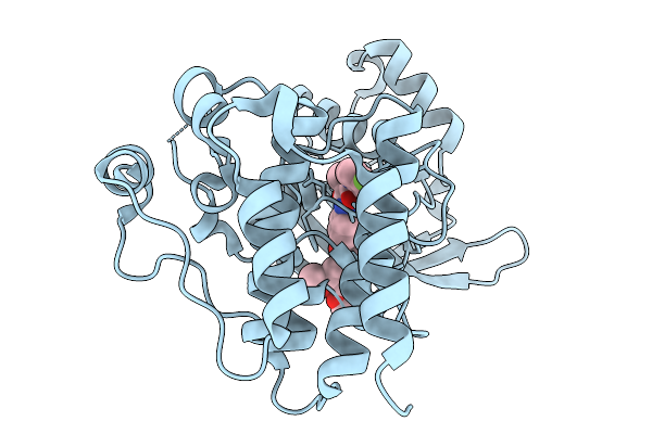
Crystal Structure Of D1228V C-Met Bound By Glesatinib.
Organism: Homo sapiens
Method: X-RAY DIFFRACTION Resolution:1.94 Å Release Date: 2026-02-18 Classification: TRANSFERASE Ligands: A1JSR |
Organism: Homo sapiens
Method: X-RAY DIFFRACTION
Release Date: 2026-02-18
Ligands: A1JSR
 |
Crystal Structure Of Wild-Type C-Met Bound By Cabozantinib.
Organism: Homo sapiens
Method: X-RAY DIFFRACTION Resolution:1.67 Å Release Date: 2026-02-18 Classification: TRANSFERASE Ligands: A1JS8 |
Organism: Homo sapiens
Method: X-RAY DIFFRACTION
Release Date: 2026-02-18
Ligands: A1JS8
 |
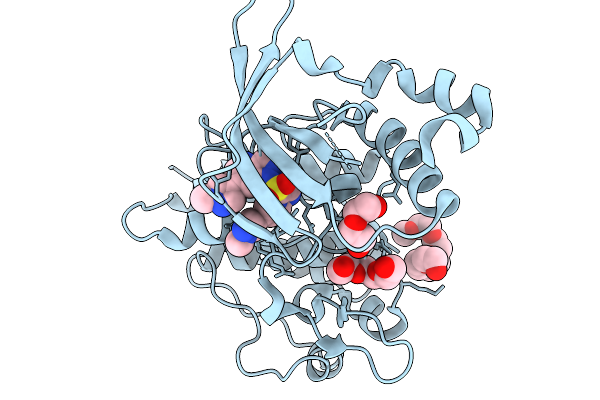
Crystal Structure Of D1228V C-Met Bound By Cabozantinib.
Organism: Homo sapiens
Method: X-RAY DIFFRACTION Resolution:1.63 Å Release Date: 2026-02-18 Classification: TRANSFERASE Ligands: A1JS8 |
Organism: Homo sapiens
Method: X-RAY DIFFRACTION
Release Date: 2026-02-18
Ligands: A1JS8
 |
Crystal Structure Of Wild-Type C-Met Bound By Glumetinib.
Organism: Homo sapiens
Method: X-RAY DIFFRACTION Resolution:1.13 Å Release Date: 2026-02-18 Classification: TRANSFERASE Ligands: A1JT6, 15P, PEG |
Organism: Homo sapiens
Method: X-RAY DIFFRACTION
Release Date: 2026-02-18
Ligands: A1JT6, 15P, PEG
 |
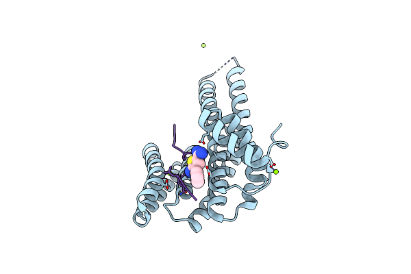
Fragment Az-006 Binding At The P53Pt387/14-3-3 Sigma Interface
Organism: Homo sapiens
Method: X-RAY DIFFRACTION Resolution:1.88 Å Release Date: 2020-06-17 Classification: PEPTIDE BINDING PROTEIN Ligands: MG, CL, GOL, JT2 |
Organism: Homo sapiens
Method: X-RAY DIFFRACTION
Release Date: 2020-06-17
Ligands: MG, CL, GOL, JT2
 |
Fragment Az-003 Binding At The Tazps89/14-3-3 Sigma Interface
Organism: Homo sapiens
Method: X-RAY DIFFRACTION Resolution:1.20 Å Release Date: 2020-06-17 Classification: PEPTIDE BINDING PROTEIN Ligands: MG, CL, K48 |
Organism: Homo sapiens
Method: X-RAY DIFFRACTION
Release Date: 2020-06-17
Ligands: MG, CL, K48
 |
Fragment Az-018 Binding At The Tazps89/14-3-3 Sigma Interface
Organism: Homo sapiens
Method: X-RAY DIFFRACTION Resolution:1.28 Å Release Date: 2020-06-17 Classification: PEPTIDE BINDING PROTEIN Ligands: K5Z |
Organism: Homo sapiens
Method: X-RAY DIFFRACTION
Release Date: 2020-06-17
Ligands: K5Z
 |
Fragment Az-006 Binding At The Tazps89/14-3-3 Sigma Interface
Organism: Homo sapiens
Method: X-RAY DIFFRACTION Resolution:1.89 Å Release Date: 2020-06-17 Classification: PEPTIDE BINDING PROTEIN Ligands: JT2 |
Organism: Homo sapiens
Method: X-RAY DIFFRACTION
Release Date: 2020-06-17
Ligands: JT2
 |
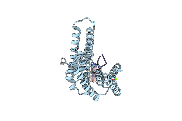
Fragment Az-015 Binding At The P53Pt387/14-3-3 Sigma Interface
Organism: Homo sapiens
Method: X-RAY DIFFRACTION Resolution:1.58 Å Release Date: 2020-06-17 Classification: PEPTIDE BINDING PROTEIN Ligands: MG, CL, GOL, K6B |
Organism: Homo sapiens
Method: X-RAY DIFFRACTION
Release Date: 2020-06-17
Ligands: MG, CL, GOL, K6B
 |
Fragment Az-014 Binding At The P53Pt387/14-3-3 Sigma Interface
Organism: Homo sapiens
Method: X-RAY DIFFRACTION Resolution:1.60 Å Release Date: 2020-06-17 Classification: PEPTIDE BINDING PROTEIN Ligands: MG, CL, K6N |
Organism: Homo sapiens
Method: X-RAY DIFFRACTION
Release Date: 2020-06-17
Ligands: MG, CL, K6N
 |
Fragment Az-023 Binding At The P53Pt387/14-3-3 Sigma Interface
Organism: Homo sapiens
Method: X-RAY DIFFRACTION Resolution:1.88 Å Release Date: 2020-06-17 Classification: PEPTIDE BINDING PROTEIN Ligands: MG, CL, K6T |
Organism: Homo sapiens
Method: X-RAY DIFFRACTION
Release Date: 2020-06-17
Ligands: MG, CL, K6T
 |
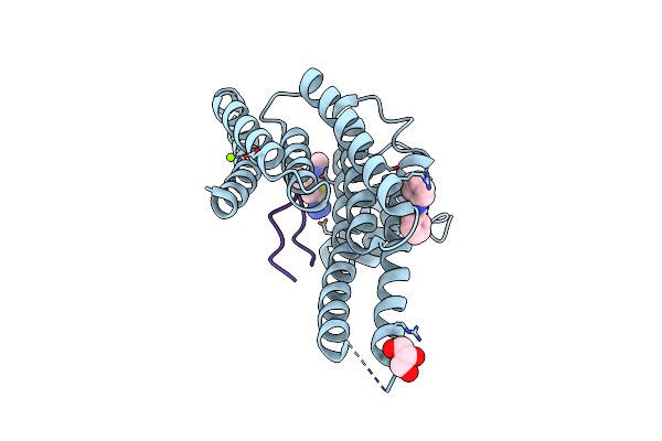
Fragment Az-021 Binding At The P53Pt387/14-3-3 Sigma Interface
Organism: Homo sapiens
Method: X-RAY DIFFRACTION Resolution:1.88 Å Release Date: 2020-06-17 Classification: PEPTIDE BINDING PROTEIN Ligands: MG, CL, GOL, K6W |
Organism: Homo sapiens
Method: X-RAY DIFFRACTION
Release Date: 2020-06-17
Ligands: MG, CL, GOL, K6W
 |
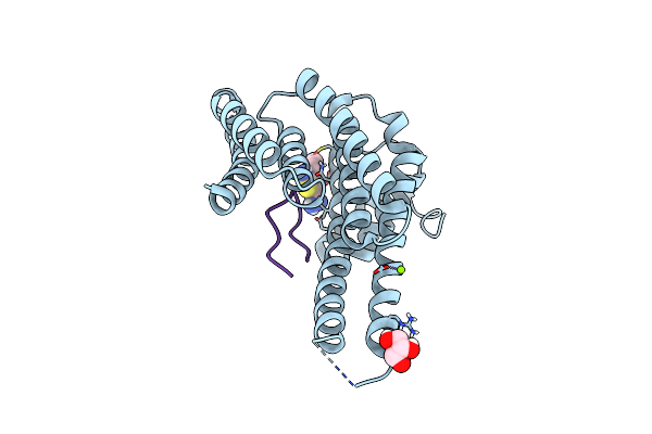
Fragment Az-022 Binding At The P53Pt387/14-3-3 Sigma Interface
Organism: Homo sapiens
Method: X-RAY DIFFRACTION Resolution:1.88 Å Release Date: 2020-06-17 Classification: PEPTIDE BINDING PROTEIN Ligands: MG, CL, GOL, K6Z |
Organism: Homo sapiens
Method: X-RAY DIFFRACTION
Release Date: 2020-06-17
Ligands: MG, CL, GOL, K6Z
 |
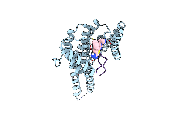
Fragment Az-003 Binding At The P53Pt387/14-3-3 Sigma Interface
Organism: Homo sapiens
Method: X-RAY DIFFRACTION Resolution:1.30 Å Release Date: 2020-06-17 Classification: PEPTIDE BINDING PROTEIN Ligands: MG, CL, GOL, K48 |
Organism: Homo sapiens
Method: X-RAY DIFFRACTION
Release Date: 2020-06-17
Ligands: MG, CL, GOL, K48
 |
Fragment Az-025 Binding At The P53Pt387/14-3-3 Sigma Interface
Organism: Homo sapiens
Method: X-RAY DIFFRACTION Resolution:1.60 Å Release Date: 2020-06-17 Classification: PEPTIDE BINDING PROTEIN Ligands: MG, CL, K7N |
Organism: Homo sapiens
Method: X-RAY DIFFRACTION
Release Date: 2020-06-17
Ligands: MG, CL, K7N