

Search Count: 60
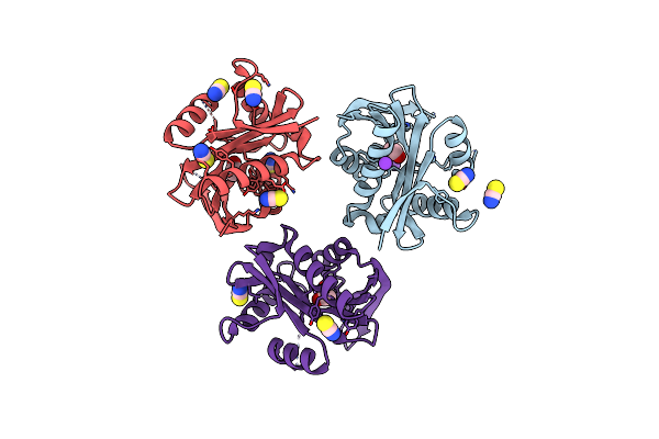 |
Crystal Structure Of A Protein Acetyltransferase, Hp0935
Organism: Helicobacter pylori 26695
Method: X-RAY DIFFRACTION Resolution:2.00 Å Release Date: 2024-04-10 Classification: TRANSFERASE Ligands: K, EDO, SCN |
Organism: Helicobacter pylori 26695
Method: X-RAY DIFFRACTION
Release Date: 2024-04-10
Ligands: K, EDO, SCN
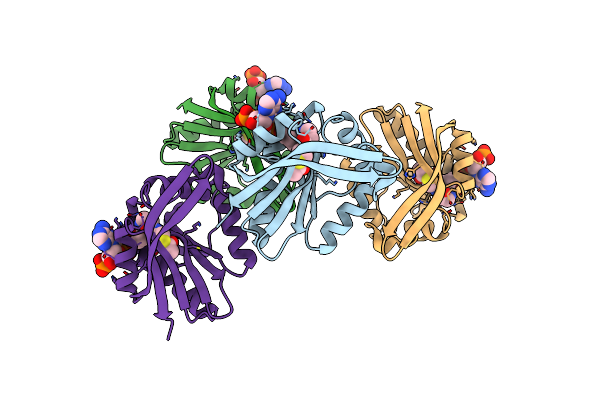 |
Crystal Structure Of A Protein Acetyltransferase, Hp0935, Acetyl-Coa Bound Form
Organism: Helicobacter pylori 26695
Method: X-RAY DIFFRACTION Resolution:2.40 Å Release Date: 2024-04-10 Classification: TRANSFERASE Ligands: ACO |
Organism: Helicobacter pylori 26695
Method: X-RAY DIFFRACTION
Release Date: 2024-04-10
Ligands: ACO
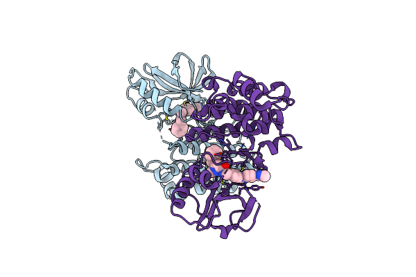 |
The Ripk1 Kinase Domain In Complex With Qy7-2B Compound
Organism: Homo sapiens
Method: X-RAY DIFFRACTION Resolution:2.29 Å Release Date: 2024-01-31 Classification: TRANSFERASE Ligands: O4U |
Organism: Homo sapiens
Method: X-RAY DIFFRACTION
Release Date: 2024-01-31
Ligands: O4U
 |
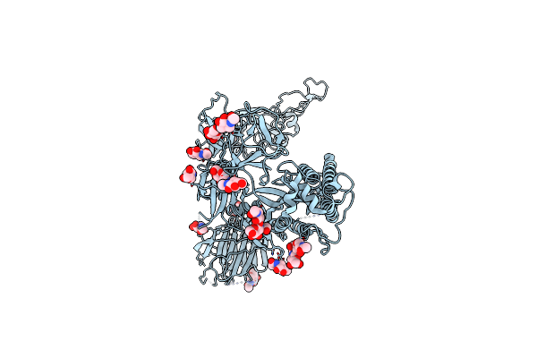
Structure Of The Sars-Cov-2 Xbb.1.5 Spike Glycoprotein (Closed State 1)
Organism: Severe acute respiratory syndrome coronavirus 2
Method: ELECTRON MICROSCOPY Release Date: 2024-01-03 Classification: VIRAL PROTEIN Ligands: NAG |
Organism: Severe acute respiratory syndrome coronavirus 2
Method: ELECTRON MICROSCOPY
Release Date: 2024-01-03
Ligands: NAG
 |
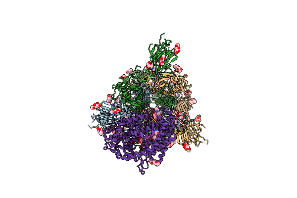
Structure Of The Sars-Cov-2 Xbb.1.5 Spike Glycoprotein (Closed State 2)
Organism: Severe acute respiratory syndrome coronavirus 2
Method: ELECTRON MICROSCOPY Release Date: 2024-01-03 Classification: VIRAL PROTEIN Ligands: NAG |
Organism: Severe acute respiratory syndrome coronavirus 2
Method: ELECTRON MICROSCOPY
Release Date: 2024-01-03
Ligands: NAG
 |
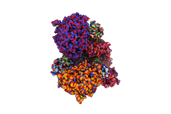
Structure Of Sars-Cov-2 Xbb.1.5 Spike Glycoprotein In Complex With Ace2 (1-Up State)
Organism: Severe acute respiratory syndrome coronavirus 2, Homo sapiens
Method: ELECTRON MICROSCOPY Release Date: 2024-01-03 Classification: VIRAL PROTEIN/PROTEIN BINDING Ligands: NAG |
Organism: Severe acute respiratory syndrome coronavirus 2, Homo sapiens
Method: ELECTRON MICROSCOPY
Release Date: 2024-01-03
Ligands: NAG
 |
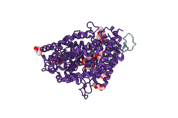
Structure Of Sars-Cov-2 Xbb.1.5 Spike Glycoprotein In Complex With Ace2 (2-Up State)
Organism: Severe acute respiratory syndrome coronavirus 2, Homo sapiens
Method: ELECTRON MICROSCOPY Release Date: 2024-01-03 Classification: VIRAL PROTEIN/PROTEIN BINDING Ligands: NAG |
Organism: Severe acute respiratory syndrome coronavirus 2, Homo sapiens
Method: ELECTRON MICROSCOPY
Release Date: 2024-01-03
Ligands: NAG
 |
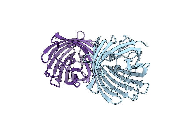
Structure Of Sars-Cov-2 Xbb.1.5 Spike Rbd In Complex With Ace2
Organism: Severe acute respiratory syndrome coronavirus 2, Homo sapiens
Method: ELECTRON MICROSCOPY Release Date: 2024-01-03 Classification: VIRAL PROTEIN/PROTEIN BINDING Ligands: NAG |
Organism: Severe acute respiratory syndrome coronavirus 2, Homo sapiens
Method: ELECTRON MICROSCOPY
Release Date: 2024-01-03
Ligands: NAG
 |
Crystal Structure Of Fluorescent Protein Rasm
Organism: Discosoma sp.
Method: X-RAY DIFFRACTION Resolution:1.01 Å Release Date: 2023-09-27 Classification: FLUORESCENT PROTEIN |
Organism: Discosoma sp.
Method: X-RAY DIFFRACTION
Release Date: 2023-09-27
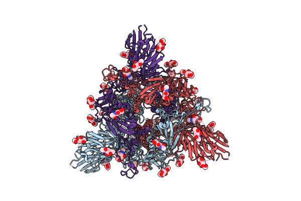 |
Structure Of The Sars-Cov-2 Xbb.1 Spike Glycoprotein (Closed-1 State)
Organism: Severe acute respiratory syndrome coronavirus 2
Method: ELECTRON MICROSCOPY Release Date: 2023-05-24 Classification: VIRAL PROTEIN Ligands: NAG |
Organism: Severe acute respiratory syndrome coronavirus 2
Method: ELECTRON MICROSCOPY
Release Date: 2023-05-24
Ligands: NAG
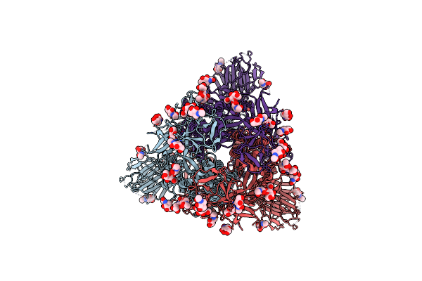 |
Structure Of The Sars-Cov-2 Xbb.1 Spike Glycoprotein (Closed-2 State)
Organism: Severe acute respiratory syndrome coronavirus 2
Method: ELECTRON MICROSCOPY Release Date: 2023-05-24 Classification: VIRAL PROTEIN Ligands: NAG |
Organism: Severe acute respiratory syndrome coronavirus 2
Method: ELECTRON MICROSCOPY
Release Date: 2023-05-24
Ligands: NAG
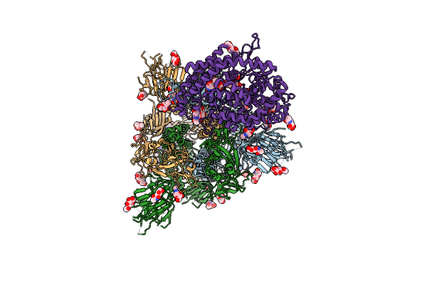 |
Structure Of Sars-Cov-2 Xbb.1 Spike Glycoprotein In Complex With Ace2 (1-Up State)
Organism: Severe acute respiratory syndrome coronavirus 2, Homo sapiens
Method: ELECTRON MICROSCOPY Release Date: 2023-05-24 Classification: VIRAL PROTEIN/PROTEIN BINDING Ligands: NAG |
Organism: Severe acute respiratory syndrome coronavirus 2, Homo sapiens
Method: ELECTRON MICROSCOPY
Release Date: 2023-05-24
Ligands: NAG
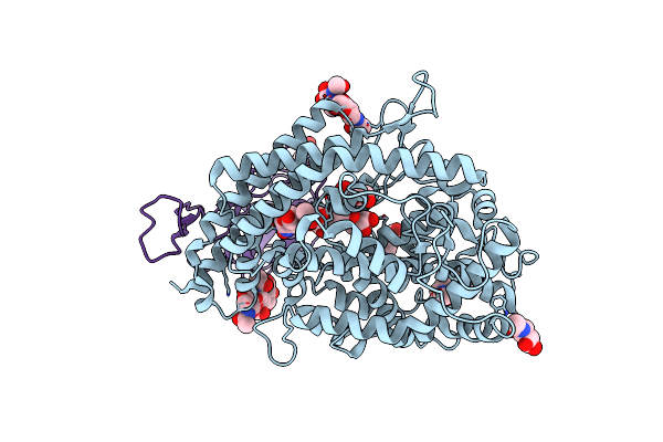 |
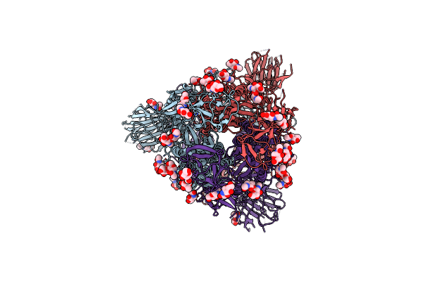
Structure Of Sars-Cov-2 Xbb.1 Spike Rbd In Complex With Ace2
Organism: Homo sapiens, Severe acute respiratory syndrome coronavirus 2
Method: ELECTRON MICROSCOPY Release Date: 2023-05-24 Classification: VIRAL PROTEIN/PROTEIN BINDING Ligands: NAG |
Organism: Homo sapiens, Severe acute respiratory syndrome coronavirus 2
Method: ELECTRON MICROSCOPY
Release Date: 2023-05-24
Ligands: NAG
 |
Structure Of The Sars-Cov-2 Ba.2.75 Spike Glycoprotein (Closed State 1)
Organism: Severe acute respiratory syndrome coronavirus 2
Method: ELECTRON MICROSCOPY Release Date: 2022-10-26 Classification: VIRAL PROTEIN Ligands: NAG |
Organism: Severe acute respiratory syndrome coronavirus 2
Method: ELECTRON MICROSCOPY
Release Date: 2022-10-26
Ligands: NAG
 |
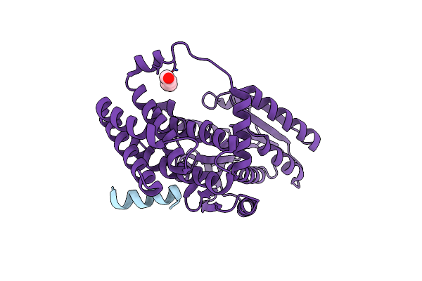
Crystal Structure Of The Receptor Binding Domain Of Sars-Cov-2 Omicron Ba.4/5 Variant Spike Protein In Complex With Its Receptor Ace2
Organism: Homo sapiens, Severe acute respiratory syndrome coronavirus 2
Method: X-RAY DIFFRACTION Resolution:3.36 Å Release Date: 2022-09-28 Classification: VIRAL PROTEIN Ligands: ZN, NAG |
Organism: Homo sapiens, Severe acute respiratory syndrome coronavirus 2
Method: X-RAY DIFFRACTION
Release Date: 2022-09-28
Ligands: ZN, NAG
 |
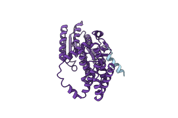
The Crystal Structure Of The Jackdaw/Idd10 Bound To The Homodimeric Scl3
Organism: Arabidopsis thaliana
Method: X-RAY DIFFRACTION Resolution:2.40 Å Release Date: 2020-09-02 Classification: TRANSCRIPTION Ligands: PEG |
Organism: Arabidopsis thaliana
Method: X-RAY DIFFRACTION
Release Date: 2020-09-02
Ligands: PEG
 |
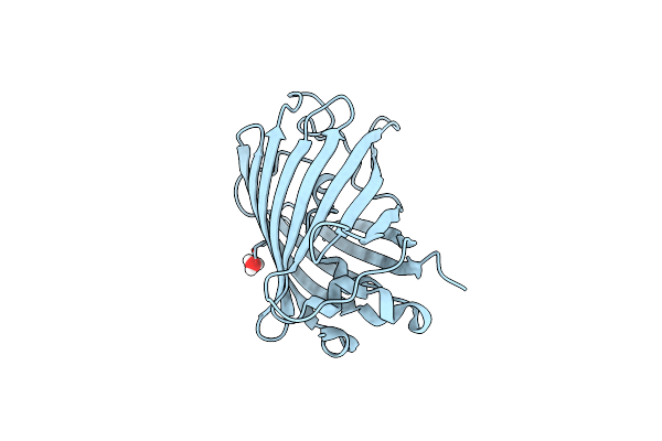
The Crystal Structure Of The Baldibis/Idd9 Bound To The Homodimeric Scl3
Organism: Arabidopsis thaliana
Method: X-RAY DIFFRACTION Resolution:3.20 Å Release Date: 2020-09-02 Classification: TRANSCRIPTION |
Organism: Arabidopsis thaliana
Method: X-RAY DIFFRACTION
Release Date: 2020-09-02
 |
Neutron Crystal Structure Of The Mutant Green Fluorescent Protein (Egfp)
Organism: Aequorea victoria
Method: NEUTRON DIFFRACTION Resolution:1.44 Å Release Date: 2020-04-08 Classification: FLUORESCENT PROTEIN Ligands: D3O, DOD |
Organism: Aequorea victoria
Method: NEUTRON DIFFRACTION
Release Date: 2020-04-08
Ligands: D3O, DOD
 |
X-Ray Crystal Structure Of The Mutant Green Fluorescent Protein
Organism: Aequorea victoria
Method: X-RAY DIFFRACTION Resolution:0.77 Å Release Date: 2020-04-01 Classification: FLUORESCENT PROTEIN Ligands: DOD |
Organism: Aequorea victoria
Method: X-RAY DIFFRACTION
Release Date: 2020-04-01
Ligands: DOD
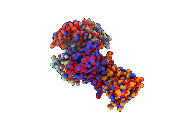 |
Rapid Crystallization Of Streptavidin Using Charged Peptides
Organism: Streptomyces avidinii
Method: X-RAY DIFFRACTION Resolution:1.80 Å Release Date: 2020-03-18 Classification: PEPTIDE BINDING PROTEIN Ligands: GOL |
Organism: Streptomyces avidinii
Method: X-RAY DIFFRACTION
Release Date: 2020-03-18
Ligands: GOL