

Search Count: 42
 |
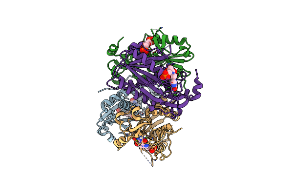
Mitoribosomal Small Subunit In Complex With Mettl15 And Mettl17
Organism: Trypanosoma brucei
Method: ELECTRON MICROSCOPY Release Date: 2025-03-26 Classification: RIBOSOME Ligands: GTP, MG, SF4, SAM, ACO, ZN, PO4 |
Organism: Trypanosoma brucei
Method: ELECTRON MICROSCOPY
Release Date: 2025-03-26
Ligands: GTP, MG, SF4, SAM, ACO, ZN, PO4
 |
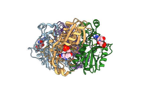
Structure Of Human Hypoxanthine Guanine Phosphoribzosyltransferase In Complex With [2S,4R]-4-Guanin-9-Yl-2-(2-Phosphonoethoxymethyl)-1-N-(3-Phosphonopropionyl)Pyrrolidine
Organism: Homo sapiens
Method: X-RAY DIFFRACTION Resolution:2.27 Å Release Date: 2024-05-08 Classification: TRANSFERASE Ligands: JEI, MG |
Organism: Homo sapiens
Method: X-RAY DIFFRACTION
Release Date: 2024-05-08
Ligands: JEI, MG
 |
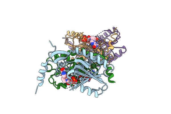
Structure Of Human Hypoxanthine Guanine Phosphoribzosyltransferase In Complex With [2S,4R] 4-Guanin-9-Yl-2-Hydroxymethyl-1-N-(3-Phosphonopropionyl)Pyrrolidine
Organism: Homo sapiens
Method: X-RAY DIFFRACTION Resolution:2.50 Å Release Date: 2024-05-08 Classification: TRANSFERASE Ligands: JG6, MG |
Organism: Homo sapiens
Method: X-RAY DIFFRACTION
Release Date: 2024-05-08
Ligands: JG6, MG
 |
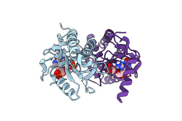
Crystal Structure Of Trypanosome Brucei Hypoxanthine Guanine Phosphopribosyltransferase In Complex With [2S,4R]-4-Guanin-9-Yl-2-(2- Phosphonoethoxymethyl)-1-N-(3-Phosphonopropionyl)Pyrrolidine
Organism: Trypanosoma brucei
Method: X-RAY DIFFRACTION Resolution:2.46 Å Release Date: 2024-05-08 Classification: TRANSFERASE Ligands: JEI |
Organism: Trypanosoma brucei
Method: X-RAY DIFFRACTION
Release Date: 2024-05-08
Ligands: JEI
 |
Crystal Structure Of T. Brucei Hypoxanthine Guanine Phosphoribosyltransferase In Complex With [2S,4S]-4-Guanin-9-Yl-2-(2-Phosphonoethoxymethyl)-1-N-(3-Phosphonopropionyl)Pyrrolidine
Organism: Trypanosoma brucei
Method: X-RAY DIFFRACTION Resolution:2.20 Å Release Date: 2024-05-08 Classification: TRANSFERASE Ligands: MG, KFF |
Organism: Trypanosoma brucei
Method: X-RAY DIFFRACTION
Release Date: 2024-05-08
Ligands: MG, KFF
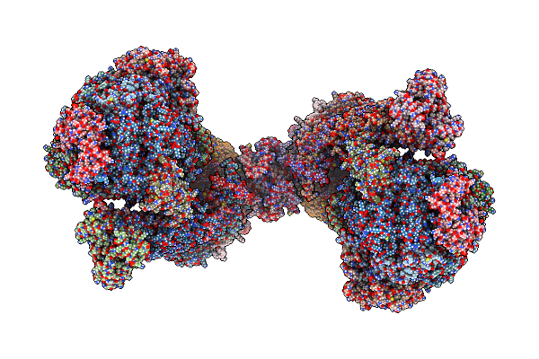 |
Trypanosoma Brucei Mitochondrial F1Fo Atp Synthase Dimer
Organism: Trypanosoma brucei brucei
Method: ELECTRON MICROSCOPY Release Date: 2022-12-07 Classification: MEMBRANE PROTEIN Ligands: ATP, MG, CDL, ADP, LMT, Q7G, PEE, PC1, UTP |
Organism: Trypanosoma brucei brucei
Method: ELECTRON MICROSCOPY
Release Date: 2022-12-07
Ligands: ATP, MG, CDL, ADP, LMT, Q7G, PEE, PC1, UTP
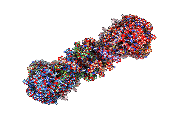 |
Membrane Region Of The Trypanosoma Brucei Mitochondrial Atp Synthase Dimer
Organism: Trypanosoma brucei brucei
Method: ELECTRON MICROSCOPY Release Date: 2022-10-26 Classification: MEMBRANE PROTEIN Ligands: CDL, LMT, Q7G, PEE, PC1 |
Organism: Trypanosoma brucei brucei
Method: ELECTRON MICROSCOPY
Release Date: 2022-10-26
Ligands: CDL, LMT, Q7G, PEE, PC1
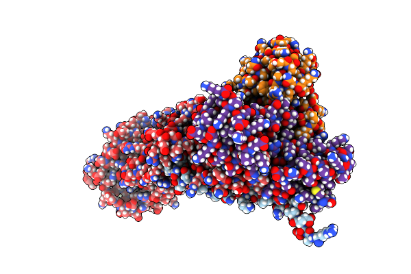 |
Peripheral Stalk Of Trypanosoma Brucei Mitochondrial Atp Synthase
Organism: Trypanosoma brucei brucei
Method: ELECTRON MICROSCOPY Release Date: 2022-10-26 Classification: MEMBRANE PROTEIN |
Organism: Trypanosoma brucei brucei
Method: ELECTRON MICROSCOPY
Release Date: 2022-10-26
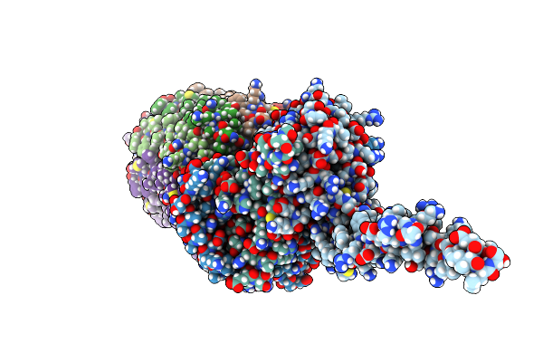 |
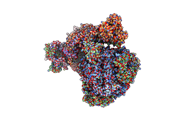
Rotor Of The Trypanosoma Brucei Mitochondrial Atp Synthase Dimer
Organism: Trypanosoma brucei brucei
Method: ELECTRON MICROSCOPY Release Date: 2022-10-26 Classification: MEMBRANE PROTEIN Ligands: UTP |
Organism: Trypanosoma brucei brucei
Method: ELECTRON MICROSCOPY
Release Date: 2022-10-26
Ligands: UTP
 |
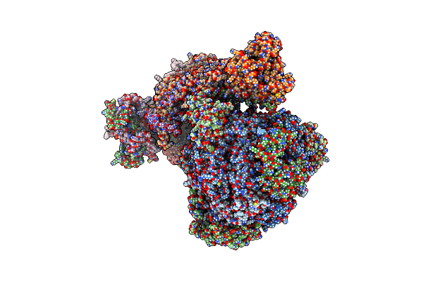
Rotational State 1A Of The Trypanosoma Brucei Mitochondrial Atp Synthase Dimer
Organism: Trypanosoma brucei brucei
Method: ELECTRON MICROSCOPY Release Date: 2022-10-26 Classification: MEMBRANE PROTEIN Ligands: ATP, MG, ADP, UTP, CDL, PEE, LMT, Q7G, PC1 |
Organism: Trypanosoma brucei brucei
Method: ELECTRON MICROSCOPY
Release Date: 2022-10-26
Ligands: ATP, MG, ADP, UTP, CDL, PEE, LMT, Q7G, PC1
 |
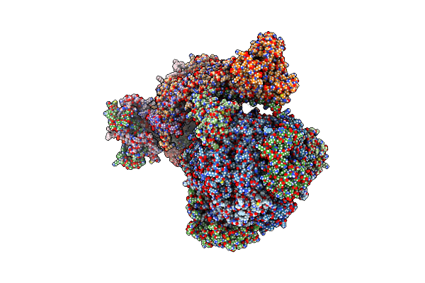
Rotational State 1B Of The Trypanosoma Brucei Mitochondrial Atp Synthase Dimer
Organism: Trypanosoma brucei brucei
Method: ELECTRON MICROSCOPY Release Date: 2022-10-26 Classification: MEMBRANE PROTEIN Ligands: ATP, MG, ADP, UTP, CDL, 3PE, PC1, LMT, Q7G |
Organism: Trypanosoma brucei brucei
Method: ELECTRON MICROSCOPY
Release Date: 2022-10-26
Ligands: ATP, MG, ADP, UTP, CDL, 3PE, PC1, LMT, Q7G
 |
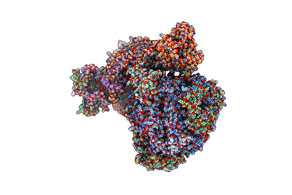
Rotational State 1C Of The Trypanosoma Brucei Mitochondrial Atp Synthase Dimer
Organism: Trypanosoma brucei brucei
Method: ELECTRON MICROSCOPY Release Date: 2022-10-26 Classification: MEMBRANE PROTEIN Ligands: ATP, MG, ADP, UTP, CDL, 3PE, LMT, Q7G, PC1 |
Organism: Trypanosoma brucei brucei
Method: ELECTRON MICROSCOPY
Release Date: 2022-10-26
Ligands: ATP, MG, ADP, UTP, CDL, 3PE, LMT, Q7G, PC1
 |
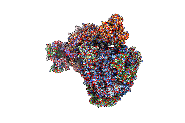
Rotational State 1D Of The Trypanosoma Brucei Mitochondrial Atp Synthase Dimer
Organism: Trypanosoma brucei brucei
Method: ELECTRON MICROSCOPY Release Date: 2022-10-26 Classification: MEMBRANE PROTEIN Ligands: CDL, PEE, PC1, LMT, Q7G, ATP, MG, ADP, UTP |
Organism: Trypanosoma brucei brucei
Method: ELECTRON MICROSCOPY
Release Date: 2022-10-26
Ligands: CDL, PEE, PC1, LMT, Q7G, ATP, MG, ADP, UTP
 |
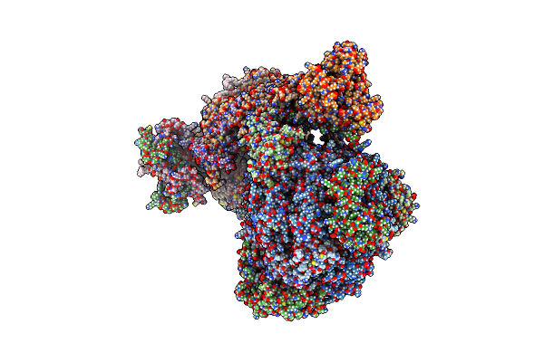
Rotational State 1E Of The Trypanosoma Brucei Mitochondrial Atp Synthase Dimer
Organism: Trypanosoma brucei brucei
Method: ELECTRON MICROSCOPY Release Date: 2022-10-26 Classification: MEMBRANE PROTEIN Ligands: ATP, MG, ADP, UTP, CDL, PEE, PC1, LMT, Q7G |
Organism: Trypanosoma brucei brucei
Method: ELECTRON MICROSCOPY
Release Date: 2022-10-26
Ligands: ATP, MG, ADP, UTP, CDL, PEE, PC1, LMT, Q7G
 |
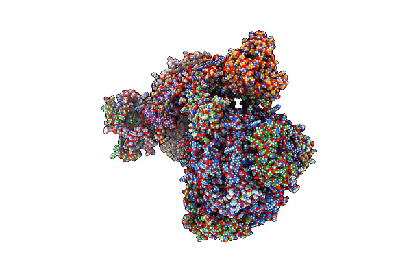
Rotational State 2A Of The Trypanosoma Brucei Mitochondrial Atp Synthase Dimer
Organism: Trypanosoma brucei brucei
Method: ELECTRON MICROSCOPY Release Date: 2022-10-26 Classification: MEMBRANE PROTEIN Ligands: CDL, PEE, LMT, Q7G, PC1, UTP, ATP, MG, ADP |
Organism: Trypanosoma brucei brucei
Method: ELECTRON MICROSCOPY
Release Date: 2022-10-26
Ligands: CDL, PEE, LMT, Q7G, PC1, UTP, ATP, MG, ADP
 |
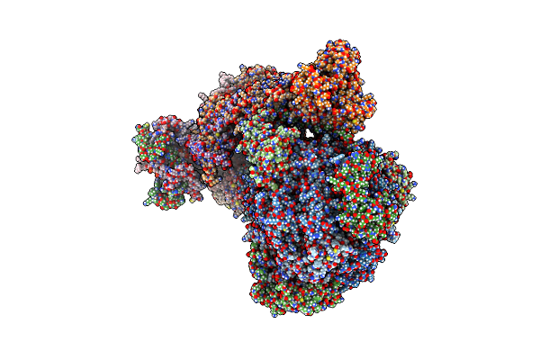
Rotational State 2B Of The Trypanosoma Brucei Mitochondrial Atp Synthase Dimer
Organism: Trypanosoma brucei brucei
Method: ELECTRON MICROSCOPY Release Date: 2022-10-26 Classification: MEMBRANE PROTEIN Ligands: CDL, PEE, LMT, Q7G, PC1, UTP, ATP, MG, ADP |
Organism: Trypanosoma brucei brucei
Method: ELECTRON MICROSCOPY
Release Date: 2022-10-26
Ligands: CDL, PEE, LMT, Q7G, PC1, UTP, ATP, MG, ADP
 |
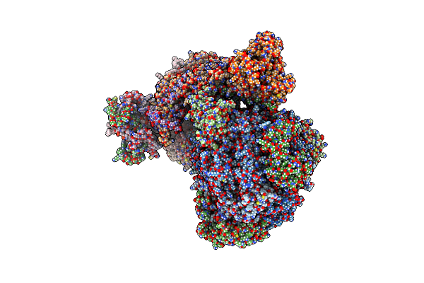
Rotational State 2C Of The Trypanosoma Brucei Mitochondrial Atp Synthase Dimer
Organism: Trypanosoma brucei brucei
Method: ELECTRON MICROSCOPY Release Date: 2022-10-26 Classification: MEMBRANE PROTEIN Ligands: CDL, PEE, LMT, Q7G, PC1, UTP, ATP, MG, ADP |
Organism: Trypanosoma brucei brucei
Method: ELECTRON MICROSCOPY
Release Date: 2022-10-26
Ligands: CDL, PEE, LMT, Q7G, PC1, UTP, ATP, MG, ADP
 |
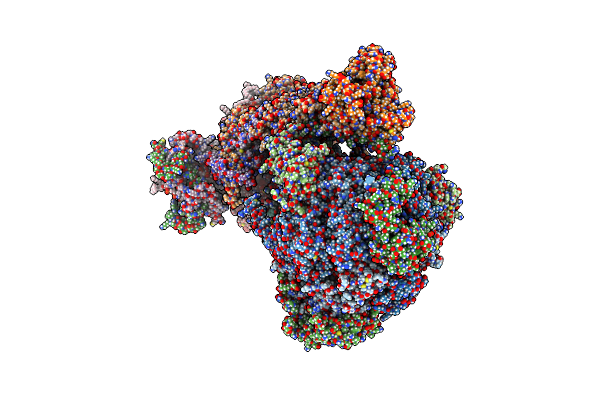
Rotational State 2D Of Trypanosoma Brucei Mitochondrial Atp Synthase
Organism: Trypanosoma brucei brucei
Method: ELECTRON MICROSCOPY Release Date: 2022-10-26 Classification: MEMBRANE PROTEIN Ligands: CDL, PEE, LMT, Q7G, PC1, UTP, ATP, MG, ADP |
Organism: Trypanosoma brucei brucei
Method: ELECTRON MICROSCOPY
Release Date: 2022-10-26
Ligands: CDL, PEE, LMT, Q7G, PC1, UTP, ATP, MG, ADP
 |
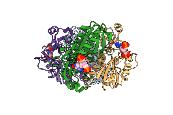
Rotational State 3 Of The Trypanosoma Brucei Mitochondrial Atp Synthase Dimer
Organism: Trypanosoma brucei brucei
Method: ELECTRON MICROSCOPY Release Date: 2022-10-26 Classification: MEMBRANE PROTEIN Ligands: CDL, 3PE, PC1, LMT, Q7G, ATP, MG, ADP, UTP |
Organism: Trypanosoma brucei brucei
Method: ELECTRON MICROSCOPY
Release Date: 2022-10-26
Ligands: CDL, 3PE, PC1, LMT, Q7G, ATP, MG, ADP, UTP
 |
Crystal Structure Of Human Hypoxanthine Guanine Phosphoribzosyltransferase In Complex With (4S,7S)-7-Hydroxy-4-((Guanin-9-Yl)Methyl)-2,5-Dioxaheptan-1,7-Diphosphonate
Organism: Homo sapiens
Method: X-RAY DIFFRACTION Resolution:2.58 Å Release Date: 2022-05-18 Classification: TRANSFERASE/TRANSFERASE INHIBITOR Ligands: 8QI |
Organism: Homo sapiens
Method: X-RAY DIFFRACTION
Release Date: 2022-05-18
Ligands: 8QI