

Search Count: 18
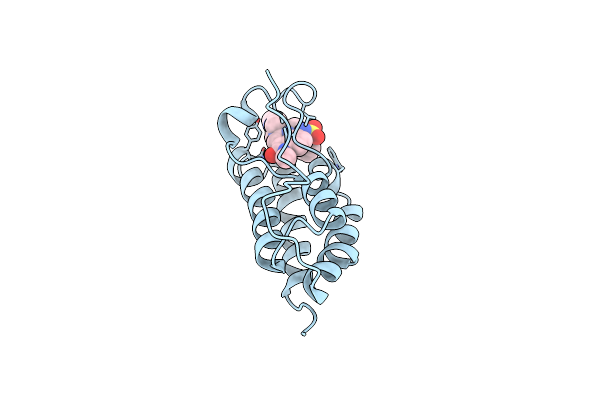 |
Brd4 In Complex With 138A
Organism: Homo sapiens
Method: X-RAY DIFFRACTION Resolution:1.72 Å Release Date: 2020-01-22 Classification: SIGNALING PROTEIN/INHIBITOR Ligands: BS6 |
Organism: Homo sapiens
Method: X-RAY DIFFRACTION
Release Date: 2020-01-22
Ligands: BS6
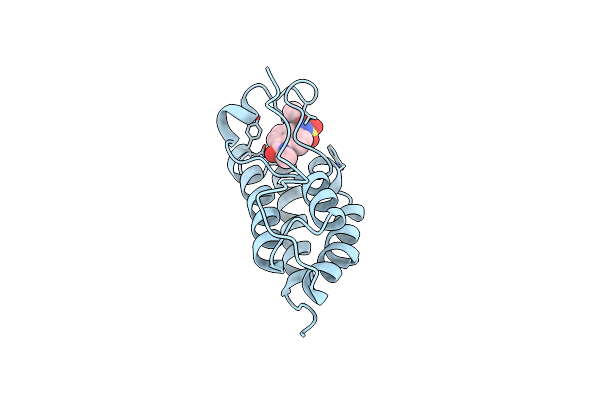 |
Brd4 In Complex With Zzm1
Organism: Homo sapiens
Method: X-RAY DIFFRACTION Resolution:1.51 Å Release Date: 2020-01-22 Classification: SIGNALING PROTEIN/INHIBITOR Ligands: BT0 |
Organism: Homo sapiens
Method: X-RAY DIFFRACTION
Release Date: 2020-01-22
Ligands: BT0
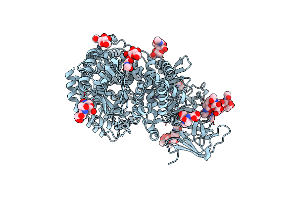 |
Crystal Structure Of Porcine Aminopeptidase N Ectodomain In Functional Form
Organism: Sus scrofa
Method: X-RAY DIFFRACTION Resolution:2.65 Å Release Date: 2019-01-02 Classification: HYDROLASE Ligands: NAG, ZN |
Organism: Sus scrofa
Method: X-RAY DIFFRACTION
Release Date: 2019-01-02
Ligands: NAG, ZN
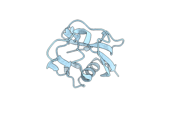 |
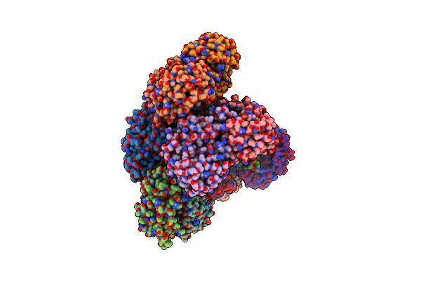
Crystal Structure Of The Scavenger Receptor Cysteine-Rich Domain 5 (Srcr5) From Porcine Cd163
Organism: Sus scrofa
Method: X-RAY DIFFRACTION Resolution:2.00 Å Release Date: 2017-03-22 Classification: ENDOCYTOSIS |
Organism: Sus scrofa
Method: X-RAY DIFFRACTION
Release Date: 2017-03-22
 |
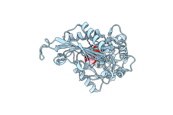
Crystal Structure Of The G136F Mutant Of The First R-Stereoselective -Transaminase Identified From Arthrobacter Sp. Knk168 (Ferm-Bp-5228)
Organism: Arthrobacter sp. knk168
Method: X-RAY DIFFRACTION Resolution:2.27 Å Release Date: 2015-08-19 Classification: TRANSFERASE Ligands: PLP |
Organism: Arthrobacter sp. knk168
Method: X-RAY DIFFRACTION
Release Date: 2015-08-19
Ligands: PLP
 |
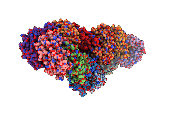
Crystal Structure Of The First R-Stereoselective -Transaminase Identified From Arthrobacter Sp. Knk168 (Ferm-Bp-5228)
Organism: Arthrobacter sp. knk168
Method: X-RAY DIFFRACTION Resolution:1.65 Å Release Date: 2015-08-12 Classification: TRANSFERASE Ligands: PLP, GOL |
Organism: Arthrobacter sp. knk168
Method: X-RAY DIFFRACTION
Release Date: 2015-08-12
Ligands: PLP, GOL
 |
Crystal Structure Of An Engineered Sitagliptin-Producing Transaminase, Ata-117-Rd11
Organism: Arthrobacter sp. knk168
Method: X-RAY DIFFRACTION Resolution:2.20 Å Release Date: 2015-08-12 Classification: TRANSFERASE Ligands: PLP |
Organism: Arthrobacter sp. knk168
Method: X-RAY DIFFRACTION
Release Date: 2015-08-12
Ligands: PLP
 |
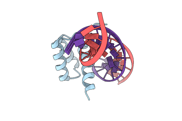
Crystal Structure Of The Dna-Binding Domain Of Adpa, The Global Transcriptional Factor, In Complex With A Target Dna
Organism: Streptomyces griseus
Method: X-RAY DIFFRACTION Resolution:2.95 Å Release Date: 2013-09-18 Classification: TRANSCRIPTION ACTIVATOR/DNA |
Organism: Streptomyces griseus
Method: X-RAY DIFFRACTION
Release Date: 2013-09-18
 |
Structure Of Aristaless Homeodomain In Complex With Dna
Organism: Drosophila melanogaster
Method: X-RAY DIFFRACTION Resolution:2.25 Å Release Date: 2010-04-28 Classification: Gene Regulation/DNA Ligands: ACT |
Organism: Drosophila melanogaster
Method: X-RAY DIFFRACTION
Release Date: 2010-04-28
Ligands: ACT
 |
Crystal Structure Of Aristaless And Clawless Homeodomains Bound To Dna
Organism: Drosophila melanogaster
Method: X-RAY DIFFRACTION Resolution:2.70 Å Release Date: 2010-03-09 Classification: Gene Regulation/DNA |
Organism: Drosophila melanogaster
Method: X-RAY DIFFRACTION
Release Date: 2010-03-09
 |
Crystal Structure Of Aristaless Homeodomain
Organism: Drosophila melanogaster
Method: X-RAY DIFFRACTION Resolution:1.00 Å Release Date: 2010-03-09 Classification: GENE REGULATION Ligands: CD, CL |
Organism: Drosophila melanogaster
Method: X-RAY DIFFRACTION
Release Date: 2010-03-09
Ligands: CD, CL
 |
Crystal Structure Of Hox11L1 Homeodomain
Organism: Homo sapiens
Method: X-RAY DIFFRACTION Resolution:1.54 Å Release Date: 2010-03-09 Classification: GENE REGULATION |
Organism: Homo sapiens
Method: X-RAY DIFFRACTION
Release Date: 2010-03-09
 |
Crystal Structure Of (+)-Aba-Bound Pyl1 In Complex With Abi1
Organism: Arabidopsis thaliana
Method: X-RAY DIFFRACTION Resolution:2.10 Å Release Date: 2009-11-03 Classification: HYDROLASE/Hormone Receptor Ligands: A8S |
Organism: Arabidopsis thaliana
Method: X-RAY DIFFRACTION
Release Date: 2009-11-03
Ligands: A8S
 |
Crystal Structure Of (+)-Aba-Bound Pyl1
Organism: Arabidopsis thaliana
Method: X-RAY DIFFRACTION Resolution:2.05 Å Release Date: 2009-11-03 Classification: Hormone Receptor Ligands: A8S |
Organism: Arabidopsis thaliana
Method: X-RAY DIFFRACTION
Release Date: 2009-11-03
Ligands: A8S
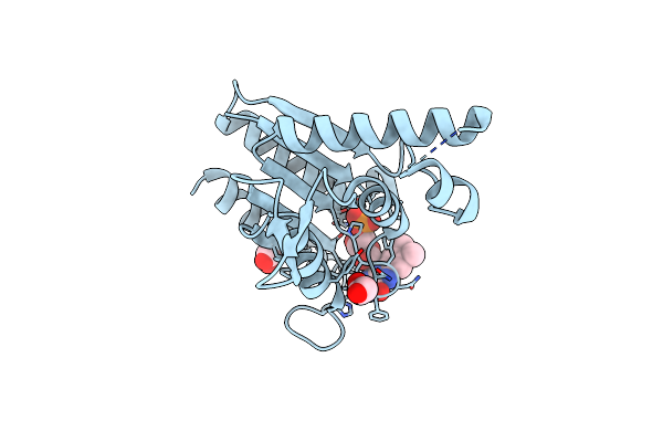 |
The Crystal Structure Of Azor (Azoreductase) From Escherichia Coli: Oxidized Azor In Tetragonal Crystals (The Resolution Has Improved From 1.8 (1V4B) To 1.4 Angstrom)
Organism: Escherichia coli
Method: X-RAY DIFFRACTION Resolution:1.40 Å Release Date: 2008-03-11 Classification: OXIDOREDUCTASE Ligands: FMN, EDO |
Organism: Escherichia coli
Method: X-RAY DIFFRACTION
Release Date: 2008-03-11
Ligands: FMN, EDO
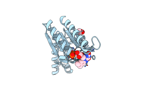 |
The Crystal Structure Of Azor (Azoreductase) From Escherichia Coli: Reduced Azor In Tetragonal Crystals
Organism: Escherichia coli
Method: X-RAY DIFFRACTION Resolution:1.70 Å Release Date: 2008-03-11 Classification: OXIDOREDUCTASE Ligands: FMN, EDO |
Organism: Escherichia coli
Method: X-RAY DIFFRACTION
Release Date: 2008-03-11
Ligands: FMN, EDO
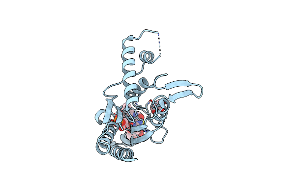 |
The Crystal Structure Of Azor (Azoreductase) From Escherichia Coli: Azor In Complex With Dicoumarol
Organism: Escherichia coli
Method: X-RAY DIFFRACTION Resolution:2.30 Å Release Date: 2008-03-11 Classification: OXIDOREDUCTASE Ligands: FMN, DTC, GOL |
Organism: Escherichia coli
Method: X-RAY DIFFRACTION
Release Date: 2008-03-11
Ligands: FMN, DTC, GOL
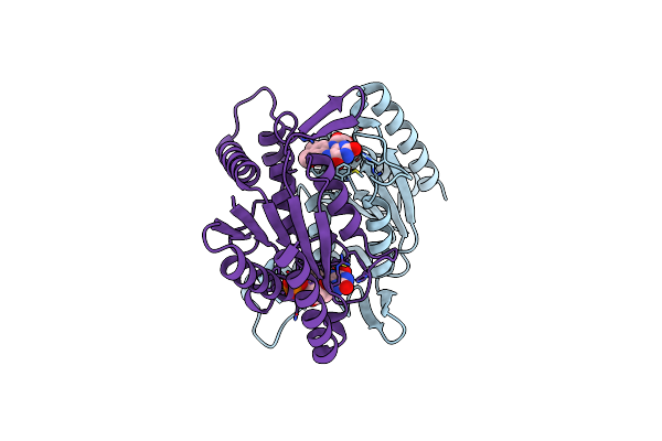 |
The Crystal Structure Of Azor (Azoreductase) From Escherichia Coli: Oxidized Azor In Orthorhombic Crystals
Organism: Escherichia coli
Method: X-RAY DIFFRACTION Resolution:2.10 Å Release Date: 2008-03-11 Classification: OXIDOREDUCTASE Ligands: FMN |
Organism: Escherichia coli
Method: X-RAY DIFFRACTION
Release Date: 2008-03-11
Ligands: FMN