

Search Count: 10
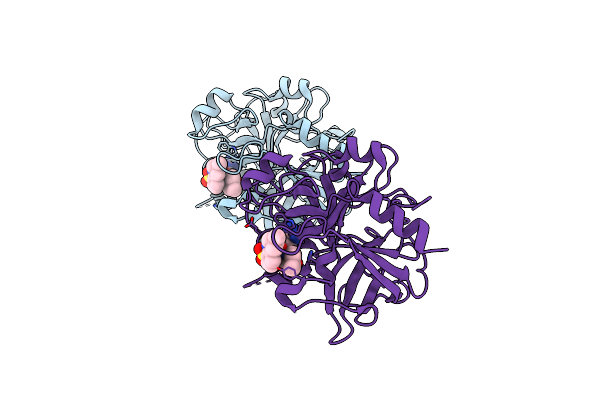 |
Structure Of Prethrombin-2 Mutant S195A Bound To The Active Site Inhibitor Argatroban
Organism: Homo sapiens
Method: X-RAY DIFFRACTION Resolution:3.00 Å Release Date: 2014-11-05 Classification: HYDROLASE/HYDROLASE INHIBITOR Ligands: 15U |
Organism: Homo sapiens
Method: X-RAY DIFFRACTION
Release Date: 2014-11-05
Ligands: 15U
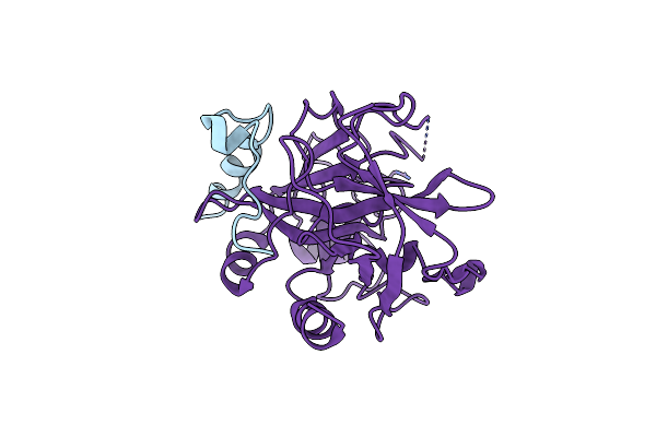 |
Crystal Structure Of Thrombin Mutant E14Ea/D14La/E18A/S195A
Organism: Homo sapiens
Method: X-RAY DIFFRACTION Resolution:2.19 Å Release Date: 2013-03-13 Classification: HYDROLASE/PEPTIDE |
Organism: Homo sapiens
Method: X-RAY DIFFRACTION
Release Date: 2013-03-13
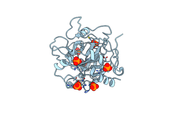 |
Crystal Structure Of Prethrombin-2 Mutant E14Ea/D14La/E18A/S195A
Organism: Homo sapiens
Method: X-RAY DIFFRACTION Resolution:2.40 Å Release Date: 2013-03-13 Classification: HYDROLASE Ligands: PO4 |
Organism: Homo sapiens
Method: X-RAY DIFFRACTION
Release Date: 2013-03-13
Ligands: PO4
 |
Structure Of Thrombin Mutant S195A Bound To The Active Site Inhibitor Argatroban
Organism: Homo sapiens
Method: X-RAY DIFFRACTION Resolution:2.40 Å Release Date: 2013-03-13 Classification: HYDROLASE/HYDROLASE INHIBITOR |
Organism: Homo sapiens
Method: X-RAY DIFFRACTION
Release Date: 2013-03-13
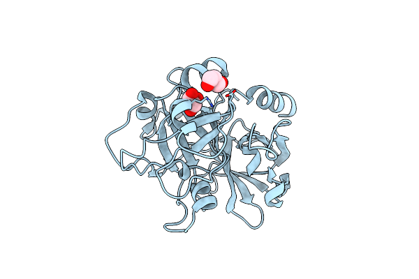 |
Crystal Structure Of Prethrombin-2 Mutant S195A In The Alternative Form
Organism: Homo sapiens
Method: X-RAY DIFFRACTION Resolution:1.90 Å Release Date: 2011-11-30 Classification: HYDROLASE Ligands: GOL |
Organism: Homo sapiens
Method: X-RAY DIFFRACTION
Release Date: 2011-11-30
Ligands: GOL
 |
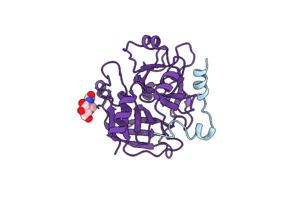
Crystal Structure Of Prethrombin-2 Mutant S195A In The The Open Form
Organism: Homo sapiens
Method: X-RAY DIFFRACTION Resolution:2.20 Å Release Date: 2011-11-30 Classification: HYDROLASE |
Organism: Homo sapiens
Method: X-RAY DIFFRACTION
Release Date: 2011-11-30
 |
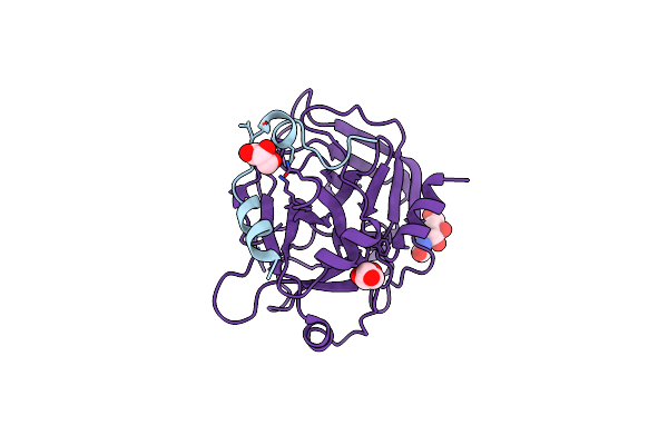
The Allosteric E*-E Equilibrium Is A Key Property Of The Trypsin Fold
Organism: Homo sapiens
Method: X-RAY DIFFRACTION Resolution:2.10 Å Release Date: 2011-07-06 Classification: HYDROLASE Ligands: IOD, NAG, CL |
Organism: Homo sapiens
Method: X-RAY DIFFRACTION
Release Date: 2011-07-06
Ligands: IOD, NAG, CL
 |
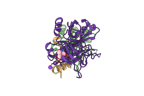
Structure Of Thrombin Mutant Y225P In The E* Form
Organism: Homo sapiens
Method: X-RAY DIFFRACTION Resolution:1.90 Å Release Date: 2011-07-06 Classification: HYDROLASE Ligands: NAG, GOL |
Organism: Homo sapiens
Method: X-RAY DIFFRACTION
Release Date: 2011-07-06
Ligands: NAG, GOL
 |
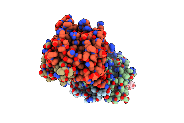
Structure Of Thrombin Mutant Y225P In The E Form
Organism: Homo sapiens
Method: X-RAY DIFFRACTION Resolution:1.90 Å Release Date: 2011-07-06 Classification: HYDROLASE Ligands: K, TRS |
Organism: Homo sapiens
Method: X-RAY DIFFRACTION
Release Date: 2011-07-06
Ligands: K, TRS
 |
Crystal Structure Of The Human Thrombin Mutant D102N In Complex With The Extracellular Fragment Of Human Par4.
Organism: Homo sapiens
Method: X-RAY DIFFRACTION Resolution:2.80 Å Release Date: 2011-06-29 Classification: HYDROLASE/HYDROLASE RECEPTOR |
Organism: Homo sapiens
Method: X-RAY DIFFRACTION
Release Date: 2011-06-29