

Search Count: 17
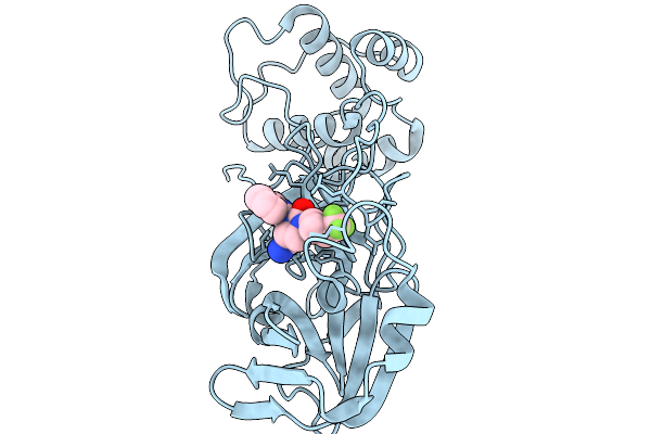 |
Co-Structure Of Sars-Cov-2 3C-Like Proteinase Nsp5 With Compound 34
Organism: Severe acute respiratory syndrome coronavirus 2
Method: X-RAY DIFFRACTION Resolution:1.87 Å Release Date: 2026-03-04 Classification: HYDROLASE Ligands: A1CAR |
Organism: Severe acute respiratory syndrome coronavirus 2
Method: X-RAY DIFFRACTION
Release Date: 2026-03-04
Ligands: A1CAR
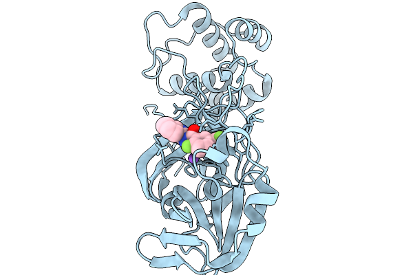 |
Co-Structure Of Main Protease Of Sars-Cov-2 With Compound 1
Organism: Severe acute respiratory syndrome coronavirus 2
Method: X-RAY DIFFRACTION Resolution:1.60 Å Release Date: 2026-02-18 Classification: HYDROLASE Ligands: A1CAS, NA |
Organism: Severe acute respiratory syndrome coronavirus 2
Method: X-RAY DIFFRACTION
Release Date: 2026-02-18
Ligands: A1CAS, NA
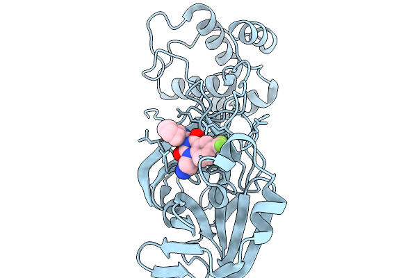 |
Co-Structure Of Main Protease Of Sars-Cov-2 With Compound 11
Organism: Severe acute respiratory syndrome coronavirus 2
Method: X-RAY DIFFRACTION Resolution:2.46 Å Release Date: 2026-02-18 Classification: HYDROLASE Ligands: A1CBN |
Organism: Severe acute respiratory syndrome coronavirus 2
Method: X-RAY DIFFRACTION
Release Date: 2026-02-18
Ligands: A1CBN
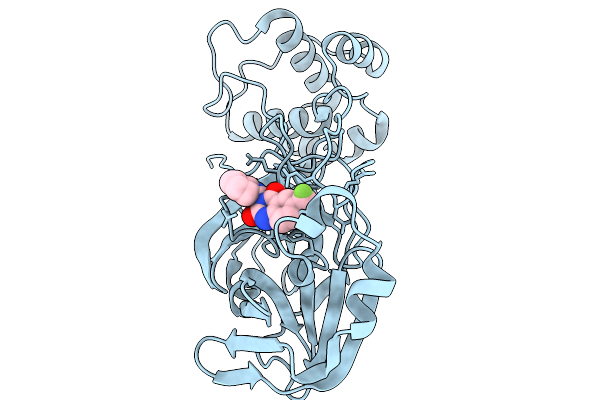 |
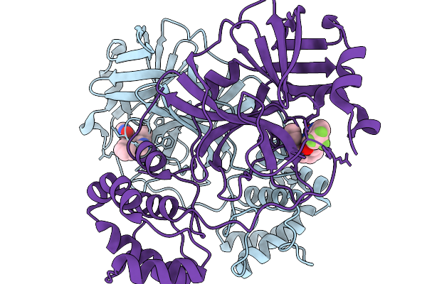
Co-Structure Of Main Protease Of Sars-Cov-2 With Compound 2
Organism: Severe acute respiratory syndrome coronavirus 2
Method: X-RAY DIFFRACTION Resolution:2.21 Å Release Date: 2026-02-18 Classification: HYDROLASE Ligands: A1CBY |
Organism: Severe acute respiratory syndrome coronavirus 2
Method: X-RAY DIFFRACTION
Release Date: 2026-02-18
Ligands: A1CBY
 |
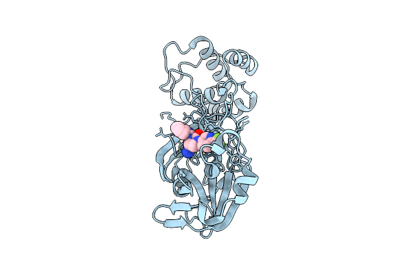
Co-Structure Of Main Protease Of Sars-Cov-2 With Compound 10
Organism: Severe acute respiratory syndrome coronavirus 2
Method: X-RAY DIFFRACTION Resolution:1.71 Å Release Date: 2026-02-18 Classification: HYDROLASE Ligands: A1CB2 |
Organism: Severe acute respiratory syndrome coronavirus 2
Method: X-RAY DIFFRACTION
Release Date: 2026-02-18
Ligands: A1CB2
 |
Co-Structure Of Main Protease Of Sars-Cov-2 With Nvp-Egt710
Organism: Severe acute respiratory syndrome coronavirus 2
Method: X-RAY DIFFRACTION Resolution:1.87 Å Release Date: 2025-12-24 Classification: HYDROLASE Ligands: A1CBU |
Organism: Severe acute respiratory syndrome coronavirus 2
Method: X-RAY DIFFRACTION
Release Date: 2025-12-24
Ligands: A1CBU
 |
Cleavage And Polyadenylation Specificity Factor Subunit 3 (Cpsf3) In Complex With Nvp-Ltm531
Organism: Homo sapiens
Method: X-RAY DIFFRACTION Resolution:2.49 Å Release Date: 2019-11-27 Classification: HYDROLASE Ligands: ZN, JBG, PO4 |
Organism: Homo sapiens
Method: X-RAY DIFFRACTION
Release Date: 2019-11-27
Ligands: ZN, JBG, PO4
 |
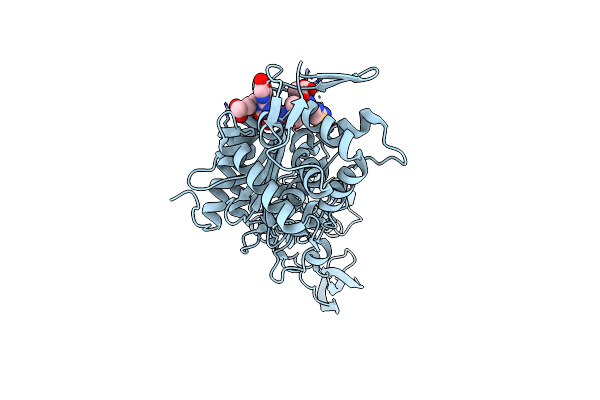
Crystal Structure Of Pseudomonas Aeruginosa Pbp3 With A R4 Substituted Allyl Monocarbam
Organism: Pseudomonas aeruginosa pa14
Method: X-RAY DIFFRACTION Resolution:2.05 Å Release Date: 2015-04-22 Classification: Penicillin-binding protein/inhibitor Ligands: 3LB |
Organism: Pseudomonas aeruginosa pa14
Method: X-RAY DIFFRACTION
Release Date: 2015-04-22
Ligands: 3LB
 |
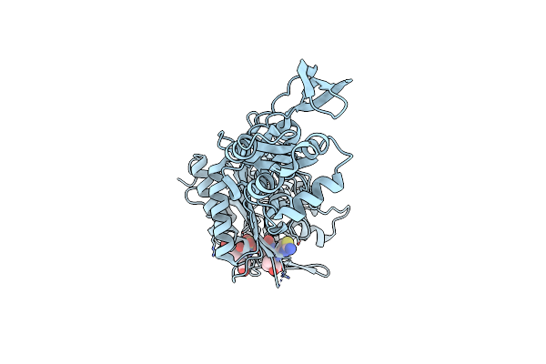
Crystal Structure Of Pseudomonas Aeruginosa Pbp3 With A R4 Substituted Vinyl Monocarbam
Organism: Pseudomonas aeruginosa atcc 14886
Method: X-RAY DIFFRACTION Resolution:1.74 Å Release Date: 2015-04-22 Classification: Penicillin-binding protein/inhibitor Ligands: 3LC |
Organism: Pseudomonas aeruginosa atcc 14886
Method: X-RAY DIFFRACTION
Release Date: 2015-04-22
Ligands: 3LC
 |
Crystal Structure Of Pseudomonas Aeruginosa Pbp3 With Smc-3176
Organism: Pseudomonas aeruginosa pa14
Method: X-RAY DIFFRACTION Resolution:1.99 Å Release Date: 2015-04-22 Classification: Penicillin-binding protein/inhibitor Ligands: 3LE |
Organism: Pseudomonas aeruginosa pa14
Method: X-RAY DIFFRACTION
Release Date: 2015-04-22
Ligands: 3LE
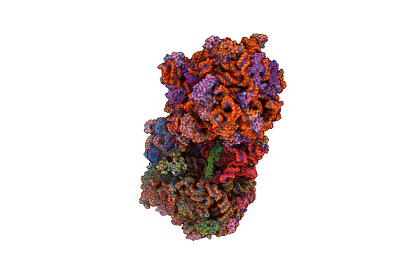 |
Crystal Structure Of The E. Coli Ribosome Bound To Quinupristin.
Organism: Synthetic construct, Escherichia coli str. k-12 substr. mds42, Escherichia coli, Thermus thermophilus
Method: X-RAY DIFFRACTION Resolution:2.95 Å Release Date: 2014-07-30 Classification: RIBOSOME Ligands: MG, ZN |
Organism: Synthetic construct, Escherichia coli str. k-12 substr. mds42, Escherichia coli, Thermus thermophilus
Method: X-RAY DIFFRACTION
Release Date: 2014-07-30
Ligands: MG, ZN
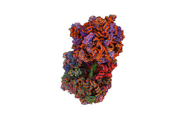 |
Crystal Structure Of The E. Coli Ribosome Bound To Linopristin.
Organism: Synthetic construct, Escherichia coli str. k-12 substr. mds42, Escherichia coli, Thermus thermophilus
Method: X-RAY DIFFRACTION Resolution:3.00 Å Release Date: 2014-07-30 Classification: RIBOSOME Ligands: MG, ZN |
Organism: Synthetic construct, Escherichia coli str. k-12 substr. mds42, Escherichia coli, Thermus thermophilus
Method: X-RAY DIFFRACTION
Release Date: 2014-07-30
Ligands: MG, ZN
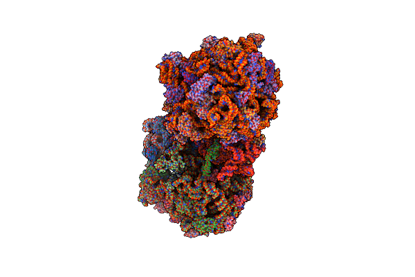 |
Crystal Structure Of The E. Coli Ribosome Bound To Flopristin.
Organism: Escherichia coli str. k-12 substr. mds42, Escherichia coli, Thermus thermophilus
Method: X-RAY DIFFRACTION Resolution:2.90 Å Release Date: 2014-07-30 Classification: RIBOSOME Ligands: MG, VIF, ZN |
Organism: Escherichia coli str. k-12 substr. mds42, Escherichia coli, Thermus thermophilus
Method: X-RAY DIFFRACTION
Release Date: 2014-07-30
Ligands: MG, VIF, ZN
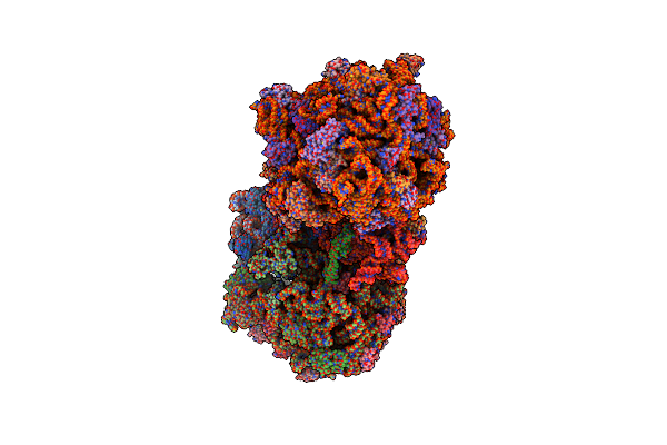 |
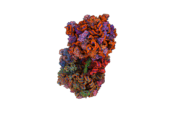
Crystal Structure Of The E. Coli Ribosome Bound To Dalfopristin.
Organism: Escherichia coli str. k-12 substr. mds42, Escherichia coli, Thermus thermophilus
Method: X-RAY DIFFRACTION Resolution:2.90 Å Release Date: 2014-07-30 Classification: RIBOSOME Ligands: MG, DOL, ZN |
Organism: Escherichia coli str. k-12 substr. mds42, Escherichia coli, Thermus thermophilus
Method: X-RAY DIFFRACTION
Release Date: 2014-07-30
Ligands: MG, DOL, ZN
 |
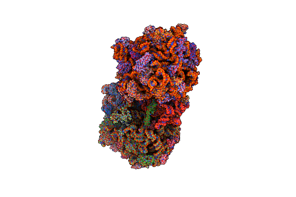
Crystal Structure Of The E. Coli Ribosome Bound To Virginiamycin M1.
Organism: Escherichia coli str. k-12 substr. mds42, Escherichia coli, Thermus thermophilus
Method: X-RAY DIFFRACTION Resolution:2.90 Å Release Date: 2014-07-30 Classification: RIBOSOME Ligands: MG, VIR, ZN |
Organism: Escherichia coli str. k-12 substr. mds42, Escherichia coli, Thermus thermophilus
Method: X-RAY DIFFRACTION
Release Date: 2014-07-30
Ligands: MG, VIR, ZN
 |
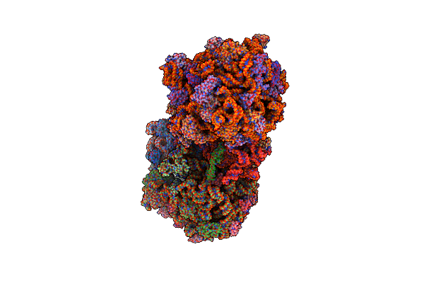
Crystal Structure Of The E. Coli Ribosome Bound To Dalfopristin And Quinupristin.
Organism: Synthetic construct, Escherichia coli str. k-12 substr. mds42, Escherichia coli, Thermus thermophilus
Method: X-RAY DIFFRACTION Resolution:2.80 Å Release Date: 2014-07-30 Classification: RIBOSOME Ligands: MG, DOL, ZN |
Organism: Synthetic construct, Escherichia coli str. k-12 substr. mds42, Escherichia coli, Thermus thermophilus
Method: X-RAY DIFFRACTION
Release Date: 2014-07-30
Ligands: MG, DOL, ZN
 |
Crystal Structure Of The E. Coli Ribosome Bound To Flopristin And Linopristin.
Organism: Synthetic construct, Escherichia coli str. k-12 substr. mds42, Escherichia coli, Thermus thermophilus
Method: X-RAY DIFFRACTION Resolution:2.80 Å Release Date: 2014-07-30 Classification: RIBOSOME Ligands: MG, VIF, ZN |
Organism: Synthetic construct, Escherichia coli str. k-12 substr. mds42, Escherichia coli, Thermus thermophilus
Method: X-RAY DIFFRACTION
Release Date: 2014-07-30
Ligands: MG, VIF, ZN