
Search Count: 67
 |
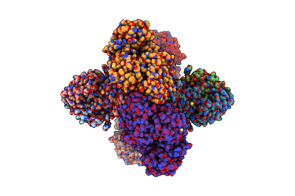
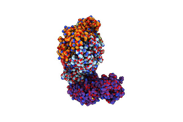
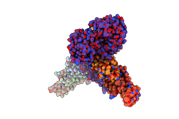
Vitamin K-Dependent Gamma-Carboxylase In Complex With Coagulation Factor Ix And Vitamin K
Organism: Homo sapiens
Method: ELECTRON MICROSCOPY Resolution:2.78 Å Release Date: 2025-10-08 Classification: MEMBRANE PROTEIN Ligands: 6PL, CLR, 1L3 |
 |
Double-Mutant (K217A & K218A) Vitamin K-Dependent Gamma-Carboxylase In Complex With Coagulation Factor Ix And Vitamin K
Organism: Homo sapiens
Method: ELECTRON MICROSCOPY Release Date: 2025-10-08 Classification: MEMBRANE PROTEIN Ligands: A1EMC, 6PL, CLR |
 |
Double-Mutant (K217A & K218A) Vitamin K-Dependent Gamma-Carboxylase In Complex With Coagulation Factors X And Vitamin K
Organism: Homo sapiens
Method: ELECTRON MICROSCOPY Resolution:2.58 Å Release Date: 2025-10-08 Classification: MEMBRANE PROTEIN Ligands: NAG, A1EMC, 6PL, CLR |
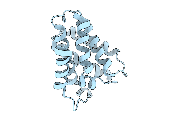 |
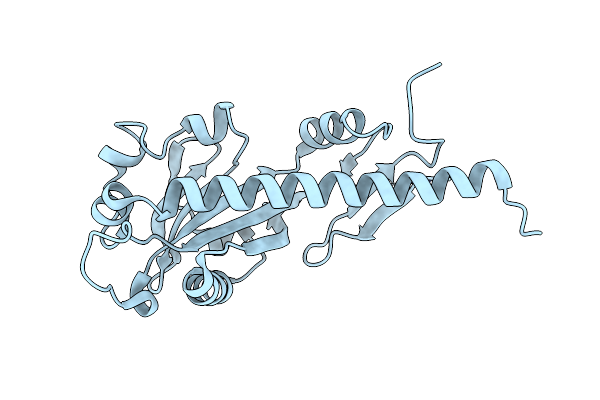
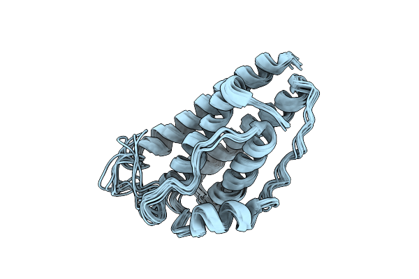
Repetitive Domain (Rp) 2 Structure Of Aciniform Spidroin From Latrodectus Hesperus
Organism: Latrodectus hesperus
Method: X-RAY DIFFRACTION Resolution:1.06 Å Release Date: 2025-10-01 Classification: STRUCTURAL PROTEIN |
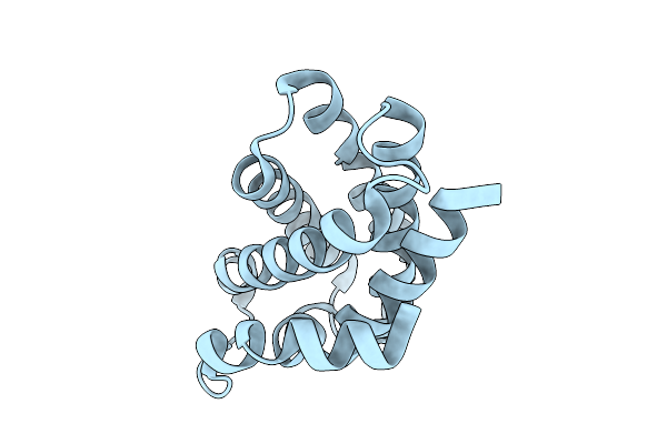 |
Repetitive Domain (Rp) 1 Structure Of Aciniform Spidroin From Latrodectus Hesperus
Organism: Latrodectus hesperus
Method: X-RAY DIFFRACTION Resolution:1.38 Å Release Date: 2025-10-01 Classification: STRUCTURAL PROTEIN |
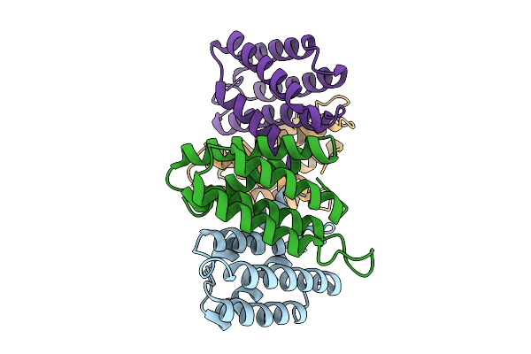 |
N-Terminal Domain (Ntd) Structure Of Aciniform Spidroin 1(Acsp1) From Latrodectus Hesperus
Organism: Latrodectus hesperus
Method: X-RAY DIFFRACTION Resolution:2.98 Å Release Date: 2025-10-01 Classification: STRUCTURAL PROTEIN |
 |
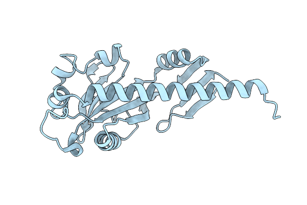
Organism: Pseudomonas aeruginosa pao1
Method: SOLUTION NMR Release Date: 2025-04-23 Classification: HYDROLASE |
 |
Organism: Pseudomonas aeruginosa pao1
Method: X-RAY DIFFRACTION Resolution:2.39 Å Release Date: 2025-04-23 Classification: SIGNALING PROTEIN |
 |
Organism: Pseudomonas aeruginosa pao1
Method: X-RAY DIFFRACTION Resolution:2.17 Å Release Date: 2025-04-23 Classification: SIGNALING PROTEIN |
 |
Organism: Bacillus cereus (strain vd045)
Method: ELECTRON MICROSCOPY Release Date: 2024-02-28 Classification: IMMUNE SYSTEM |
 |
Organism: Bacillus cereus vd045
Method: ELECTRON MICROSCOPY Release Date: 2024-02-28 Classification: IMMUNE SYSTEM |
 |
Organism: Bacillus cereus (strain vd045)
Method: ELECTRON MICROSCOPY Release Date: 2024-02-28 Classification: IMMUNE SYSTEM |
 |
Organism: Bacillus cereus vd045
Method: ELECTRON MICROSCOPY Release Date: 2024-02-28 Classification: DNA BINDING PROTEIN Ligands: ATP |
 |
Organism: Bacillus cereus vd045, Synthetic construct
Method: ELECTRON MICROSCOPY Release Date: 2024-02-28 Classification: DNA BINDING PROTEIN/DNA Ligands: CA |
 |
Organism: Bacillus cereus vd045, Synthetic construct
Method: ELECTRON MICROSCOPY Release Date: 2024-02-28 Classification: DNA BINDING PROTEIN/DNA Ligands: CA |
 |
Organism: Bacillus cereus vd045
Method: ELECTRON MICROSCOPY Release Date: 2024-02-28 Classification: DNA BINDING PROTEIN Ligands: ATP, MG |
 |
Organism: Bacillus cereus vd045
Method: ELECTRON MICROSCOPY Release Date: 2024-02-28 Classification: DNA BINDING PROTEIN Ligands: ATP, MG |
 |
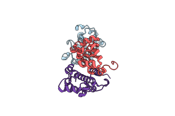 |
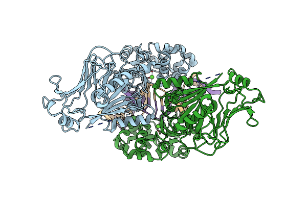
Structure Of Vpa0770 Toxin Bound To Vpa0769 Antitoxin In Vibrio Parahaemolyticus
Organism: Vibrio parahaemolyticus serotype o3:k6 (strain rimd 2210633)
Method: X-RAY DIFFRACTION Resolution:2.85 Å Release Date: 2023-07-26 Classification: TOXIN |
 |
Organism: Sulfolobus spindle-shaped virus
Method: ELECTRON MICROSCOPY Release Date: 2022-08-10 Classification: VIRAL PROTEIN |