
Search Count: 111
All
Selected
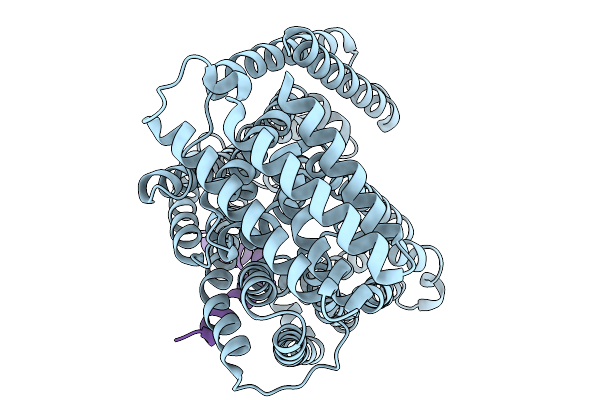 |
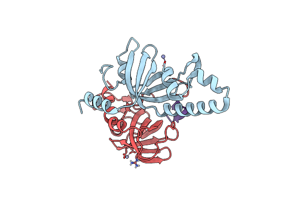
Organism: Escherichia coli k-12, Enterobacteria phage m
Method: ELECTRON MICROSCOPY Resolution:3.60 Å Release Date: 2026-02-11 Classification: TRANSPORT PROTEIN |
 |
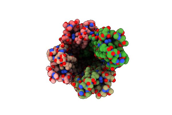
Organism: Escherichia coli k-12, Pseudomonas phage pp7
Method: ELECTRON MICROSCOPY Resolution:3.70 Å Release Date: 2026-02-11 Classification: TRANSPORT PROTEIN |
 |
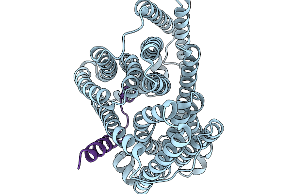
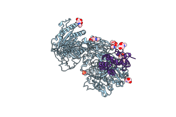
Structure Of Murj In Complex With Single Gene Lysis Protein From Phage Changjiang3
Organism: Escherichia coli k-12, Changjiang levi-like virus 3
Method: ELECTRON MICROSCOPY Resolution:3.60 Å Release Date: 2026-02-11 Classification: TRANSPORT PROTEIN |
 |
Organism: Homo sapiens, Synthetic construct
Method: X-RAY DIFFRACTION Resolution:2.01 Å Release Date: 2026-02-04 Classification: NUCLEAR PROTEIN |
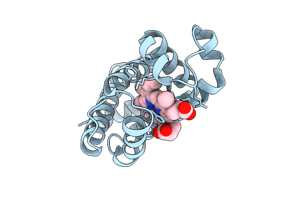 |
Organism: Physeter catodon
Method: X-RAY DIFFRACTION Resolution:1.80 Å Release Date: 2025-10-15 Classification: OXIDOREDUCTASE Ligands: HEM, O |
 |
Organism: Physeter catodon
Method: X-RAY DIFFRACTION Resolution:1.68 Å Release Date: 2025-10-15 Classification: OXIDOREDUCTASE Ligands: HEM, PEG, SO4, CL |
 |
Organism: Severe acute respiratory syndrome coronavirus 2
Method: X-RAY DIFFRACTION Resolution:2.69 Å Release Date: 2025-07-16 Classification: VIRAL PROTEIN, HYDROLASE Ligands: WZK |
 |
Organism: Severe acute respiratory syndrome coronavirus 2
Method: X-RAY DIFFRACTION Resolution:1.81 Å Release Date: 2025-07-16 Classification: VIRAL PROTEIN, HYDROLASE |
 |
Organism: Homo sapiens, Synthetic construct
Method: X-RAY DIFFRACTION Resolution:2.35 Å Release Date: 2025-07-02 Classification: NUCLEAR PROTEIN |
 |
Organism: Homo sapiens, Synthetic construct
Method: X-RAY DIFFRACTION Resolution:2.70 Å Release Date: 2025-07-02 Classification: NUCLEAR PROTEIN Ligands: ZN |
 |
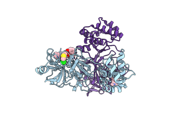
Organism: Mus musculus
Method: X-RAY DIFFRACTION Resolution:2.72 Å Release Date: 2025-04-02 Classification: SIGNALING PROTEIN Ligands: NAG, SO4 |
 |
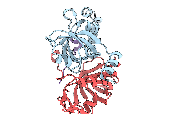
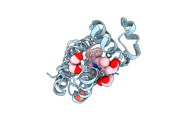
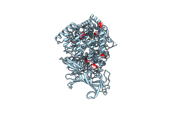
Erap1 In Complex With 1-[2-(3-Oxo-3,4-Dihydro-2H-1,4-Benzothiazin-4-Yl)Acetamido]Cyclohexane-1-Carboxylic Acid
Organism: Homo sapiens
Method: X-RAY DIFFRACTION Resolution:1.72 Å Release Date: 2025-01-22 Classification: PEPTIDE BINDING PROTEIN Ligands: ZN, B3P, EDO, BR, A1IMK |
 |
Erap1 In Complex With 1-[2-(6-Bromo-3-Oxo-3,4-Dihydro-2H-1,4-Benzoxazin-4-Yl)Acetamido]-4,4-Difluorocyclohexane-1-Carboxylic Acid
Organism: Homo sapiens
Method: X-RAY DIFFRACTION Resolution:1.35 Å Release Date: 2025-01-22 Classification: PEPTIDE BINDING PROTEIN Ligands: ZN, PO4, EDO, A1IMJ |
 |
Erap1 In Complex With 1-[2-(6-Chloro-3-Oxo-3,4-Dihydro-2H-1,4-Benzothiazin-4-Yl)Acetamido]Cyclohexane-1-Carboxylic Acid
Organism: Homo sapiens
Method: X-RAY DIFFRACTION Resolution:1.33 Å Release Date: 2025-01-22 Classification: PEPTIDE BINDING PROTEIN Ligands: ZN, MLT, EDO, A1IMM |
 |
Erap1 In Complex With 1-[2-(2-Oxo-5-Phenyl-2,3-Dihydro-1,3-Benzothiazol-3-Yl)Acetamido]Cyclohexane-1-Carboxylic Acid
Organism: Homo sapiens
Method: X-RAY DIFFRACTION Resolution:1.37 Å Release Date: 2025-01-22 Classification: PEPTIDE BINDING PROTEIN Ligands: ZN, PO4, EDO, A1IML |
 |
Organism: Homo sapiens
Method: ELECTRON MICROSCOPY Release Date: 2024-07-24 Classification: CELL CYCLE |
 |
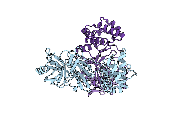
Organism: Homo sapiens, Xenopus laevis
Method: ELECTRON MICROSCOPY Release Date: 2024-07-24 Classification: CELL CYCLE |
 |
Organism: Mus musculus, Homo sapiens
Method: X-RAY DIFFRACTION Resolution:4.70 Å Release Date: 2024-04-24 Classification: SIGNALING PROTEIN Ligands: NAG |
 |
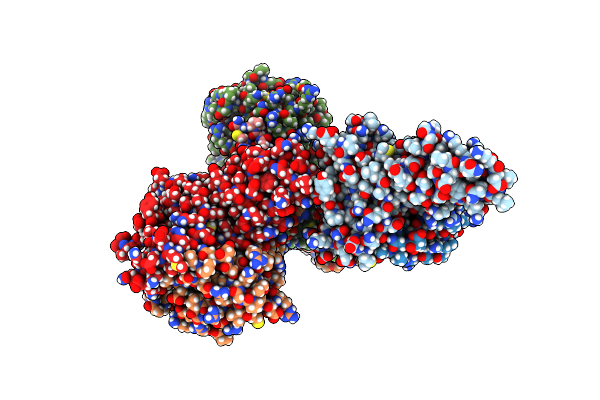
Organism: Acinetobacter genomosp. 16bj
Method: ELECTRON MICROSCOPY Release Date: 2024-03-06 Classification: CELL ADHESION |
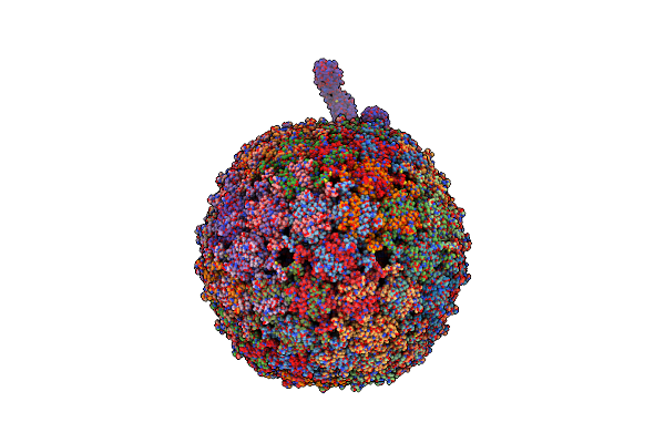 |
Organism: Bacteria abnormis, Acinetobacter phage ap205
Method: ELECTRON MICROSCOPY Release Date: 2024-03-06 Classification: VIRUS/RNA |