

Search Count: 12
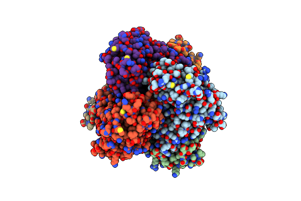 |
Mycobacterium Tuberculosis Coax Homohexamer
Organism: Mycobacterium tuberculosis
Method: ELECTRON MICROSCOPY Release Date: 2024-11-13 Classification: CYTOSOLIC PROTEIN |
Organism: Mycobacterium tuberculosis
Method: ELECTRON MICROSCOPY
Release Date: 2024-11-13
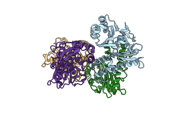 |
Mycobacterium Tuberculosis Coax Homotetramer
Organism: Mycobacterium tuberculosis
Method: ELECTRON MICROSCOPY Release Date: 2024-11-13 Classification: CYTOSOLIC PROTEIN |
Organism: Mycobacterium tuberculosis
Method: ELECTRON MICROSCOPY
Release Date: 2024-11-13
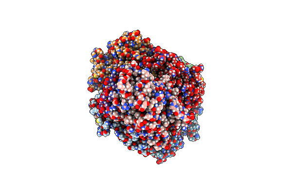 |
Complex Of M. Smegmatis Dop With M. Tuberculosis Coax And Pup91
Organism: Mycobacterium tuberculosis, Mycolicibacterium smegmatis
Method: ELECTRON MICROSCOPY Release Date: 2024-11-13 Classification: CYTOSOLIC PROTEIN |
Organism: Mycobacterium tuberculosis, Mycolicibacterium smegmatis
Method: ELECTRON MICROSCOPY
Release Date: 2024-11-13
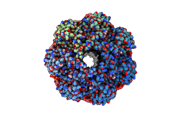 |
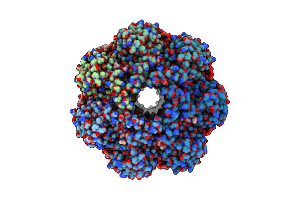
Structure Of The Mycobacterium Tuberculosis 20S Proteasome Bound To The C-Terminal Gqyl Motif Of The Atp-Bound Mpa Atpase
Organism: Mycobacterium tuberculosis
Method: ELECTRON MICROSCOPY Release Date: 2022-08-03 Classification: ANTIMICROBIAL PROTEIN |
Organism: Mycobacterium tuberculosis
Method: ELECTRON MICROSCOPY
Release Date: 2022-08-03
 |
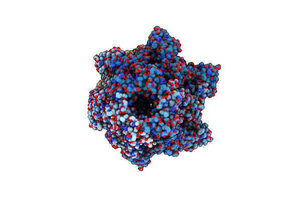
Structure Of The Mycobacterium Tuberculosis 20S Proteasome Bound To The C-Terminal Gqyl Motif Of The Adp-Bound Mpa Atpase
Organism: Mycobacterium tuberculosis
Method: ELECTRON MICROSCOPY Release Date: 2022-08-03 Classification: ANTIMICROBIAL PROTEIN |
Organism: Mycobacterium tuberculosis
Method: ELECTRON MICROSCOPY
Release Date: 2022-08-03
 |
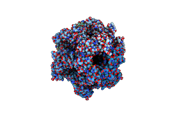
Structure Of The Mycobacterium Tuberculosis 20S Proteasome Bound To The Atp-Bound Mpa Atpase
Organism: Mycobacterium tuberculosis
Method: ELECTRON MICROSCOPY Release Date: 2022-08-03 Classification: ANTIMICROBIAL PROTEIN |
Organism: Mycobacterium tuberculosis
Method: ELECTRON MICROSCOPY
Release Date: 2022-08-03
 |
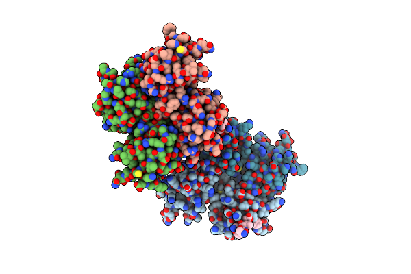
Structure Of The Mycobacterium Tuberculosis 20S Proteasome Bound To The Adp-Bound Mpa Atpase
Organism: Mycobacterium tuberculosis
Method: ELECTRON MICROSCOPY Release Date: 2022-08-03 Classification: ANTIMICROBIAL PROTEIN |
Organism: Mycobacterium tuberculosis
Method: ELECTRON MICROSCOPY
Release Date: 2022-08-03
 |
The Complex Structure Of Pdz Domains In Syntenin-1 With 4L Peptide
Organism: Rattus norvegicus
Method: X-RAY DIFFRACTION Resolution:2.50 Å Release Date: 2016-06-01 Classification: SIGNALING PROTEIN |
Organism: Rattus norvegicus
Method: X-RAY DIFFRACTION
Release Date: 2016-06-01
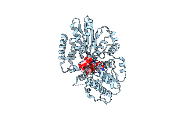 |
Crystal Structure Of The Sialyltransferase Pm0188 With Cmp-3Fneuac And Lactose
Organism: Pasteurella multocida
Method: X-RAY DIFFRACTION Resolution:2.50 Å Release Date: 2007-09-18 Classification: TRANSFERASE Ligands: CSF |
Organism: Pasteurella multocida
Method: X-RAY DIFFRACTION
Release Date: 2007-09-18
Ligands: CSF
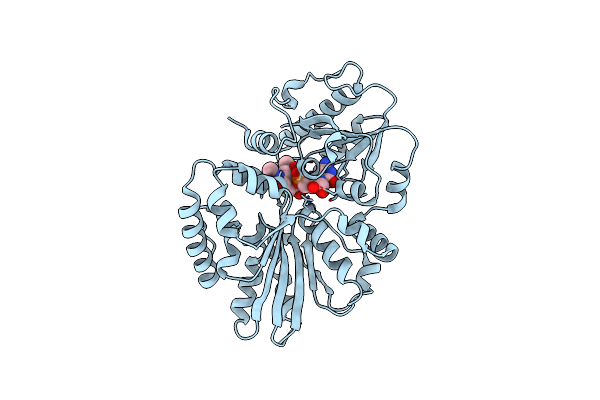 |
Crystal Structure Of The Sialyltransferase Pm0188 With Cmp-3Fneuac
Organism: Pasteurella multocida
Method: X-RAY DIFFRACTION Resolution:1.85 Å Release Date: 2007-08-21 Classification: TRANSFERASE Ligands: CSF |
Organism: Pasteurella multocida
Method: X-RAY DIFFRACTION
Release Date: 2007-08-21
Ligands: CSF
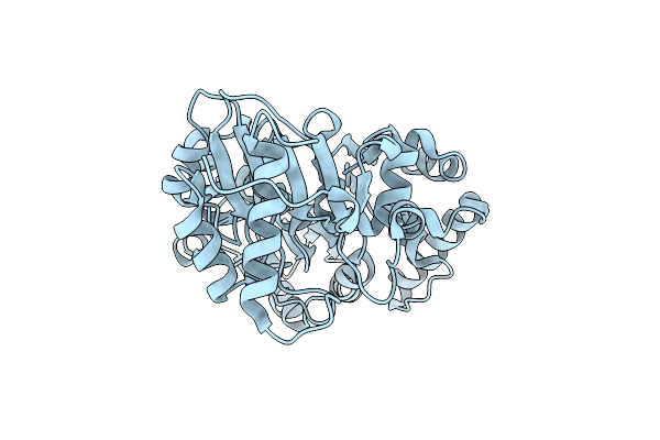 |
Crystal Structure Of The Sialyltransferase Pm0188
Organism: Pasteurella multocida
Method: X-RAY DIFFRACTION Resolution:1.90 Å Release Date: 2007-03-27 Classification: TRANSFERASE |
Organism: Pasteurella multocida
Method: X-RAY DIFFRACTION
Release Date: 2007-03-27
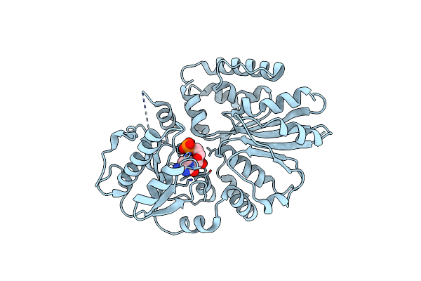 |
Crystal Structure Of The Sialyltransferase Pm0188 With Cmp
Organism: pasteurella multocida
Method: X-RAY DIFFRACTION Resolution:2.31 Å Release Date: 2007-03-27 Classification: TRANSFERASE Ligands: C5P |
Organism: pasteurella multocida
Method: X-RAY DIFFRACTION
Release Date: 2007-03-27
Ligands: C5P