

Search Count: 13
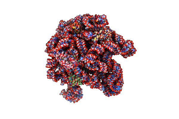 |
E. Coli 50S Ribosome Bound To Antibiotic Analog Slc09
Organism: Escherichia coli
Method: ELECTRON MICROSCOPY Release Date: 2023-06-28 Classification: RIBOSOME Ligands: UI9 |
Organism: Escherichia coli
Method: ELECTRON MICROSCOPY
Release Date: 2023-06-28
Ligands: UI9
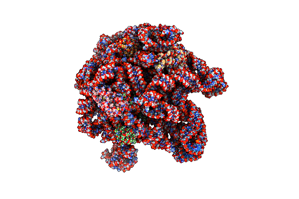 |
E. Coli 50S Ribosome Bound To Antibiotic Analog Slc17
Organism: Escherichia coli
Method: ELECTRON MICROSCOPY Release Date: 2023-06-28 Classification: RIBOSOME Ligands: UI6 |
Organism: Escherichia coli
Method: ELECTRON MICROSCOPY
Release Date: 2023-06-28
Ligands: UI6
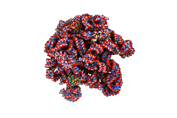 |
E. Coli 50S Ribosome Bound To Antibiotic Analog Slc21
Organism: Escherichia coli
Method: ELECTRON MICROSCOPY Release Date: 2023-06-28 Classification: RIBOSOME Ligands: UHP |
Organism: Escherichia coli
Method: ELECTRON MICROSCOPY
Release Date: 2023-06-28
Ligands: UHP
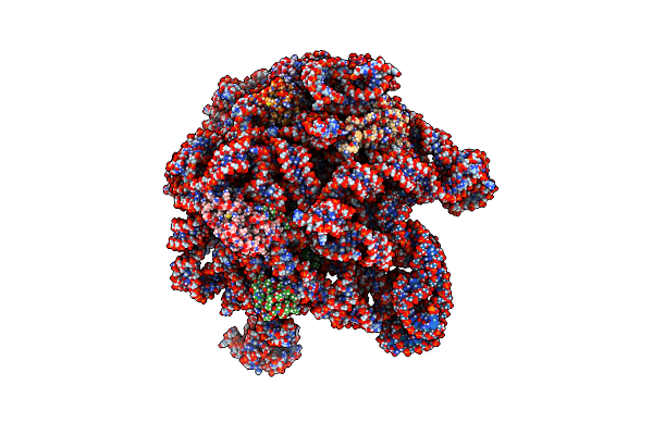 |
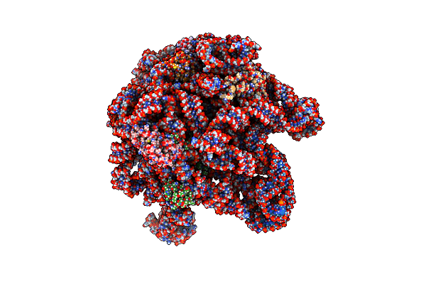
E. Coli 50S Ribosome Bound To Antibiotic Analog Slc26
Organism: Escherichia coli
Method: ELECTRON MICROSCOPY Release Date: 2023-06-28 Classification: RIBOSOME Ligands: UH6 |
Organism: Escherichia coli
Method: ELECTRON MICROSCOPY
Release Date: 2023-06-28
Ligands: UH6
 |
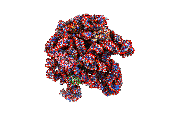
E. Coli 50S Ribosome Bound To Antibiotic Analog Slc30
Organism: Escherichia coli
Method: ELECTRON MICROSCOPY Release Date: 2023-06-28 Classification: RIBOSOME Ligands: UIF |
Organism: Escherichia coli
Method: ELECTRON MICROSCOPY
Release Date: 2023-06-28
Ligands: UIF
 |
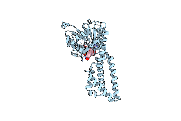
E. Coli 50S Ribosome Bound To Antibiotic Analog Slc31
Organism: Escherichia coli
Method: ELECTRON MICROSCOPY Release Date: 2023-06-28 Classification: RIBOSOME Ligands: UH0 |
Organism: Escherichia coli
Method: ELECTRON MICROSCOPY
Release Date: 2023-06-28
Ligands: UH0
 |
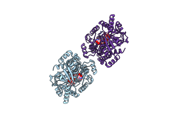
Crystal Structure Of Prephenate Dehydrogenase From Streptococcus Mutans
Organism: Streptococcus mutans
Method: X-RAY DIFFRACTION Resolution:2.10 Å Release Date: 2011-10-26 Classification: OXIDOREDUCTASE Ligands: NAD |
Organism: Streptococcus mutans
Method: X-RAY DIFFRACTION
Release Date: 2011-10-26
Ligands: NAD
 |
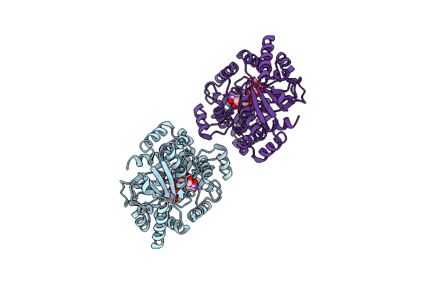
Crystal Structure Of Trehalose Synthase Tret Mutant E326A From P. Horikoshii In Complex With Udpg
Organism: Pyrococcus horikoshii
Method: X-RAY DIFFRACTION Resolution:2.50 Å Release Date: 2011-03-16 Classification: BIOSYNTHETIC PROTEIN Ligands: UPG |
Organism: Pyrococcus horikoshii
Method: X-RAY DIFFRACTION
Release Date: 2011-03-16
Ligands: UPG
 |
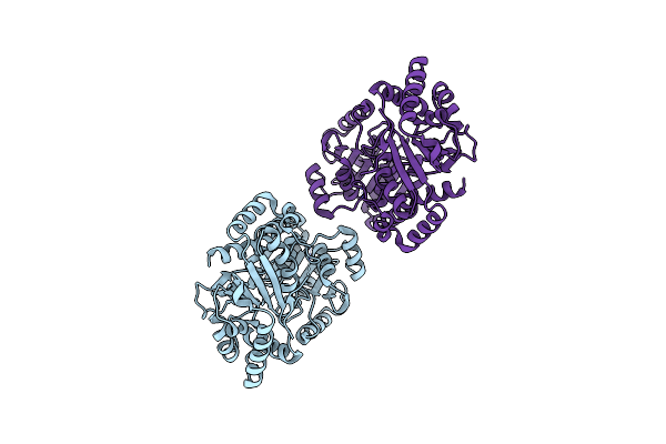
Crystal Structure Of Trehalose Synthase Tret Mutant E326A From P. Horikoshii In Complex With Udpg
Organism: Pyrococcus horikoshii
Method: X-RAY DIFFRACTION Resolution:2.50 Å Release Date: 2011-03-16 Classification: BIOSYNTHETIC PROTEIN Ligands: UPG |
Organism: Pyrococcus horikoshii
Method: X-RAY DIFFRACTION
Release Date: 2011-03-16
Ligands: UPG
 |
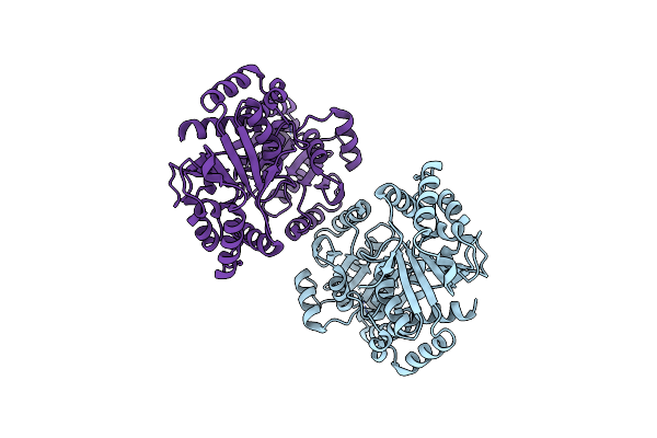
Crystal Structure Of Trehalose Synthase Tret From P.Horikoshi
Organism: Pyrococcus horikoshii
Method: X-RAY DIFFRACTION Resolution:2.20 Å Release Date: 2010-10-13 Classification: BIOSYNTHETIC PROTEIN |
Organism: Pyrococcus horikoshii
Method: X-RAY DIFFRACTION
Release Date: 2010-10-13
 |
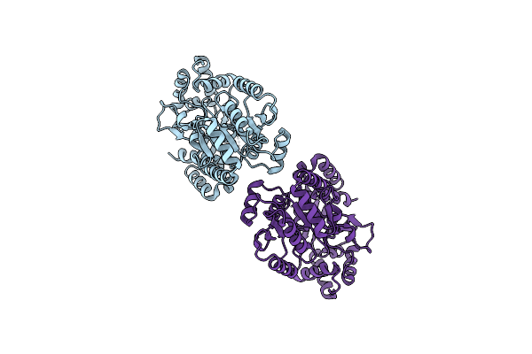
Crystal Structure Of Trehalose Synthase Tret From P.Horikoshi Produced By Soaking In Trehalose
Organism: Pyrococcus horikoshii
Method: X-RAY DIFFRACTION Resolution:2.20 Å Release Date: 2010-10-13 Classification: ISOMERASE |
Organism: Pyrococcus horikoshii
Method: X-RAY DIFFRACTION
Release Date: 2010-10-13
 |
Crystal Structure Of Trehalose Synthase Tret From P.Horikoshii (Seleno Derivative)
Organism: Pyrococcus horikoshii
Method: X-RAY DIFFRACTION Resolution:2.47 Å Release Date: 2010-10-13 Classification: BIOSYNTHETIC PROTEIN |
Organism: Pyrococcus horikoshii
Method: X-RAY DIFFRACTION
Release Date: 2010-10-13
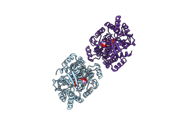 |
Crystal Structure Of Trehalose Synthase Tret Mutant E326A From P. Horishiki In Complex With Udp
Organism: Pyrococcus horikoshii
Method: X-RAY DIFFRACTION Resolution:2.50 Å Release Date: 2010-10-13 Classification: SUGAR BINDING PROTEIN Ligands: UDP |
Organism: Pyrococcus horikoshii
Method: X-RAY DIFFRACTION
Release Date: 2010-10-13
Ligands: UDP