

Search Count: 75
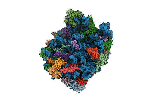 |
Cryo-Em Structure Of Chaetomium Thermophilum Ribosome-Bound Snd3 Translocon
Organism: Thermochaetoides thermophila dsm 1495
Method: ELECTRON MICROSCOPY Release Date: 2025-11-12 Classification: RIBOSOME Ligands: MG |
Organism: Thermochaetoides thermophila dsm 1495
Method: ELECTRON MICROSCOPY
Release Date: 2025-11-12
Ligands: MG
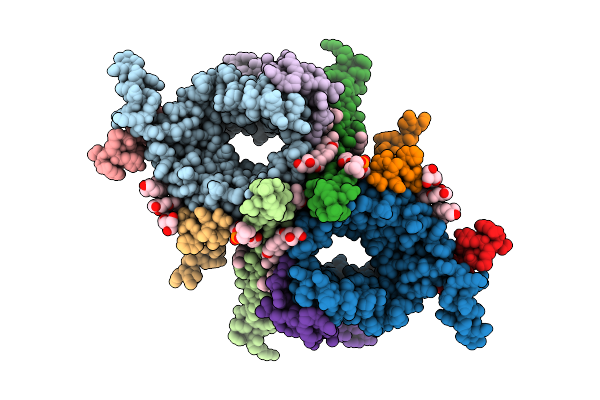 |
Cryoem Structure Of The Chaetomium Thermophilum Tom Core Complex At 2.7 Angstrom Resolution (Paldh Treated)
Organism: Thermochaetoides thermophila dsm 1495
Method: ELECTRON MICROSCOPY Resolution:2.70 Å Release Date: 2025-07-09 Classification: MEMBRANE PROTEIN Ligands: PC1, DU0, PLC |
Organism: Thermochaetoides thermophila dsm 1495
Method: ELECTRON MICROSCOPY
Release Date: 2025-07-09
Ligands: PC1, DU0, PLC
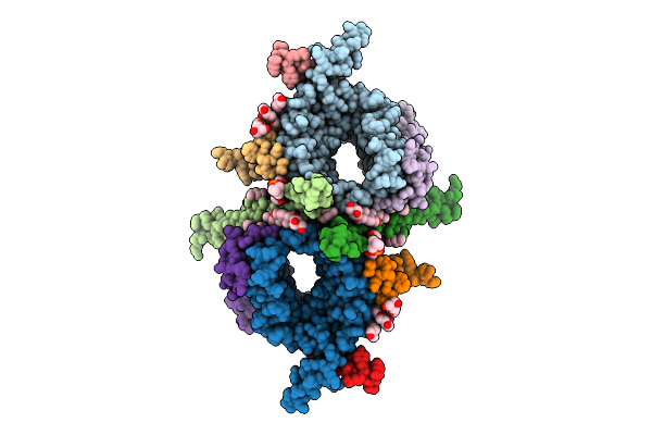 |
Cryoem Structure Of The Chaetomium Thermophilum Tom Core Complex At 3.2 Angstrom Resolution
Organism: Thermochaetoides thermophila dsm 1495
Method: ELECTRON MICROSCOPY Resolution:3.20 Å Release Date: 2025-07-09 Classification: MEMBRANE PROTEIN Ligands: PC1, DU0, PLC |
Organism: Thermochaetoides thermophila dsm 1495
Method: ELECTRON MICROSCOPY
Release Date: 2025-07-09
Ligands: PC1, DU0, PLC
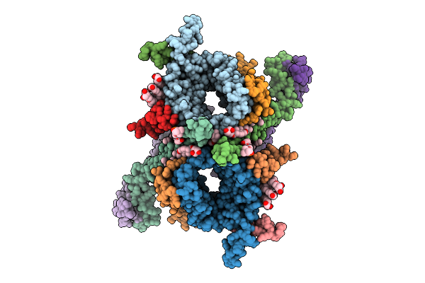 |
Cryoem Structure Of The Chaetomium Thermophilum Tom Holo Complex At 3.2 Angstrom Resolution (Paldh Treated)
Organism: Thermochaetoides thermophila dsm 1495
Method: ELECTRON MICROSCOPY Resolution:3.20 Å Release Date: 2025-07-09 Classification: MEMBRANE PROTEIN Ligands: PC1, DU0, PLC |
Organism: Thermochaetoides thermophila dsm 1495
Method: ELECTRON MICROSCOPY
Release Date: 2025-07-09
Ligands: PC1, DU0, PLC
 |
Cryoem Structure Of The Chaetomium Thermophilum Tom Holo Complex At 3.8 Angstrom Resolution
Organism: Thermochaetoides thermophila dsm 1495
Method: ELECTRON MICROSCOPY Resolution:3.80 Å Release Date: 2025-07-09 Classification: MEMBRANE PROTEIN Ligands: PC1, DU0, PLC |
Organism: Thermochaetoides thermophila dsm 1495
Method: ELECTRON MICROSCOPY
Release Date: 2025-07-09
Ligands: PC1, DU0, PLC
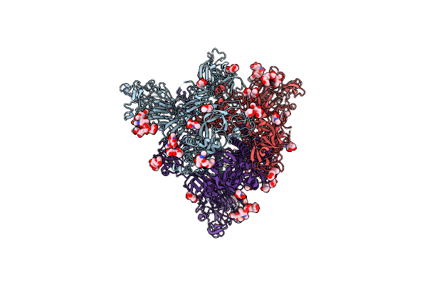 |
Cryo-Em Structure Of Mers-Cov Spike Protein, Two Rbd-Up Conformation 2
Organism: Human betacoronavirus 2c emc/2012
Method: ELECTRON MICROSCOPY Release Date: 2023-08-09 Classification: VIRAL PROTEIN Ligands: NAG |
Organism: Human betacoronavirus 2c emc/2012
Method: ELECTRON MICROSCOPY
Release Date: 2023-08-09
Ligands: NAG
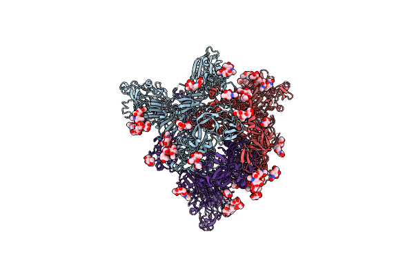 |
Cryo-Em Structure Of Mers-Cov Spike Protein, Two Rbd-Up Conformation 1
Organism: Human betacoronavirus 2c emc/2012
Method: ELECTRON MICROSCOPY Release Date: 2023-08-09 Classification: VIRAL PROTEIN Ligands: NAG |
Organism: Human betacoronavirus 2c emc/2012
Method: ELECTRON MICROSCOPY
Release Date: 2023-08-09
Ligands: NAG
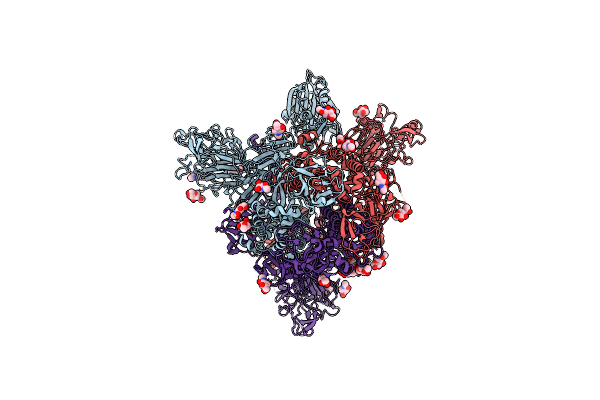 |
Cryo-Em Structure Of Mers-Cov Spike Protein, One Rbd-Up Conformation 4
Organism: Human betacoronavirus 2c emc/2012
Method: ELECTRON MICROSCOPY Release Date: 2023-08-09 Classification: VIRAL PROTEIN Ligands: NAG |
Organism: Human betacoronavirus 2c emc/2012
Method: ELECTRON MICROSCOPY
Release Date: 2023-08-09
Ligands: NAG
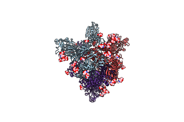 |
Cryo-Em Structure Of Mers-Cov Spike Protein, One Rbd-Up Conformation 2
Organism: Human betacoronavirus 2c emc/2012
Method: ELECTRON MICROSCOPY Release Date: 2023-08-09 Classification: VIRAL PROTEIN Ligands: NAG |
Organism: Human betacoronavirus 2c emc/2012
Method: ELECTRON MICROSCOPY
Release Date: 2023-08-09
Ligands: NAG
 |
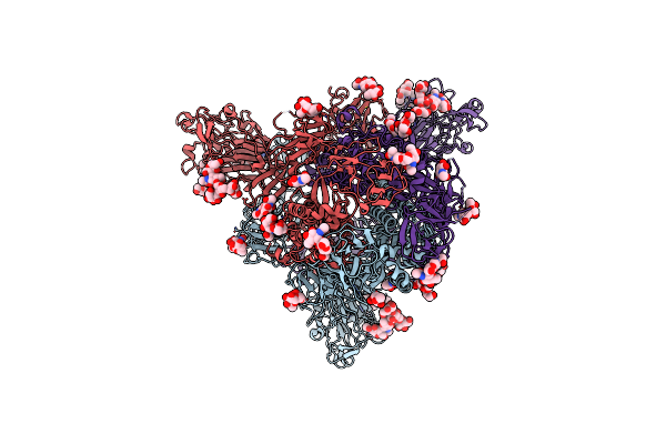
Cryo-Em Structure Of Mers-Cov Spike Protein, One Rbd-Up Conformation 1
Organism: Human betacoronavirus 2c emc/2012
Method: ELECTRON MICROSCOPY Release Date: 2023-08-09 Classification: VIRAL PROTEIN Ligands: NAG |
Organism: Human betacoronavirus 2c emc/2012
Method: ELECTRON MICROSCOPY
Release Date: 2023-08-09
Ligands: NAG
 |
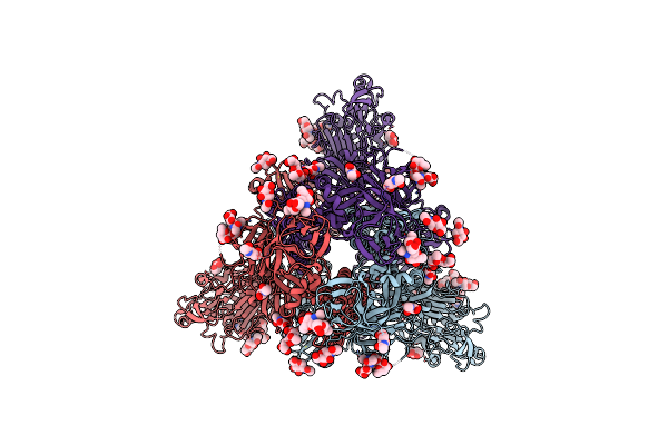
Cryo-Em Structure Of Mers-Cov Spike Protein, Intermediate Conformation
Organism: Human betacoronavirus 2c emc/2012
Method: ELECTRON MICROSCOPY Release Date: 2023-08-09 Classification: VIRAL PROTEIN Ligands: NAG |
Organism: Human betacoronavirus 2c emc/2012
Method: ELECTRON MICROSCOPY
Release Date: 2023-08-09
Ligands: NAG
 |
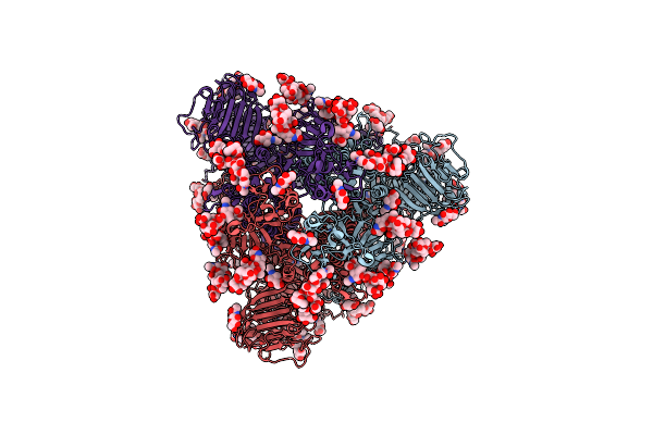
Cryo-Em Structure Of Mers-Cov Spike Protein, All Rbd-Down Conformation
Organism: Human betacoronavirus 2c emc/2012
Method: ELECTRON MICROSCOPY Release Date: 2023-08-09 Classification: VIRAL PROTEIN Ligands: NAG |
Organism: Human betacoronavirus 2c emc/2012
Method: ELECTRON MICROSCOPY
Release Date: 2023-08-09
Ligands: NAG
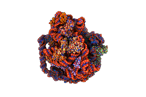 |
Unmethylated Mtb Ribosome 50S With Seq-9
Organism: Mycobacterium tuberculosis
Method: ELECTRON MICROSCOPY Release Date: 2022-10-12 Classification: RIBOSOME/ANTIBIOTIC Ligands: ZN, WDP, MG |
Organism: Mycobacterium tuberculosis
Method: ELECTRON MICROSCOPY
Release Date: 2022-10-12
Ligands: ZN, WDP, MG
 |
Cryo-Em Map Of Pedv (Pintung 52) S Protein With All Three Protomers In The D0-Down Conformation Determined In Situ On Intact Viral Particles.
Organism: Porcine epidemic diarrhea virus
Method: ELECTRON MICROSCOPY Release Date: 2022-08-03 Classification: VIRAL PROTEIN Ligands: NAG |
Organism: Porcine epidemic diarrhea virus
Method: ELECTRON MICROSCOPY
Release Date: 2022-08-03
Ligands: NAG
 |
Cryo-Em Map Of Pedv S Protein With One Protomer In The D0-Up Conformation While The Other Two In The D0-Down Conformation
Organism: Porcine epidemic diarrhea virus
Method: ELECTRON MICROSCOPY Release Date: 2022-08-03 Classification: VIRAL PROTEIN Ligands: NAG |
Organism: Porcine epidemic diarrhea virus
Method: ELECTRON MICROSCOPY
Release Date: 2022-08-03
Ligands: NAG
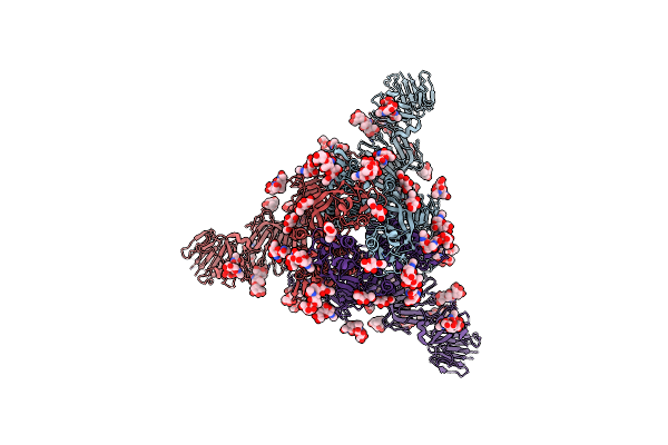 |
Cryo-Em Map Of Ipec-J2 Cell-Derived Pedv Pt52 S Protein With Three D0-Up
Organism: Porcine epidemic diarrhea virus
Method: ELECTRON MICROSCOPY Release Date: 2022-08-03 Classification: VIRAL PROTEIN Ligands: NAG |
Organism: Porcine epidemic diarrhea virus
Method: ELECTRON MICROSCOPY
Release Date: 2022-08-03
Ligands: NAG
 |
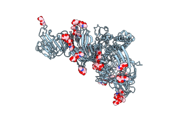
Cryo-Em Map Of Ipec-J2 Cell-Derived Pedv Pt52 S Protein One D0-Down And Two D0-Up
Organism: Porcine epidemic diarrhea virus
Method: ELECTRON MICROSCOPY Release Date: 2022-08-03 Classification: VIRAL PROTEIN Ligands: NAG |
Organism: Porcine epidemic diarrhea virus
Method: ELECTRON MICROSCOPY
Release Date: 2022-08-03
Ligands: NAG
 |
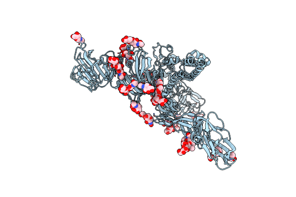
Symmetry-Expanded And Locally Refined Protomer Structure Of Ipec-J2 Cell-Derived Pedv Pt52 S With A Ctd-Close Conformation
Organism: Porcine epidemic diarrhea virus
Method: ELECTRON MICROSCOPY Release Date: 2022-08-03 Classification: VIRAL PROTEIN Ligands: NAG |
Organism: Porcine epidemic diarrhea virus
Method: ELECTRON MICROSCOPY
Release Date: 2022-08-03
Ligands: NAG
 |
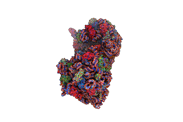
Symmetry-Expanded And Locally Refined Protomer Structure Of Ipec-J2 Cell-Derived Pedv Pt52 S With A Ctd-Open Conformation
Organism: Porcine epidemic diarrhea virus
Method: ELECTRON MICROSCOPY Resolution:3.30 Å Release Date: 2022-08-03 Classification: VIRAL PROTEIN Ligands: NAG |
Organism: Porcine epidemic diarrhea virus
Method: ELECTRON MICROSCOPY
Release Date: 2022-08-03
Ligands: NAG
 |
70S Thermus Thermophilus Ribosome With Bound Antibiotic Lead Seq-569
Organism: Thermus thermophilus (strain hb8 / atcc 27634 / dsm 579), Escherichia coli
Method: X-RAY DIFFRACTION Resolution:3.10 Å Release Date: 2022-06-08 Classification: RIBOSOME Ligands: MG, K, OHX, SJH |
Organism: Thermus thermophilus (strain hb8 / atcc 27634 / dsm 579), Escherichia coli
Method: X-RAY DIFFRACTION
Release Date: 2022-06-08
Ligands: MG, K, OHX, SJH