

Search Count: 24
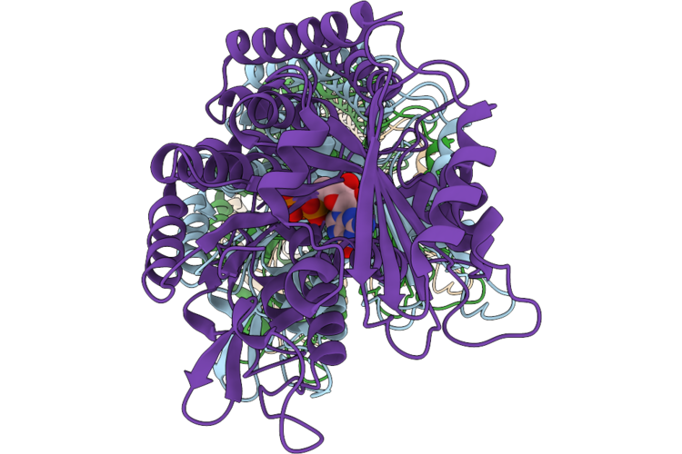 |
Averaged Protofilament Of Heimdallarchaeales Alpha/Beta Tubulin Microtubule With A Single Seam
Organism: Candidatus heimdallarchaeaceae
Method: ELECTRON MICROSCOPY Release Date: 2026-06-24 Classification: PROTEIN FIBRIL Ligands: GDP, MG |
Organism: Candidatus heimdallarchaeaceae
Method: ELECTRON MICROSCOPY
Release Date: 2026-06-24
Ligands: GDP, MG
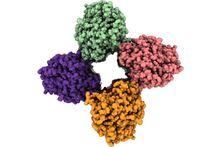 |
Heimdallarchaeales Alpha/Beta Tubulin Microtubule With A Single Seam
Organism: Candidatus heimdallarchaeaceae
Method: ELECTRON MICROSCOPY Release Date: 2026-06-10 Classification: PROTEIN FIBRIL Ligands: GDP, MG |
Organism: Candidatus heimdallarchaeaceae
Method: ELECTRON MICROSCOPY
Release Date: 2026-06-10
Ligands: GDP, MG
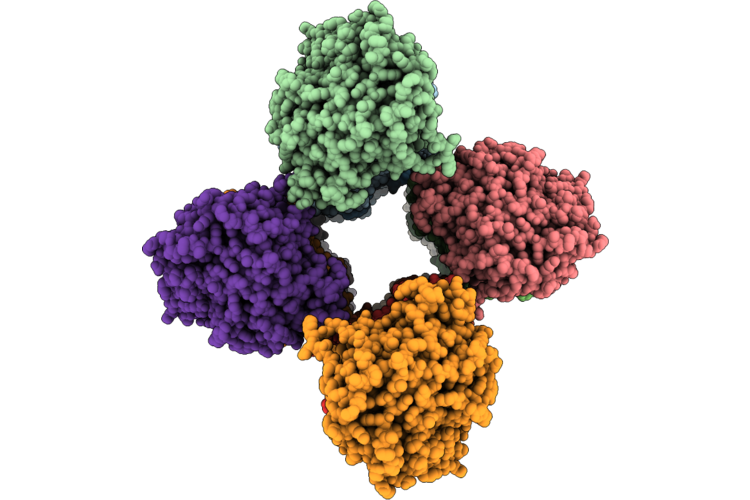 |
Heimdallarchaeales Alpha/Beta Tubulin Microtubule With Three Seams
Organism: Candidatus heimdallarchaeaceae
Method: ELECTRON MICROSCOPY Release Date: 2026-06-10 Classification: PROTEIN FIBRIL Ligands: GDP, MG |
Organism: Candidatus heimdallarchaeaceae
Method: ELECTRON MICROSCOPY
Release Date: 2026-06-10
Ligands: GDP, MG
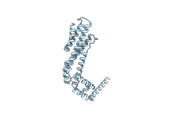 |
Crystal Structure Of The C-Terminal Fragment (Residues 756-982 With The C864S Mutation) Of Arabidopsis Thaliana Chup1
Organism: Arabidopsis thaliana
Method: X-RAY DIFFRACTION Resolution:2.80 Å Release Date: 2024-01-17 Classification: PLANT PROTEIN |
Organism: Arabidopsis thaliana
Method: X-RAY DIFFRACTION
Release Date: 2024-01-17
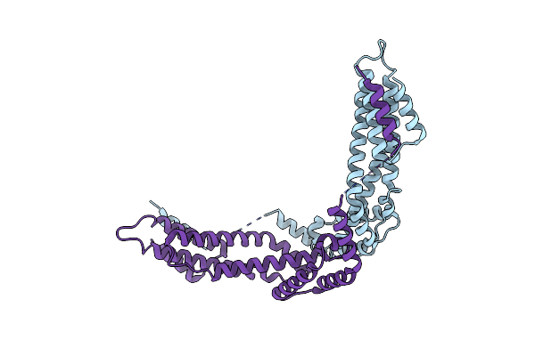 |
Crystal Structure Of The C-Terminal Fragment (Residues 716-982) Of Arabidopsis Thaliana Chup1
Organism: Arabidopsis thaliana
Method: X-RAY DIFFRACTION Resolution:3.00 Å Release Date: 2024-01-17 Classification: PLANT PROTEIN |
Organism: Arabidopsis thaliana
Method: X-RAY DIFFRACTION
Release Date: 2024-01-17
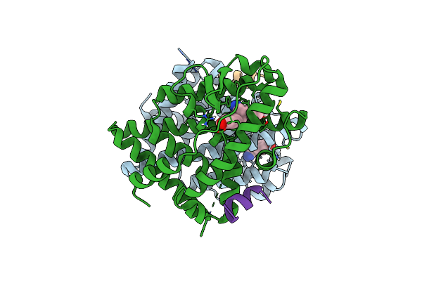 |
Human Estrogen Receptor Beta Ligand-Binding Domain In Complex With S-Dpn
Organism: Homo sapiens
Method: X-RAY DIFFRACTION Resolution:1.54 Å Release Date: 2022-07-20 Classification: TRANSCRIPTION/INHIBITOR Ligands: I0K |
Organism: Homo sapiens
Method: X-RAY DIFFRACTION
Release Date: 2022-07-20
Ligands: I0K
 |
Human Estrogen Receptor Beta Ligand-Binding Domain In Complex With (R)-3-(2-Chloro-4-Hydroxyphenyl)-2-(4-Hydroxyphenyl)Propanenitrile
Organism: Homo sapiens
Method: X-RAY DIFFRACTION Resolution:2.08 Å Release Date: 2022-07-20 Classification: TRANSCRIPTION/INHIBITOR Ligands: I1D |
Organism: Homo sapiens
Method: X-RAY DIFFRACTION
Release Date: 2022-07-20
Ligands: I1D
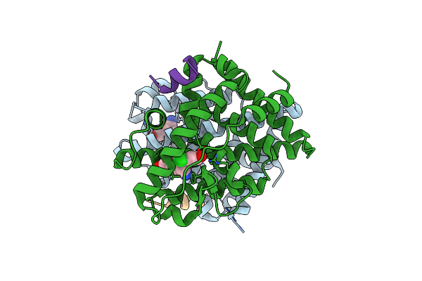 |
Human Estrogen Receptor Beta Ligand-Binding Domain In Complex With (S)-3-(2-Chloro-4-Hydroxyphenyl)-2-(4-Hydroxyphenyl)Propanenitrile
Organism: Homo sapiens
Method: X-RAY DIFFRACTION Resolution:1.92 Å Release Date: 2022-07-20 Classification: TRANSCRIPTION/INHIBITOR Ligands: I1I |
Organism: Homo sapiens
Method: X-RAY DIFFRACTION
Release Date: 2022-07-20
Ligands: I1I
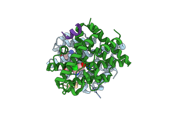 |
Human Estrogen Receptor Beta Ligand-Binding Domain In Complex With (R)-2-(2-Chloro-4-Hydroxyphenyl)-3-(4-Hydroxyphenyl)Propanenitrile
Organism: Homo sapiens
Method: X-RAY DIFFRACTION Resolution:1.89 Å Release Date: 2022-07-20 Classification: TRANSCRIPTION/INHIBITOR Ligands: I1M |
Organism: Homo sapiens
Method: X-RAY DIFFRACTION
Release Date: 2022-07-20
Ligands: I1M
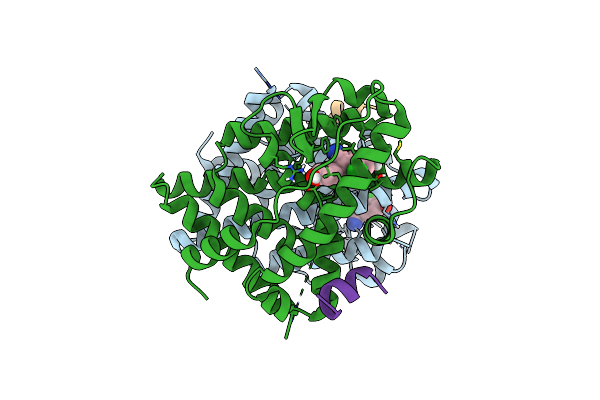 |
Human Estrogen Receptor Beta Ligand-Binding Domain In Complex With (S)-2-(2-Chloro-4-Hydroxyphenyl)-3-(4-Hydroxyphenyl)Propanenitrile
Organism: Homo sapiens
Method: X-RAY DIFFRACTION Resolution:2.16 Å Release Date: 2022-07-20 Classification: TRANSCRIPTION/INHIBITOR Ligands: I2B |
Organism: Homo sapiens
Method: X-RAY DIFFRACTION
Release Date: 2022-07-20
Ligands: I2B
 |
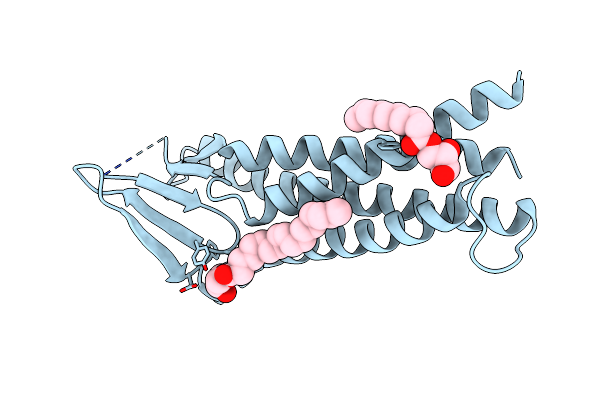
Pyp Mutant - E46Q
Organism: Halorhodospira halophila
Method: NEUTRON DIFFRACTION Resolution:1.49 Å Release Date: 2017-08-30 Classification: SIGNALING PROTEIN Ligands: HC4, DOD |
Organism: Halorhodospira halophila
Method: NEUTRON DIFFRACTION
Release Date: 2017-08-30
Ligands: HC4, DOD
 |
Crystal Structure Of Mouse Claudin-15
Organism: Mus musculus
Method: X-RAY DIFFRACTION Resolution:2.40 Å Release Date: 2014-04-30 Classification: CELL ADHESION Ligands: OLC |
Organism: Mus musculus
Method: X-RAY DIFFRACTION
Release Date: 2014-04-30
Ligands: OLC
 |
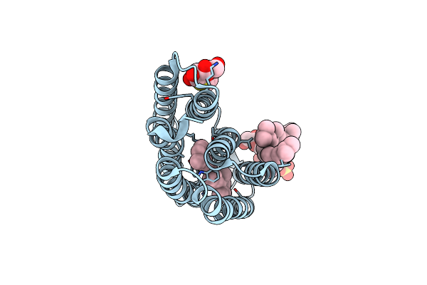
Crystal Structure Of The Trans Isomer Of The L93A Mutant Of Bacteriorhodopsin
Organism: Halobacterium
Method: X-RAY DIFFRACTION Resolution:2.30 Å Release Date: 2012-06-13 Classification: PROTON TRANSPORT Ligands: RET, SOG, L2P |
Organism: Halobacterium
Method: X-RAY DIFFRACTION
Release Date: 2012-06-13
Ligands: RET, SOG, L2P
 |
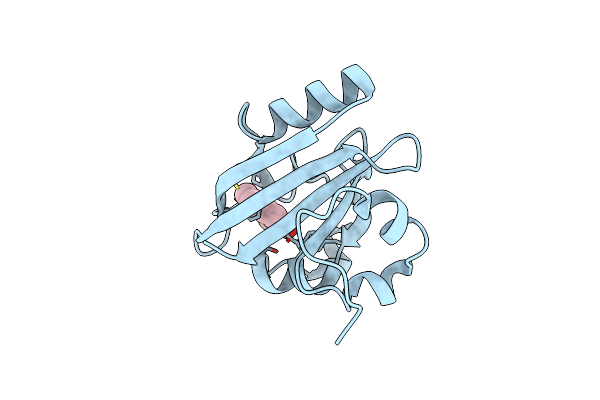
Crystal Structure Of The O Intermediate Of The L93A Mutant Of Bacteriorhodopsin
Organism: Halobacterium
Method: X-RAY DIFFRACTION Resolution:2.30 Å Release Date: 2012-06-13 Classification: PROTON TRANSPORT Ligands: RET, SOG, L2P |
Organism: Halobacterium
Method: X-RAY DIFFRACTION
Release Date: 2012-06-13
Ligands: RET, SOG, L2P
 |
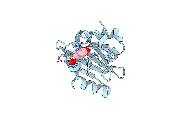
X-Ray Crystal Structure Of Photoactive Yellow Protein, Wild Type, At 295K
Organism: Halorhodospira halophila
Method: X-RAY DIFFRACTION Resolution:1.25 Å Release Date: 2009-03-24 Classification: SIGNALING PROTEIN Ligands: HC4 |
Organism: Halorhodospira halophila
Method: X-RAY DIFFRACTION
Release Date: 2009-03-24
Ligands: HC4
 |
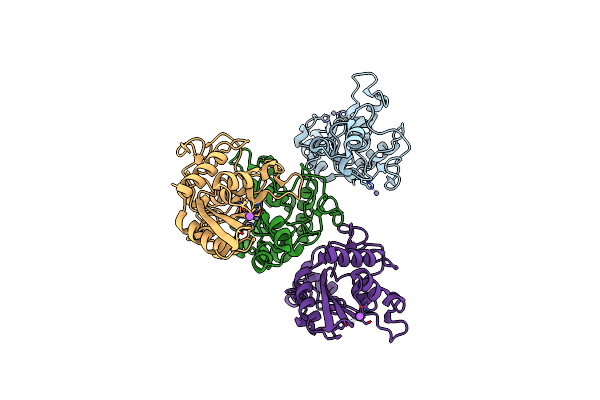
Neutron Crystal Structure Of Photoactive Yellow Protein, Wild Type, At 295K
Organism: Halorhodospira halophila
Method: NEUTRON DIFFRACTION Resolution:1.50 Å Release Date: 2009-03-24 Classification: SIGNALING PROTEIN Ligands: HC4, DOD |
Organism: Halorhodospira halophila
Method: NEUTRON DIFFRACTION
Release Date: 2009-03-24
Ligands: HC4, DOD
 |
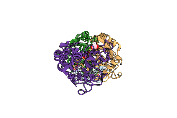
Crystal Structure Of Zinc-Bound Pseudecin From Pseudechis Porphyriacus
Organism: Pseudechis porphyriacus
Method: X-RAY DIFFRACTION Resolution:2.30 Å Release Date: 2008-03-11 Classification: TOXIN Ligands: ZN, NA |
Organism: Pseudechis porphyriacus
Method: X-RAY DIFFRACTION
Release Date: 2008-03-11
Ligands: ZN, NA
 |
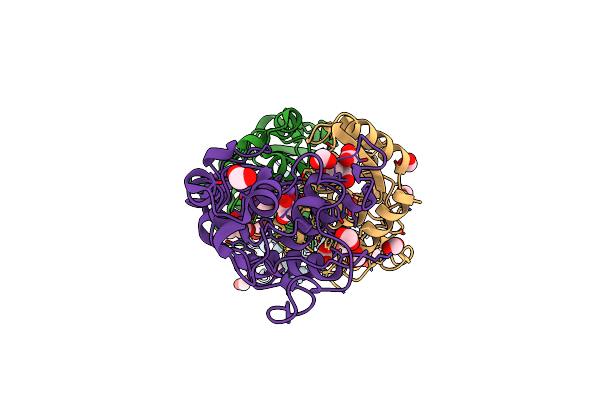
Crystal Structure Of Pseudechetoxin From Pseudechis Australis
Organism: Pseudechis australis
Method: X-RAY DIFFRACTION Resolution:2.25 Å Release Date: 2007-01-30 Classification: TOXIN |
Organism: Pseudechis australis
Method: X-RAY DIFFRACTION
Release Date: 2007-01-30
 |
Crystal Structure Of Pseudecin From Pseudechis Porphyriacus
Organism: Pseudechis porphyriacus
Method: X-RAY DIFFRACTION Resolution:1.90 Å Release Date: 2007-01-30 Classification: TOXIN |
Organism: Pseudechis porphyriacus
Method: X-RAY DIFFRACTION
Release Date: 2007-01-30
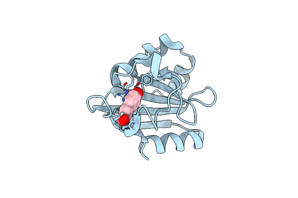 |
Wild Type Photoactive Yellow Protein, P65 Form
Organism: Halorhodospira halophila
Method: X-RAY DIFFRACTION Resolution:1.34 Å Release Date: 2006-04-04 Classification: SIGNALING PROTEIN Ligands: HC4 |
Organism: Halorhodospira halophila
Method: X-RAY DIFFRACTION
Release Date: 2006-04-04
Ligands: HC4