

Search Count: 10
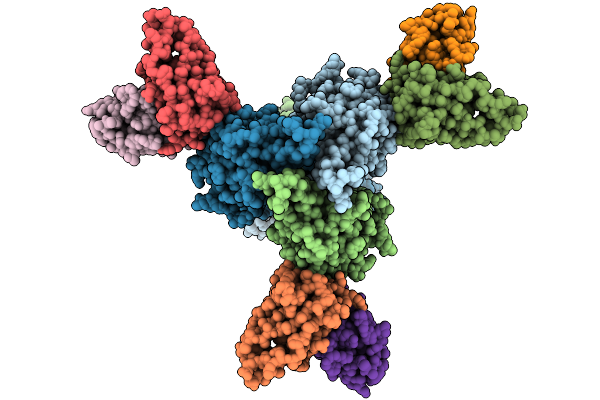 |
Structure Of Mhv68 Glycoprotein B In Complex With Fab5
Organism: Mus musculus, Murid herpesvirus 68
Method: ELECTRON MICROSCOPY Release Date: 2026-01-21 Classification: VIRAL PROTEIN/IMMUNE SYSTEM |
Organism: Mus musculus, Murid herpesvirus 68
Method: ELECTRON MICROSCOPY
Release Date: 2026-01-21
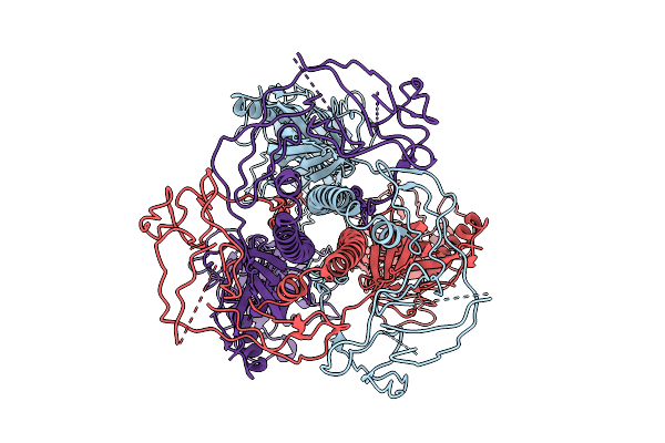 |
Structure Of Mhv68 Glycoprotein B
Organism: Murid gammaherpesvirus 4
Method: ELECTRON MICROSCOPY Resolution:3.30 Å Release Date: 2026-01-21 Classification: VIRAL PROTEIN |
Organism: Murid gammaherpesvirus 4
Method: ELECTRON MICROSCOPY
Release Date: 2026-01-21
 |
Macacine Gammaherpesvirus 4 Glycoprotein B In Complex With Fab5
Organism: Macacine gammaherpesvirus 4, Mus musculus
Method: ELECTRON MICROSCOPY Release Date: 2026-01-21 Classification: VIRAL PROTEIN/IMMUNE SYSTEM |
Organism: Macacine gammaherpesvirus 4, Mus musculus
Method: ELECTRON MICROSCOPY
Release Date: 2026-01-21
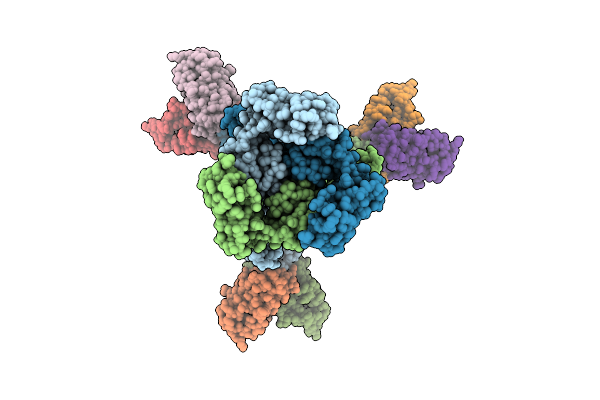 |
Structure Of A Murine Monoclonal Antibody Fab5 Targeting Epstein-Barr Virus Gb
Organism: Homo sapiens, Human gammaherpesvirus 4
Method: ELECTRON MICROSCOPY Release Date: 2025-10-15 Classification: VIRAL PROTEIN/IMMUNE SYSTEM Ligands: NAG |
Organism: Homo sapiens, Human gammaherpesvirus 4
Method: ELECTRON MICROSCOPY
Release Date: 2025-10-15
Ligands: NAG
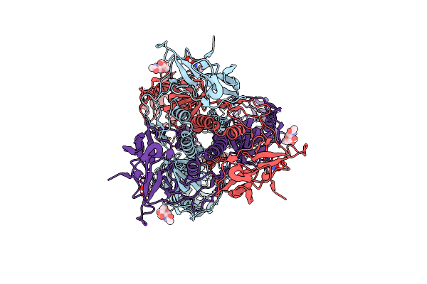 |
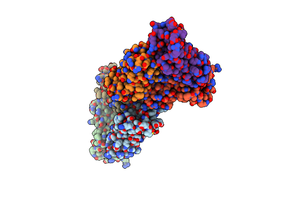
Structure Of Hhv6B Glycoprotein B
Organism: Human herpesvirus 6 strain z29
Method: ELECTRON MICROSCOPY Release Date: 2025-06-25 Classification: VIRAL PROTEIN Ligands: NAG |
Organism: Human herpesvirus 6 strain z29
Method: ELECTRON MICROSCOPY
Release Date: 2025-06-25
Ligands: NAG
 |
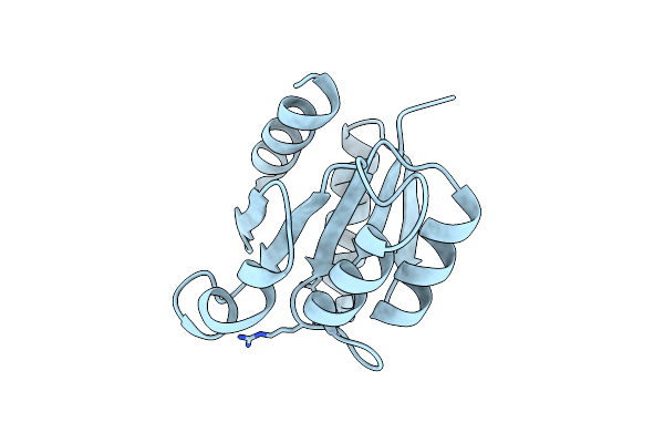
Crystal Structure Of Histidine-Containing Phosphotransfer Protein B (Hptb) From Pseudomonas Aeruginosa Pao1
Organism: Pseudomonas aeruginosa
Method: X-RAY DIFFRACTION Resolution:1.58 Å Release Date: 2020-11-04 Classification: TRANSFERASE |
Organism: Pseudomonas aeruginosa
Method: X-RAY DIFFRACTION
Release Date: 2020-11-04
 |
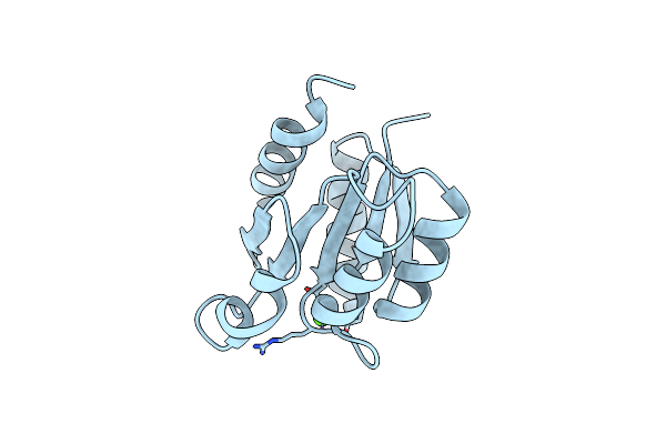
Crystal Structure Of The Receiver Domain Of Sensor Histidine Kinase Pa1611 (Pa1611Rec) From Pseudomonas Aeruginosa Pao1 With Magnesium Ion Coordinated In The Active Site Cleft
Organism: Pseudomonas aeruginosa
Method: X-RAY DIFFRACTION Resolution:1.35 Å Release Date: 2020-11-04 Classification: TRANSFERASE Ligands: MG |
Organism: Pseudomonas aeruginosa
Method: X-RAY DIFFRACTION
Release Date: 2020-11-04
Ligands: MG
 |
Crystal Structure Of The Receiver Domain Of Sensor Histidine Kinase Pa1611 (Pa1611Rec) From Pseudomonas Aeruginosa Pao1 With Calcium Ion Coordinated In The Active Site Cleft
Organism: Pseudomonas aeruginosa
Method: X-RAY DIFFRACTION Resolution:1.31 Å Release Date: 2020-11-04 Classification: TRANSFERASE Ligands: CA |
Organism: Pseudomonas aeruginosa
Method: X-RAY DIFFRACTION
Release Date: 2020-11-04
Ligands: CA
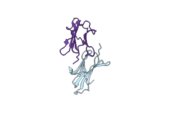 |
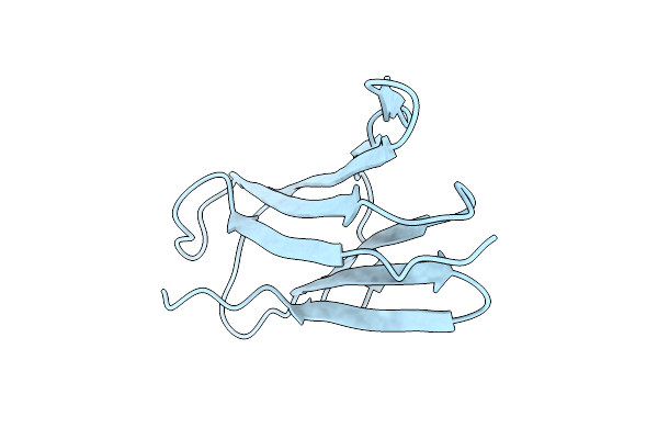
Crystal Structure Of Human Double Mutant Beta2-Microglobulin Q8H-L65T
Organism: Homo sapiens
Method: X-RAY DIFFRACTION Resolution:1.95 Å Release Date: 2014-10-15 Classification: IMMUNE SYSTEM |
Organism: Homo sapiens
Method: X-RAY DIFFRACTION
Release Date: 2014-10-15
 |
Crystal Structure Of Monomeric Zebrafish Beta-2-Microglobulin
Organism: Danio rerio
Method: X-RAY DIFFRACTION Resolution:2.14 Å Release Date: 2014-09-24 Classification: IMMUNE SYSTEM |
Organism: Danio rerio
Method: X-RAY DIFFRACTION
Release Date: 2014-09-24