
Search Count: 33
All
Selected
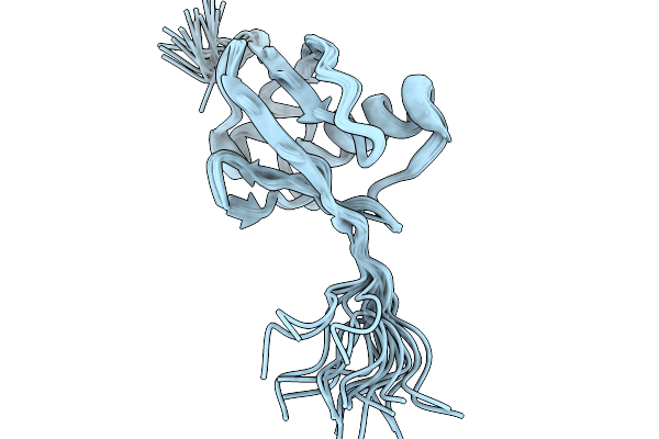 |
Nmr Structure Of Proteinmpnn-Desighed Ubiquitin Variant R4 At Ph 3 With 8 M Urea
Organism: Escherichia coli bl21(de3)
Method: SOLUTION NMR Release Date: 2026-02-11 Classification: DE NOVO PROTEIN |
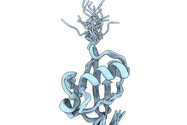 |
Nmr Structure Of Proteinmpnn-Desighed Ubiquitin Variant R4 At Ph 6.3 With 8 M Urea
Organism: Escherichia coli bl21(de3)
Method: SOLUTION NMR Release Date: 2026-02-11 Classification: DE NOVO PROTEIN |
 |
Organism: Escherichia coli bl21(de3)
Method: SOLUTION NMR Release Date: 2026-02-11 Classification: DE NOVO PROTEIN |
 |
Organism: Escherichia coli bl21(de3)
Method: SOLUTION NMR Release Date: 2026-02-11 Classification: DE NOVO PROTEIN |
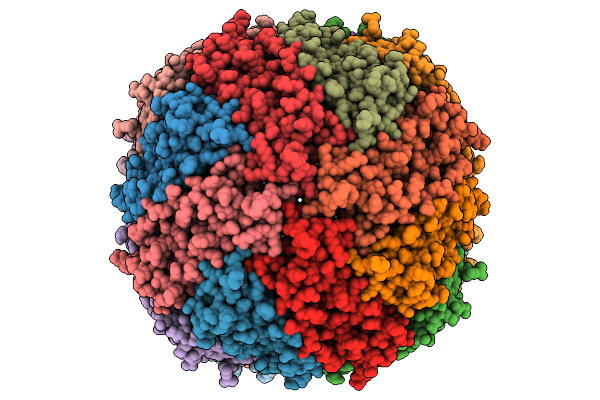 |
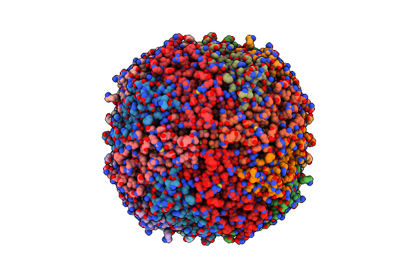
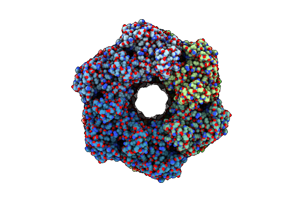
Cryo-Et Subtomogram-Averaged Structure Of Mouse Heavy-Chain Apoferritin Resolved At 2.71 Angstroms
Organism: Mus musculus
Method: ELECTRON MICROSCOPY Release Date: 2026-01-28 Classification: OXIDOREDUCTASE Ligands: MG |
 |
Crystal Structure Of Synthetic Ubiquitin Variant R10 Designed By Proteinmpnn
Organism: Synthetic construct
Method: X-RAY DIFFRACTION Resolution:1.55 Å Release Date: 2025-02-12 Classification: DE NOVO PROTEIN Ligands: GOL |
 |
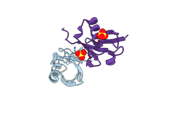
Crystal Structure Of Synthetic Ubiquitin Variant R4 Designed By Proteinmpnn
Organism: Synthetic construct
Method: X-RAY DIFFRACTION Resolution:1.39 Å Release Date: 2025-02-12 Classification: DE NOVO PROTEIN Ligands: SO4 |
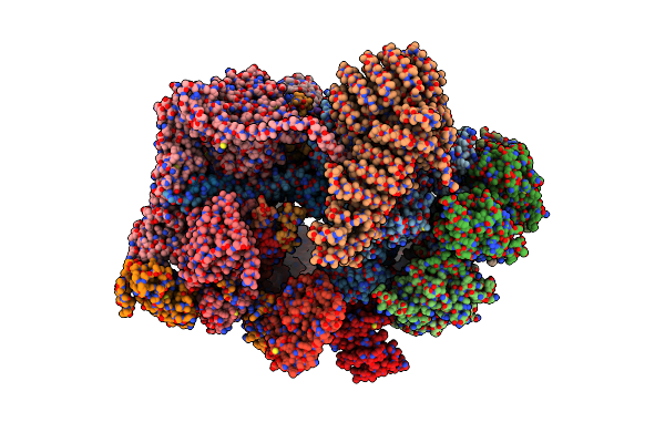 |
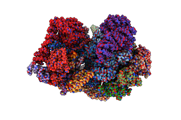
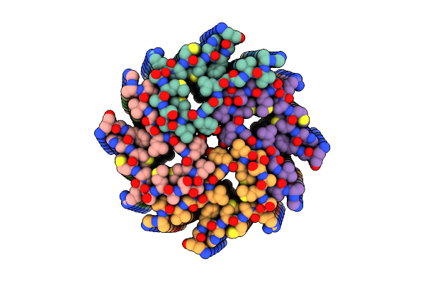
Cryo-Em Structure Of Human 26S Rp (Ed State) Bound To K11/K48-Branched Ubiquitin (Ub) Chain Composed Of Four Ub.
Organism: Homo sapiens, Saccharomyces cerevisiae
Method: ELECTRON MICROSCOPY Release Date: 2025-01-22 Classification: CYTOSOLIC PROTEIN Ligands: ATP, MG, ADP |
 |
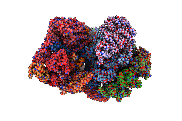
Cryo-Em Structure Of Human 26S Rp (Eb State) Bound To K11/K48-Branched Ubiquitin (Ub) Chain Composed Of Four Ub.
Organism: Homo sapiens
Method: ELECTRON MICROSCOPY Release Date: 2024-12-25 Classification: CYTOSOLIC PROTEIN Ligands: ATP, MG, ADP |
 |
Cryo-Em Structure Of Human 26S Proteasomal Rp Subcomplex (Ea State) Without Any Bound Substrate.
Organism: Homo sapiens
Method: ELECTRON MICROSCOPY Release Date: 2024-12-18 Classification: CYTOSOLIC PROTEIN Ligands: ATP, MG, ADP |
 |
Cryo-Em Structure Of Human 26S Proteasomal Rp Subcomplex (Ea State) Bound To K11/K48-Branched Ubiquitin (Ub) Chain Composed Of Three Ub.
Organism: Homo sapiens
Method: ELECTRON MICROSCOPY Release Date: 2024-12-18 Classification: CYTOSOLIC PROTEIN Ligands: ATP, MG, ADP |
 |
Cryo-Em Structure Of Mouse Heavy-Chain Apoferritin Resolved At 1.51 Angstroms
Organism: Mus musculus
Method: ELECTRON MICROSCOPY Release Date: 2024-08-21 Classification: OXIDOREDUCTASE Ligands: MG, K, FE |
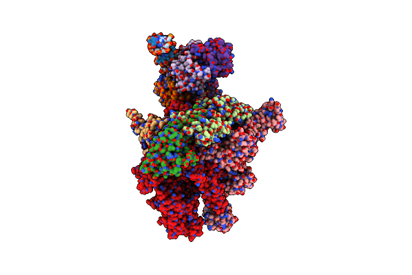 |
Structural Basis Of Transcriptional Activation By The Ompr/Phob-Family Response Regulator Pmra
Organism: Escherichia coli bl21(de3), Klebsiella pneumoniae jm45, Synthetic construct
Method: ELECTRON MICROSCOPY Release Date: 2023-08-30 Classification: TRANSCRIPTION |
 |
Robust Design Of Effective Allosteric Activator Ubv R4 For Rsp5 E3 Ligase Using The Machine-Learning Tool Proteinmpnn
Organism: Synthetic construct
Method: X-RAY DIFFRACTION Resolution:3.00 Å Release Date: 2023-08-09 Classification: BIOSYNTHETIC PROTEIN Ligands: SO4 |
 |
Organism: Escherichia coli dh5[alpha]
Method: ELECTRON MICROSCOPY Release Date: 2023-03-29 Classification: BIOSYNTHETIC PROTEIN |
 |
Organism: Escherichia coli dh5[alpha]
Method: X-RAY DIFFRACTION Resolution:1.60 Å Release Date: 2023-03-29 Classification: BIOSYNTHETIC PROTEIN Ligands: CL, NA, BME, GOL |
 |
Organism: Mesocricetus auratus
Method: ELECTRON MICROSCOPY Release Date: 2022-07-27 Classification: PROTEIN FIBRIL |
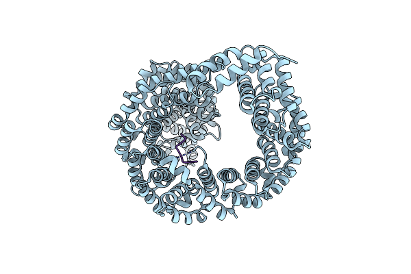 |
Crystal Structure Of Transportin-1 In Complex With Bap1 Py-Nls (Residues 706-724)
Organism: Homo sapiens
Method: X-RAY DIFFRACTION Resolution:3.76 Å Release Date: 2022-04-20 Classification: PROTEIN TRANSPORT |
 |
Organism: Escherichia coli bl21(de3)
Method: ELECTRON MICROSCOPY Release Date: 2022-04-06 Classification: BIOSYNTHETIC PROTEIN Ligands: NI |
 |
Cryo-Em Structure Of Spike Protein Of Feline Infectious Peritonitis Virus Strain Uu4
Organism: Feline infectious peritonitis virus
Method: ELECTRON MICROSCOPY Release Date: 2020-01-15 Classification: VIRAL PROTEIN Ligands: NAG, MAN |