

Search Count: 30
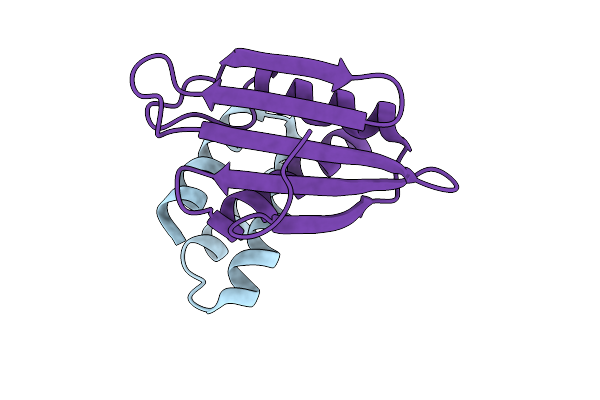 |
Crystal Structure Of Flpp3-Minibinder687 Complex
Organism: Francisella tularensis, Synthetic construct
Method: X-RAY DIFFRACTION Resolution:2.37 Å Release Date: 2025-10-22 Classification: APOPTOSIS Ligands: IOD |
Organism: Francisella tularensis, Synthetic construct
Method: X-RAY DIFFRACTION
Release Date: 2025-10-22
Ligands: IOD
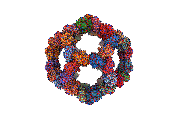 |
Structure Of A Gp140 Spytag-Spycatcher Mi3 Nanoparticle Including Mi3 Density Only.
Organism: Thermotoga maritima
Method: ELECTRON MICROSCOPY Release Date: 2024-10-09 Classification: IMMUNE SYSTEM |
Organism: Thermotoga maritima
Method: ELECTRON MICROSCOPY
Release Date: 2024-10-09
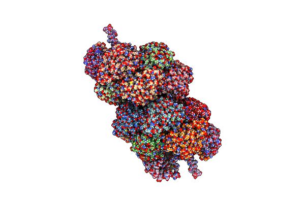 |
Cyanide Dihydratase From Bacillus Pumilus C1 E155R Variant With Altered Helical Twist.
Organism: Bacillus pumilus
Method: ELECTRON MICROSCOPY Release Date: 2024-04-10 Classification: HYDROLASE |
Organism: Bacillus pumilus
Method: ELECTRON MICROSCOPY
Release Date: 2024-04-10
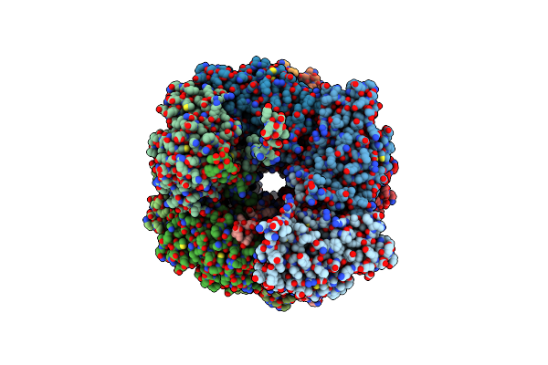 |
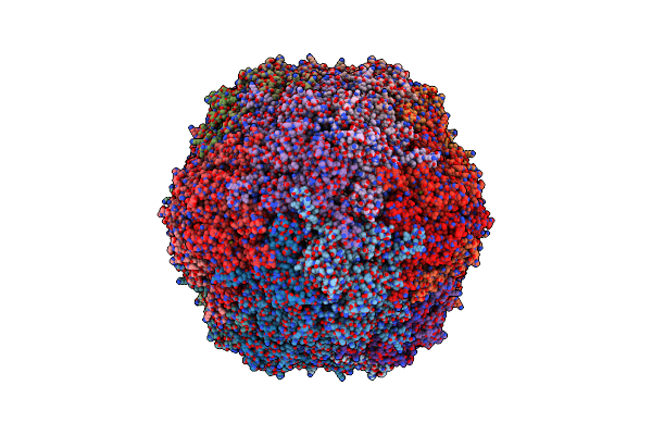
Cyanide Dihydratase From Bacillus Pumilus C1
Organism: Bacillus pumilus
Method: ELECTRON MICROSCOPY Release Date: 2023-08-16 Classification: HYDROLASE |
Organism: Bacillus pumilus
Method: ELECTRON MICROSCOPY
Release Date: 2023-08-16
 |
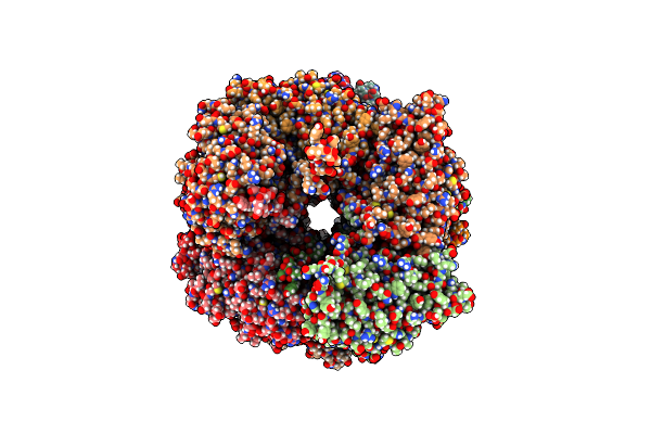
Cryo-Em Structure Of The Dyp Peroxidase-Loaded Encapsulin Nanocompartment From Mycobacterium Tuberculosis With Icosahedral Symmetry Imposed.
Organism: Mycobacterium tuberculosis h37rv
Method: ELECTRON MICROSCOPY Release Date: 2023-08-09 Classification: VIRUS LIKE PARTICLE |
Organism: Mycobacterium tuberculosis h37rv
Method: ELECTRON MICROSCOPY
Release Date: 2023-08-09
 |
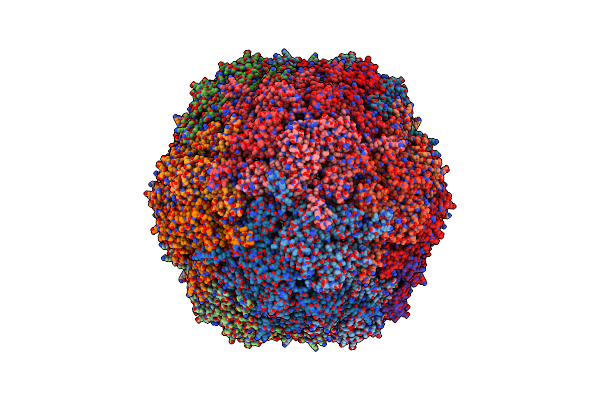
Cyanide Dihydratase From Bacillus Pumilus C1 Variant - Q86R,H305K,H308K,H323K
Organism: Bacillus pumilus
Method: ELECTRON MICROSCOPY Release Date: 2023-01-18 Classification: HYDROLASE |
Organism: Bacillus pumilus
Method: ELECTRON MICROSCOPY
Release Date: 2023-01-18
 |
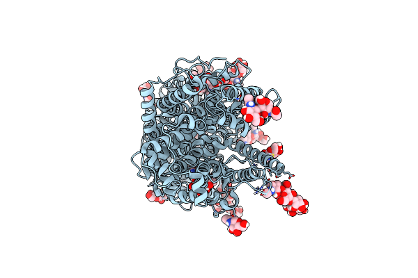
Cryo-Em Structure Of Mycobacterium Tuberculosis Encapsulin
Organism: Mycobacterium tuberculosis h37rv
Method: ELECTRON MICROSCOPY Release Date: 2022-07-20 Classification: STRUCTURAL PROTEIN |
Organism: Mycobacterium tuberculosis h37rv
Method: ELECTRON MICROSCOPY
Release Date: 2022-07-20
 |
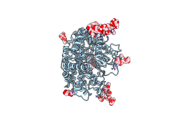
Structure Of Full-Length, Monomeric, Soluble Somatic Angiotensin I-Converting Enzyme Showing The N- And C-Terminal Ellipsoid Domains
Organism: Homo sapiens
Method: ELECTRON MICROSCOPY Release Date: 2022-07-20 Classification: HYDROLASE Ligands: ZN, CL, NAG |
Organism: Homo sapiens
Method: ELECTRON MICROSCOPY
Release Date: 2022-07-20
Ligands: ZN, CL, NAG
 |
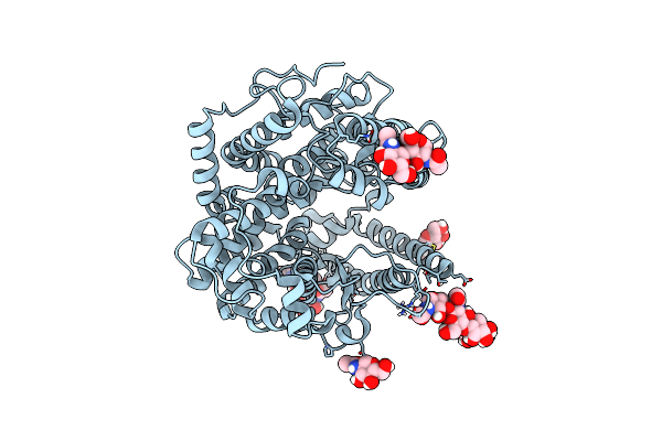
Local Refinement Structure Of The N-Domain Of Full-Length, Monomeric, Soluble Somatic Angiotensin I-Converting Enzyme
Organism: Homo sapiens
Method: ELECTRON MICROSCOPY Release Date: 2022-07-20 Classification: HYDROLASE Ligands: ZN, NAG |
Organism: Homo sapiens
Method: ELECTRON MICROSCOPY
Release Date: 2022-07-20
Ligands: ZN, NAG
 |
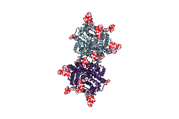
Local Refinement Structure Of The C-Domain Of Full-Length, Monomeric, Soluble Somatic Angiotensin I-Converting Enzyme
Organism: Homo sapiens
Method: ELECTRON MICROSCOPY Release Date: 2022-07-20 Classification: HYDROLASE Ligands: CL, NAG |
Organism: Homo sapiens
Method: ELECTRON MICROSCOPY
Release Date: 2022-07-20
Ligands: CL, NAG
 |
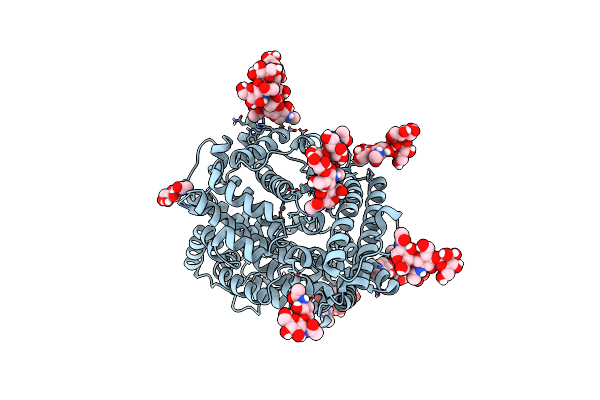
Local Refinement Structure Of The Two Interacting N-Domains Of Full-Length, Dimeric, Soluble Somatic Angiotensin I-Converting Enzyme
Organism: Homo sapiens
Method: ELECTRON MICROSCOPY Release Date: 2022-07-20 Classification: HYDROLASE Ligands: ZN, NAG |
Organism: Homo sapiens
Method: ELECTRON MICROSCOPY
Release Date: 2022-07-20
Ligands: ZN, NAG
 |
Local Refinement Structure Of A Single N-Domain Of Full-Length, Dimeric, Soluble Somatic Angiotensin I-Converting Enzyme
Organism: Homo sapiens
Method: ELECTRON MICROSCOPY Release Date: 2022-07-20 Classification: HYDROLASE Ligands: ZN, NAG |
Organism: Homo sapiens
Method: ELECTRON MICROSCOPY
Release Date: 2022-07-20
Ligands: ZN, NAG
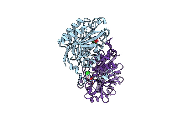 |
The C146A Variant Of An Amidase From Pyrococcus Horikoshii With Bound Acetamide
Organism: Pyrococcus horikoshii (strain atcc 700860 / dsm 12428 / jcm 9974 / nbrc 100139 / ot-3)
Method: X-RAY DIFFRACTION Resolution:1.65 Å Release Date: 2021-07-21 Classification: HYDROLASE Ligands: ACM, CL |
Organism: Pyrococcus horikoshii (strain atcc 700860 / dsm 12428 / jcm 9974 / nbrc 100139 / ot-3)
Method: X-RAY DIFFRACTION
Release Date: 2021-07-21
Ligands: ACM, CL
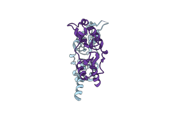 |
Crystal Structure Of Cbpb Protein (Lmo1009) From Listeria Monocytogenes
Organism: Listeria monocytogenes
Method: X-RAY DIFFRACTION Resolution:1.62 Å Release Date: 2020-09-09 Classification: CYTOSOLIC PROTEIN Ligands: CL |
Organism: Listeria monocytogenes
Method: X-RAY DIFFRACTION
Release Date: 2020-09-09
Ligands: CL
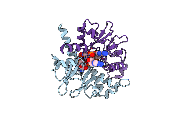 |
Crystal Structure Of Listeria Monocytogenes Cbpb Protein (Lmo1009) In Complex With C-Di-Amp
Organism: Listeria monocytogenes
Method: X-RAY DIFFRACTION Resolution:2.40 Å Release Date: 2020-09-09 Classification: CYTOSOLIC PROTEIN Ligands: 2BA |
Organism: Listeria monocytogenes
Method: X-RAY DIFFRACTION
Release Date: 2020-09-09
Ligands: 2BA
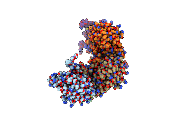 |
Cryo-Em Informed Directed Evolution Of Nitrilase 4 Leads To A Change In Quaternary Structure.
Organism: Arabidopsis thaliana
Method: ELECTRON MICROSCOPY Release Date: 2019-11-20 Classification: HYDROLASE |
Organism: Arabidopsis thaliana
Method: ELECTRON MICROSCOPY
Release Date: 2019-11-20
 |
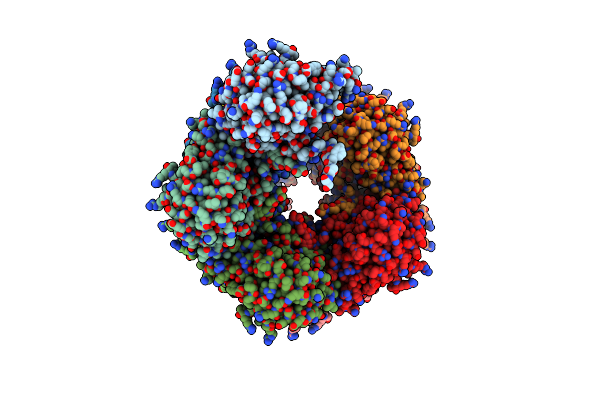
Cryo-Em Informed Directed Evolution Of Nitrilase 4 Leads To A Change In Quaternary Structure.
Organism: Arabidopsis thaliana
Method: ELECTRON MICROSCOPY Release Date: 2019-11-20 Classification: HYDROLASE |
Organism: Arabidopsis thaliana
Method: ELECTRON MICROSCOPY
Release Date: 2019-11-20
 |
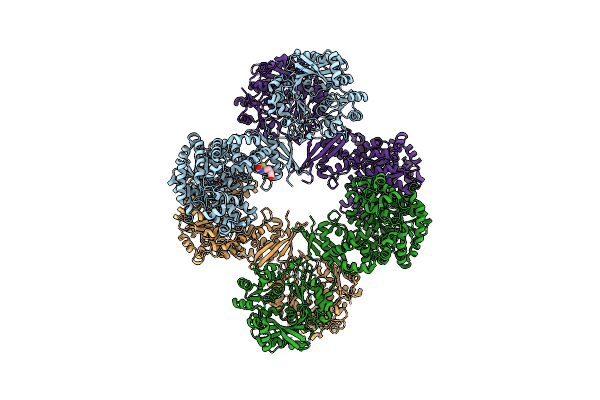
Cryo-Em Informed Directed Evolution Of Nitrilase 4 Leads To A Change In Quaternary Structure.
Organism: Arabidopsis thaliana
Method: ELECTRON MICROSCOPY Release Date: 2019-07-24 Classification: HYDROLASE |
Organism: Arabidopsis thaliana
Method: ELECTRON MICROSCOPY
Release Date: 2019-07-24
 |
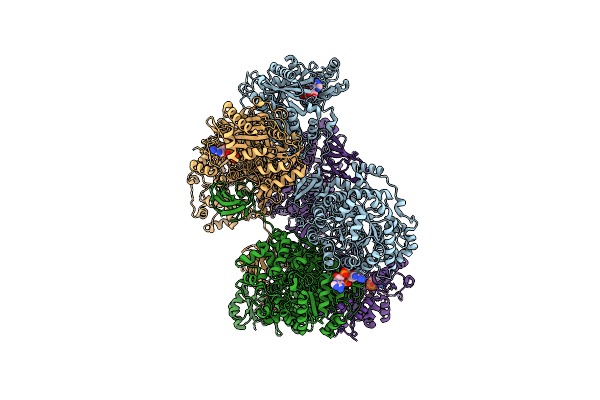
Crystal Structure Of Lactococcus Lactis Pyruvate Carboxylase
Organism: Lactococcus lactis
Method: X-RAY DIFFRACTION Resolution:3.10 Å Release Date: 2017-08-16 Classification: LIGASE Ligands: MN, BTN |
Organism: Lactococcus lactis
Method: X-RAY DIFFRACTION
Release Date: 2017-08-16
Ligands: MN, BTN
 |
Crystal Structure Of Lactococcus Lactis Pyruvate Carboxylase In Complex With Cyclic-Di-Amp
Organism: Lactococcus lactis
Method: X-RAY DIFFRACTION Resolution:2.30 Å Release Date: 2017-08-16 Classification: LIGASE Ligands: MN, ADP, MG, 2BA |
Organism: Lactococcus lactis
Method: X-RAY DIFFRACTION
Release Date: 2017-08-16
Ligands: MN, ADP, MG, 2BA