

Search Count: 81
 |
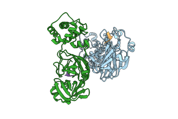
Discovery Of An Orally Bioavailable Reversible Covalent Sars-Cov-2 Mpro Inhibitor With Pan-Coronavirus Activity
Organism: Severe acute respiratory syndrome coronavirus 2, Synthetic construct
Method: X-RAY DIFFRACTION Resolution:1.37 Å Release Date: 2025-08-20 Classification: HYDROLASE/INHIBOTOR Ligands: A1L7M |
Organism: Severe acute respiratory syndrome coronavirus 2, Synthetic construct
Method: X-RAY DIFFRACTION
Release Date: 2025-08-20
Ligands: A1L7M
 |
Discovery Of An Orally Bioavailable Reversible Covalent Sars-Cov-2 Mpro Inhibitor With Pan-Coronavirus Activity
Organism: Severe acute respiratory syndrome coronavirus 2, Synthetic construct
Method: X-RAY DIFFRACTION Resolution:1.89 Å Release Date: 2025-08-20 Classification: HYDROLASE/INHIBITOR |
Organism: Severe acute respiratory syndrome coronavirus 2, Synthetic construct
Method: X-RAY DIFFRACTION
Release Date: 2025-08-20
 |
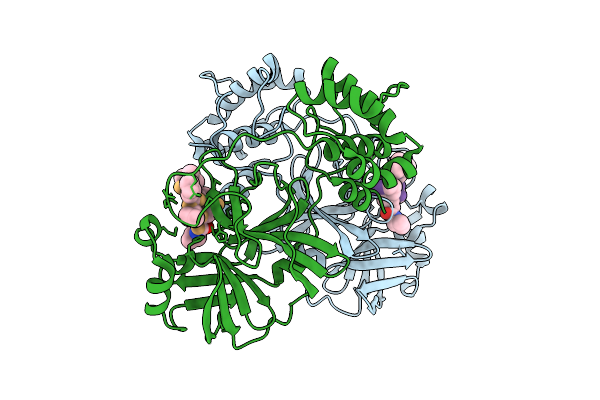
Discovery Of An Orally Bioavailable Reversible Covalent Sars-Cov-2 Mpro Inhibitor With Pan-Coronavirus Activity
Organism: Severe acute respiratory syndrome coronavirus 2, Synthetic construct
Method: X-RAY DIFFRACTION Resolution:1.50 Å Release Date: 2025-08-20 Classification: HYDROLASE/INHIBITOR |
Organism: Severe acute respiratory syndrome coronavirus 2, Synthetic construct
Method: X-RAY DIFFRACTION
Release Date: 2025-08-20
 |
Discovery Of An Orally Bioavailable Reversible Covalent Sars-Cov-2 Mpro Inhibitor With Pan-Coronavirus Activity
Organism: Severe acute respiratory syndrome coronavirus 2, Synthetic construct
Method: X-RAY DIFFRACTION Resolution:1.47 Å Release Date: 2025-08-20 Classification: HYDROLASE/INHIBITOR Ligands: A1L7M |
Organism: Severe acute respiratory syndrome coronavirus 2, Synthetic construct
Method: X-RAY DIFFRACTION
Release Date: 2025-08-20
Ligands: A1L7M
 |
Discovery Of An Orally Bioavailable Reversible Covalent Sars-Cov-2 Mpro Inhibitor With Pan-Coronavirus Activity
Organism: Severe acute respiratory syndrome coronavirus 2, Synthetic construct
Method: X-RAY DIFFRACTION Resolution:2.20 Å Release Date: 2025-08-20 Classification: HYDROLASE/INHIBITOR Ligands: A1L7M |
Organism: Severe acute respiratory syndrome coronavirus 2, Synthetic construct
Method: X-RAY DIFFRACTION
Release Date: 2025-08-20
Ligands: A1L7M
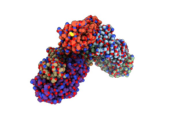 |
Crystal Structure Of Sars-Cov-2 Spike Receptor Binding Domain Bound With P5S-2B10
Organism: Homo sapiens, Severe acute respiratory syndrome coronavirus 2
Method: X-RAY DIFFRACTION Resolution:2.88 Å Release Date: 2023-05-24 Classification: VIRAL PROTEIN/IMMUNE SYSTEM |
Organism: Homo sapiens, Severe acute respiratory syndrome coronavirus 2
Method: X-RAY DIFFRACTION
Release Date: 2023-05-24
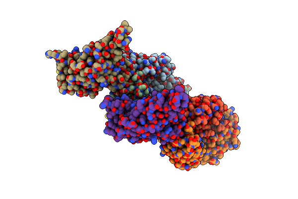 |
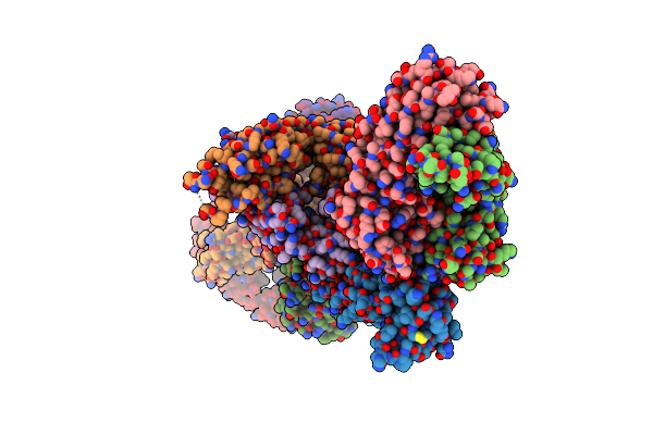
Crystal Structure Of Sars-Cov-2 Spike Receptor Binding Domain Bound With P2S-2E9 Fab
Organism: Homo sapiens, Severe acute respiratory syndrome coronavirus 2
Method: X-RAY DIFFRACTION Resolution:2.20 Å Release Date: 2023-05-10 Classification: VIRAL PROTEIN/IMMUNE SYSTEM |
Organism: Homo sapiens, Severe acute respiratory syndrome coronavirus 2
Method: X-RAY DIFFRACTION
Release Date: 2023-05-10
 |
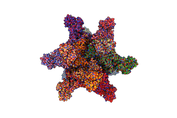
Crystal Structure Of Sars-Cov-2 Spike Receptor Binding Domain Bound With P5S-3B11 Fab
Organism: Severe acute respiratory syndrome coronavirus 2, Homo sapiens
Method: X-RAY DIFFRACTION Resolution:3.20 Å Release Date: 2023-05-10 Classification: VIRAL PROTEIN/IMMUNE SYSTEM |
Organism: Severe acute respiratory syndrome coronavirus 2, Homo sapiens
Method: X-RAY DIFFRACTION
Release Date: 2023-05-10
 |
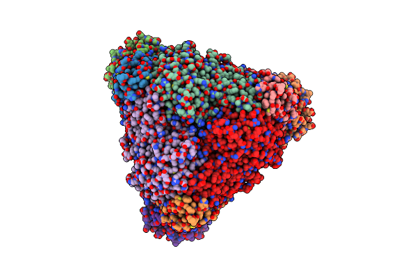
Cryoem Structure Of Helicobacter Pylori Urefd/Urease Complex
Organism: Helicobacter pylori 26695
Method: ELECTRON MICROSCOPY Release Date: 2023-05-03 Classification: HYDROLASE |
Organism: Helicobacter pylori 26695
Method: ELECTRON MICROSCOPY
Release Date: 2023-05-03
 |
Cryoem Structure Of Klebsiella Pneumoniae Ured/Urease Complex
Organism: Klebsiella pneumoniae
Method: ELECTRON MICROSCOPY Release Date: 2023-05-03 Classification: HYDROLASE |
Organism: Klebsiella pneumoniae
Method: ELECTRON MICROSCOPY
Release Date: 2023-05-03
 |
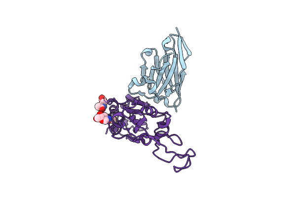
Crystal Structure Of Sars-Cov-2 Spike Receptor Binding Domain Bound With P5S-1H1 Fab
Organism: Homo sapiens, Severe acute respiratory syndrome coronavirus 2
Method: X-RAY DIFFRACTION Resolution:2.80 Å Release Date: 2023-04-26 Classification: VIRAL PROTEIN/IMMUNE SYSTEM Ligands: NAG |
Organism: Homo sapiens, Severe acute respiratory syndrome coronavirus 2
Method: X-RAY DIFFRACTION
Release Date: 2023-04-26
Ligands: NAG
 |
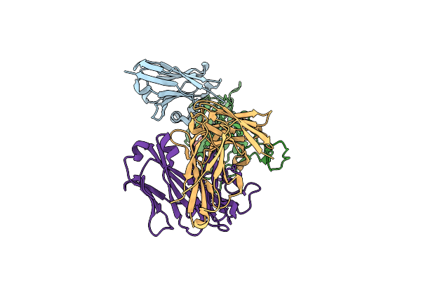
Crystal Structure Of Nanobody Nb70 With Sars-Cov Rbd
Organism: Severe acute respiratory syndrome-related coronavirus, Vicugna pacos
Method: X-RAY DIFFRACTION Resolution:2.40 Å Release Date: 2022-12-21 Classification: VIRAL PROTEIN/IMMUNE SYSTEM Ligands: NAG |
Organism: Severe acute respiratory syndrome-related coronavirus, Vicugna pacos
Method: X-RAY DIFFRACTION
Release Date: 2022-12-21
Ligands: NAG
 |
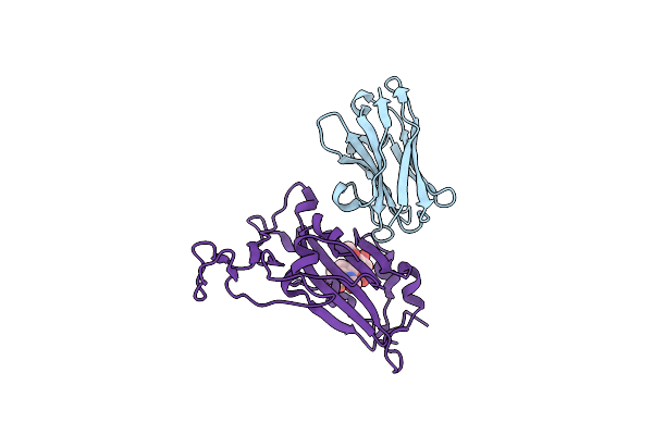
Crystal Structure Of Nanobody Nb70 With Antibody 1F11 Fab And Sars-Cov-2 Rbd
Organism: Severe acute respiratory syndrome coronavirus 2, Homo sapiens, Vicugna pacos
Method: X-RAY DIFFRACTION Resolution:2.40 Å Release Date: 2022-12-21 Classification: VIRAL PROTEIN/IMMUNE SYSTEM Ligands: NAG |
Organism: Severe acute respiratory syndrome coronavirus 2, Homo sapiens, Vicugna pacos
Method: X-RAY DIFFRACTION
Release Date: 2022-12-21
Ligands: NAG
 |
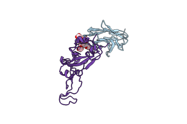
Crystal Structure Of Nanobody 1-2C7 With Sars-Cov-2 Rbd
Organism: Severe acute respiratory syndrome coronavirus 2, Vicugna pacos
Method: X-RAY DIFFRACTION Resolution:1.80 Å Release Date: 2022-12-14 Classification: VIRAL PROTEIN/IMMUNE SYSTEM |
Organism: Severe acute respiratory syndrome coronavirus 2, Vicugna pacos
Method: X-RAY DIFFRACTION
Release Date: 2022-12-14
 |
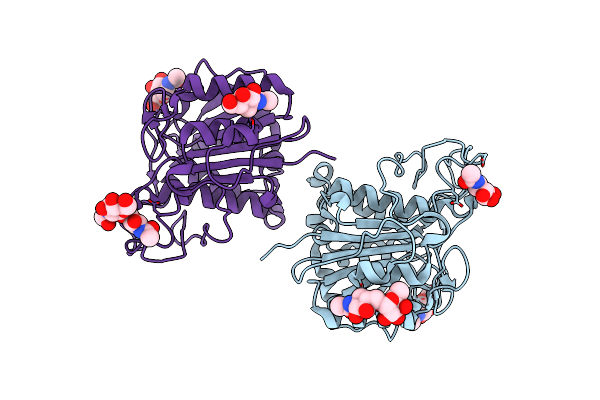
Crystal Structure Of Nanobody 3-2A2-4 With Sars-Cov-2 Rbd
Organism: Severe acute respiratory syndrome coronavirus 2, Vicugna pacos
Method: X-RAY DIFFRACTION Resolution:2.40 Å Release Date: 2022-12-07 Classification: VIRAL PROTEIN/IMMUNE SYSTEM |
Organism: Severe acute respiratory syndrome coronavirus 2, Vicugna pacos
Method: X-RAY DIFFRACTION
Release Date: 2022-12-07
 |
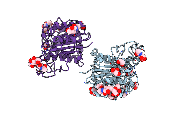
The Crystal Structure Of Vypal2-C214A, A Dead Mutant Of Vypal2 Peptide Asparaginyl Ligase In Form Ii
Organism: Viola philippica
Method: X-RAY DIFFRACTION Resolution:1.80 Å Release Date: 2022-07-13 Classification: PLANT PROTEIN Ligands: NAG |
Organism: Viola philippica
Method: X-RAY DIFFRACTION
Release Date: 2022-07-13
Ligands: NAG
 |
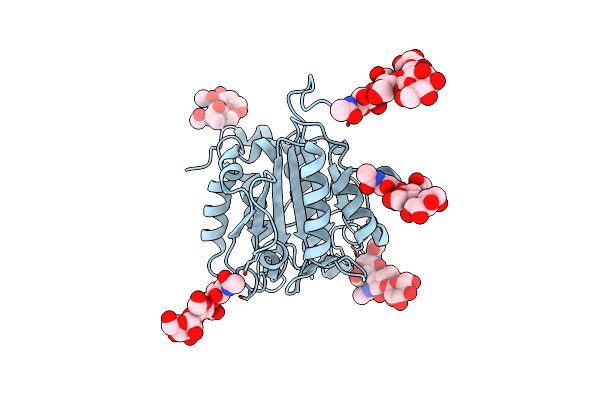
The Crystal Structure Of Vypal2-I244V, A More Efficient Mutant Of Vypal2 Peptide Asparaginyl Ligase In Its Active Enzyme Form
Organism: Viola philippica
Method: X-RAY DIFFRACTION Resolution:1.59 Å Release Date: 2022-06-29 Classification: PLANT PROTEIN Ligands: NAG, EDO, GOL |
Organism: Viola philippica
Method: X-RAY DIFFRACTION
Release Date: 2022-06-29
Ligands: NAG, EDO, GOL
 |
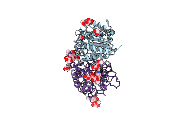
The Crystal Structure Of Vypal2-C214A, A Dead Mutant Of Vypal2 Peptide Asparaginyl Ligase In Form I
Organism: Viola philippica
Method: X-RAY DIFFRACTION Resolution:1.90 Å Release Date: 2022-06-29 Classification: PLANT PROTEIN |
Organism: Viola philippica
Method: X-RAY DIFFRACTION
Release Date: 2022-06-29
 |
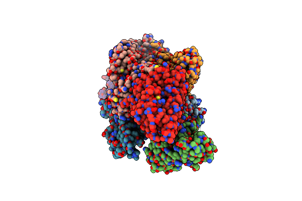
The Crystal Structure Of Vypal2 Peptide Asparaginyl Ligase In Its Active Enzyme Form
Organism: Viola philippica
Method: X-RAY DIFFRACTION Resolution:2.30 Å Release Date: 2022-06-29 Classification: PLANT PROTEIN Ligands: NAG, EDO |
Organism: Viola philippica
Method: X-RAY DIFFRACTION
Release Date: 2022-06-29
Ligands: NAG, EDO
 |
Crystal Structure Of Eif2Balpha With A Ligand.
Organism: Homo sapiens
Method: X-RAY DIFFRACTION Resolution:2.70 Å Release Date: 2021-07-21 Classification: SUGAR BINDING PROTEIN Ligands: M6P, ACT |
Organism: Homo sapiens
Method: X-RAY DIFFRACTION
Release Date: 2021-07-21
Ligands: M6P, ACT