

Search Count: 14
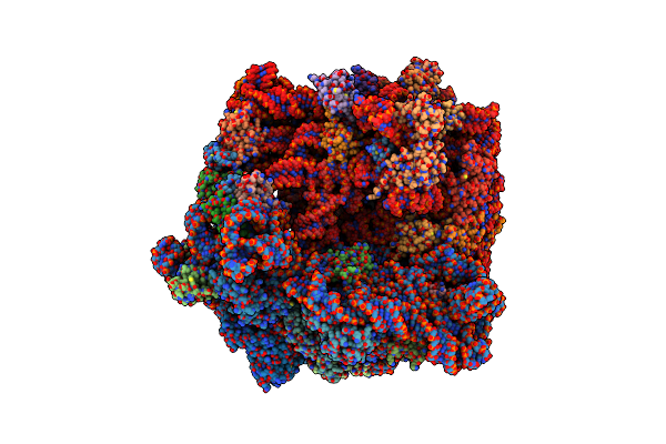 |
E. Coli 70S Ribosome Complexed With P. Putida Trnaile2 At The A-Site And P-Site
Organism: Escherichia coli, Pseudomonas putida nbrc 14164
Method: ELECTRON MICROSCOPY Release Date: 2024-11-06 Classification: RIBOSOME Ligands: MG |
Organism: Escherichia coli, Pseudomonas putida nbrc 14164
Method: ELECTRON MICROSCOPY
Release Date: 2024-11-06
Ligands: MG
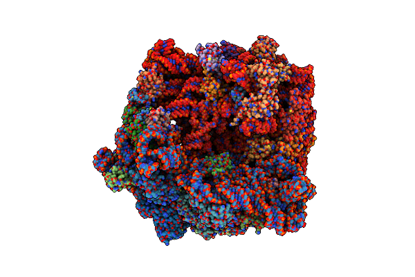 |
E. Coli 70S Ribosome Complexed With P. Putida Trnaile2 And A4 Mrna
Organism: Escherichia coli, Escherichia coli bw25113, Pseudomonas putida nbrc 14164
Method: ELECTRON MICROSCOPY Release Date: 2024-11-06 Classification: RIBOSOME Ligands: MG |
Organism: Escherichia coli, Escherichia coli bw25113, Pseudomonas putida nbrc 14164
Method: ELECTRON MICROSCOPY
Release Date: 2024-11-06
Ligands: MG
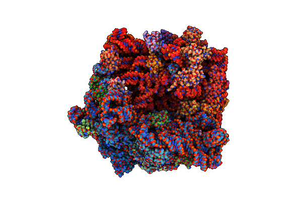 |
E. Coli 70S Ribosome Complexed With P. Putida Trnaile2 And Da4 Mrna
Organism: Escherichia coli, Escherichia coli bw25113, Pseudomonas putida nbrc 14164
Method: ELECTRON MICROSCOPY Release Date: 2024-11-06 Classification: RIBOSOME Ligands: MG |
Organism: Escherichia coli, Escherichia coli bw25113, Pseudomonas putida nbrc 14164
Method: ELECTRON MICROSCOPY
Release Date: 2024-11-06
Ligands: MG
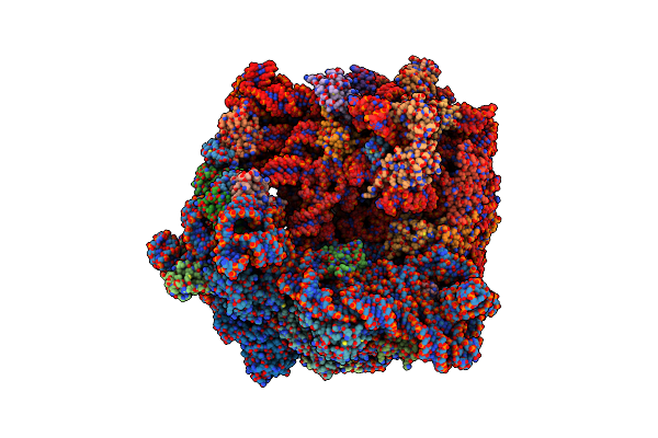 |
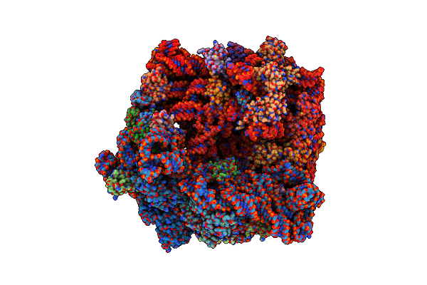
E. Coli 70S Ribosome Complexed With P. Putida Trnaile2 And Am4 Mrna
Organism: Escherichia coli, Escherichia coli bw25113, Pseudomonas putida nbrc 14164
Method: ELECTRON MICROSCOPY Release Date: 2024-11-06 Classification: RIBOSOME Ligands: MG |
Organism: Escherichia coli, Escherichia coli bw25113, Pseudomonas putida nbrc 14164
Method: ELECTRON MICROSCOPY
Release Date: 2024-11-06
Ligands: MG
 |
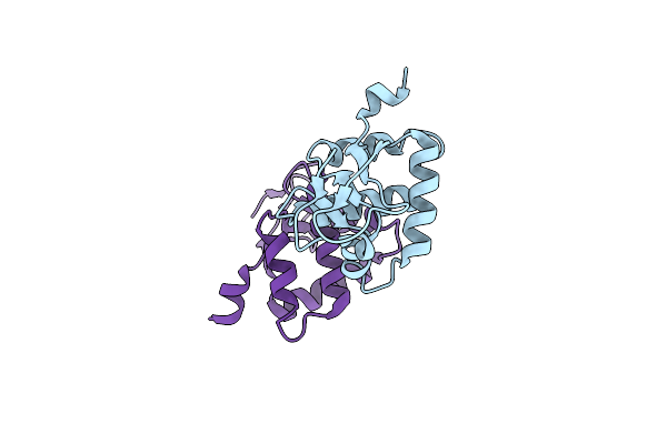
E. Coli 70S Ribosome Complexed With P.Putida Trnaile2 And A(F)4 Mrna
Organism: Escherichia coli, Escherichia coli bw25113, Pseudomonas putida nbrc 14164
Method: ELECTRON MICROSCOPY Release Date: 2024-11-06 Classification: RIBOSOME Ligands: MG |
Organism: Escherichia coli, Escherichia coli bw25113, Pseudomonas putida nbrc 14164
Method: ELECTRON MICROSCOPY
Release Date: 2024-11-06
Ligands: MG
 |
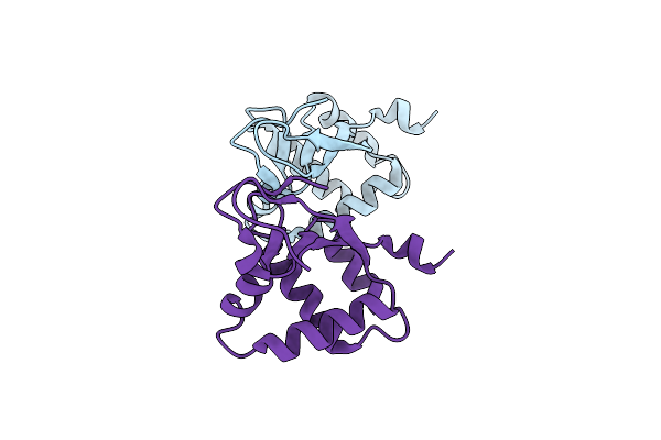
Crystal Structure Of The N-Terminal Domain (Domain 1) Of Rctb, Rctb-1-124-L48M
Organism: Vibrio cholerae
Method: X-RAY DIFFRACTION Resolution:2.00 Å Release Date: 2017-01-11 Classification: DNA BINDING PROTEIN |
Organism: Vibrio cholerae
Method: X-RAY DIFFRACTION
Release Date: 2017-01-11
 |
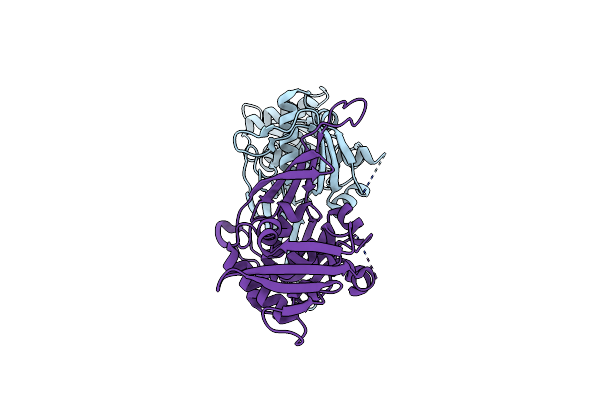
Crystal Structure Of The N-Terminal Domain (Domain 1) Of Rctb
Organism: Vibrio cholerae
Method: X-RAY DIFFRACTION Resolution:2.00 Å Release Date: 2017-01-11 Classification: DNA BINDING PROTEIN |
Organism: Vibrio cholerae
Method: X-RAY DIFFRACTION
Release Date: 2017-01-11
 |
Crystal Structure Of The Rctb Domains 2-3, Rctb-155-483
Organism: Vibrio cholerae
Method: X-RAY DIFFRACTION Resolution:2.60 Å Release Date: 2017-01-11 Classification: DNA BINDING PROTEIN |
Organism: Vibrio cholerae
Method: X-RAY DIFFRACTION
Release Date: 2017-01-11
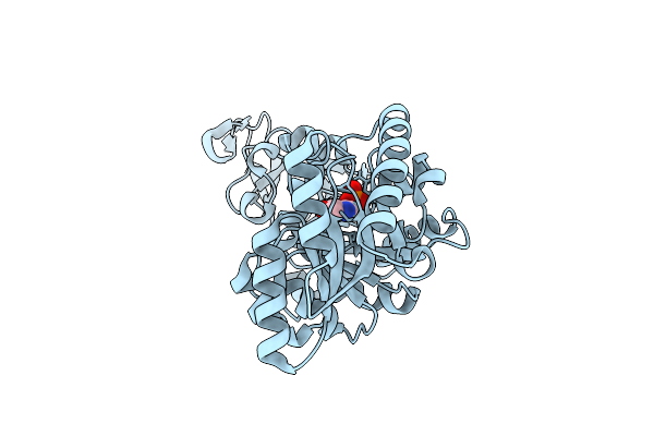 |
Structure Of Vibrio Cholerae Broad Spectrum Racemase Double Mutant R173A, N174A
Organism: Vibrio cholerae
Method: X-RAY DIFFRACTION Resolution:1.50 Å Release Date: 2014-01-15 Classification: ISOMERASE Ligands: PLP |
Organism: Vibrio cholerae
Method: X-RAY DIFFRACTION
Release Date: 2014-01-15
Ligands: PLP
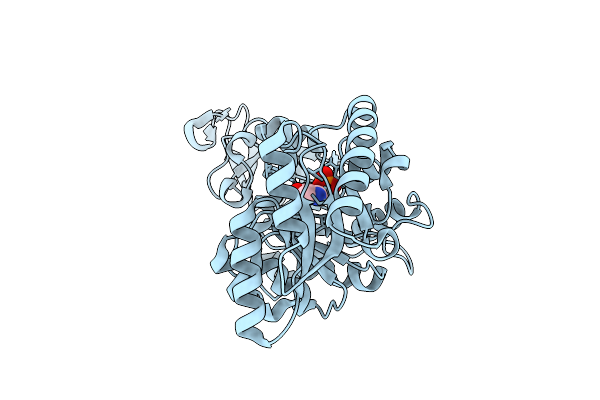 |
Structure Of Vibrio Cholerae Broad Spectrum Racemase
Organism: Vibrio cholerae
Method: X-RAY DIFFRACTION Resolution:1.15 Å Release Date: 2014-01-15 Classification: TRANSFERASE Ligands: CL, PLP |
Organism: Vibrio cholerae
Method: X-RAY DIFFRACTION
Release Date: 2014-01-15
Ligands: CL, PLP
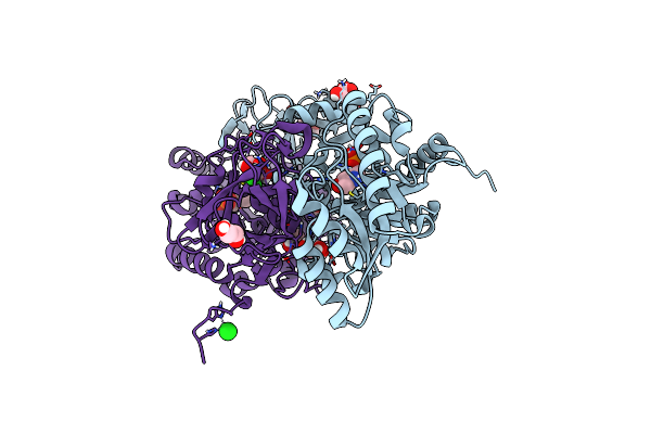 |
Structure Of Broad Spectrum Racemase From Aeromonas Hydrophila
Organism: Aeromonas hydrophila subsp. hydrophila
Method: X-RAY DIFFRACTION Resolution:1.45 Å Release Date: 2014-01-15 Classification: ISOMERASE Ligands: PLP, CL, GOL, NI |
Organism: Aeromonas hydrophila subsp. hydrophila
Method: X-RAY DIFFRACTION
Release Date: 2014-01-15
Ligands: PLP, CL, GOL, NI
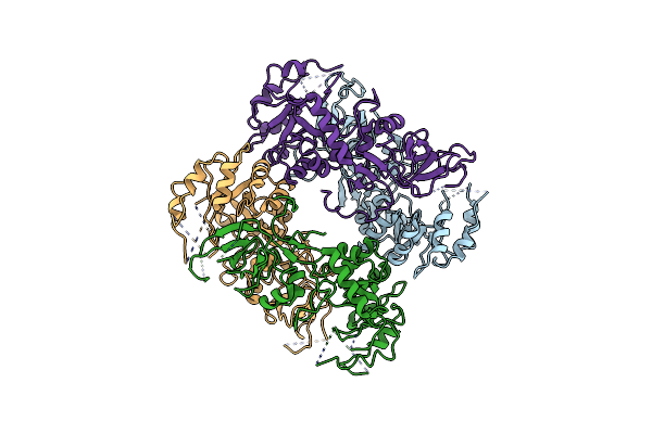 |
Structure Of Alanine Racemase From Aeromonas Hydrophila
Organism: Aeromonas hydrophila subsp. hydrophila
Method: X-RAY DIFFRACTION Resolution:3.25 Å Release Date: 2014-01-15 Classification: ISOMERASE |
Organism: Aeromonas hydrophila subsp. hydrophila
Method: X-RAY DIFFRACTION
Release Date: 2014-01-15
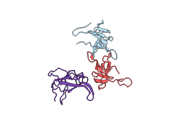 |
Crystal Structure Of The N-Terminal Domain Of The Minor Coat Protein Piii From Ctxphi
Organism: Vibrio cholerae
Method: X-RAY DIFFRACTION Resolution:2.90 Å Release Date: 2012-08-29 Classification: PROTEIN BINDING |
Organism: Vibrio cholerae
Method: X-RAY DIFFRACTION
Release Date: 2012-08-29
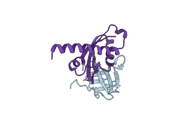 |
Crystal Structure Of A Complex Between The Ctxphi Piii N-Terminal Domain And The Vibrio Cholerae Tola C-Terminal Domain
Organism: Vibrio cholerae
Method: X-RAY DIFFRACTION Resolution:1.44 Å Release Date: 2012-08-29 Classification: PROTEIN BINDING/PROTEIN BINDING |
Organism: Vibrio cholerae
Method: X-RAY DIFFRACTION
Release Date: 2012-08-29