

Search Count: 21
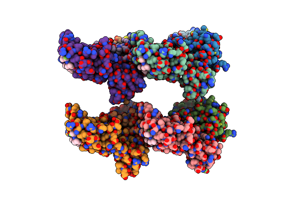 |
Vcb In Complex With Vhl-Binding Compound 114
Organism: Homo sapiens
Method: X-RAY DIFFRACTION Resolution:2.28 Å Release Date: 2025-04-23 Classification: LIGASE Ligands: A1I57 |
Organism: Homo sapiens
Method: X-RAY DIFFRACTION
Release Date: 2025-04-23
Ligands: A1I57
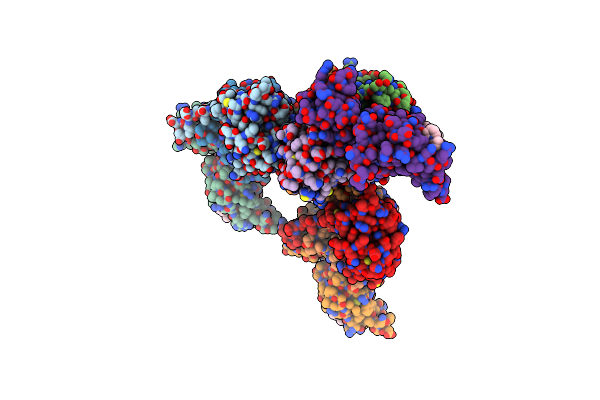 |
Vcb In Complex With Vhl-Binding Compound 82
Organism: Homo sapiens
Method: X-RAY DIFFRACTION Resolution:2.50 Å Release Date: 2025-04-23 Classification: LIGASE Ligands: A1I58 |
Organism: Homo sapiens
Method: X-RAY DIFFRACTION
Release Date: 2025-04-23
Ligands: A1I58
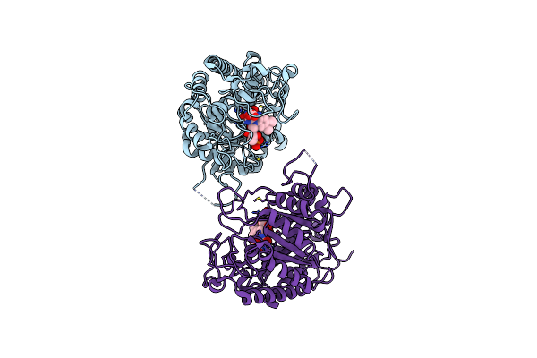 |
Crystal Structure Of Ene-Reductase Oye4 From Botryotinia Fuckeliana (Bfoye4)
Organism: Botryotinia fuckeliana (strain b05.10)
Method: X-RAY DIFFRACTION Resolution:2.15 Å Release Date: 2022-07-13 Classification: OXIDOREDUCTASE Ligands: FMN, FMT |
Organism: Botryotinia fuckeliana (strain b05.10)
Method: X-RAY DIFFRACTION
Release Date: 2022-07-13
Ligands: FMN, FMT
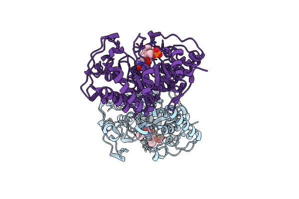 |
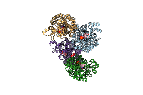
Crystal Structure Of G. Sulphuraria Ene-Reductase Gsoye In Complex With B-Angelica Lactone
Organism: Galdieria sulphuraria
Method: X-RAY DIFFRACTION Resolution:1.50 Å Release Date: 2022-07-13 Classification: OXIDOREDUCTASE Ligands: U5N, FMN |
Organism: Galdieria sulphuraria
Method: X-RAY DIFFRACTION
Release Date: 2022-07-13
Ligands: U5N, FMN
 |
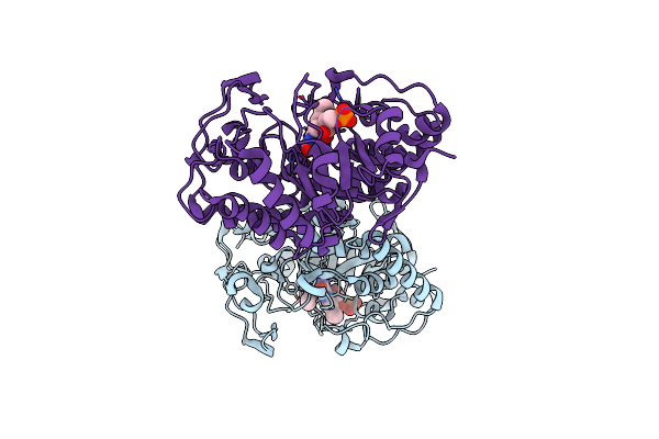
Crystal Structure Of Ene-Reductase Oye2 From S. Cerevisiae
Organism: Saccharomyces cerevisiae
Method: X-RAY DIFFRACTION Resolution:2.45 Å Release Date: 2022-07-13 Classification: OXIDOREDUCTASE Ligands: FMN, GOL |
Organism: Saccharomyces cerevisiae
Method: X-RAY DIFFRACTION
Release Date: 2022-07-13
Ligands: FMN, GOL
 |
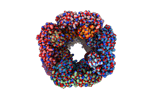
Crystal Structure Of Ene-Reductase Gsoye From Galdieria Sulphuraria In Complex With Alpha-Angelica Lactone
Organism: Galdieria sulphuraria
Method: X-RAY DIFFRACTION Resolution:1.63 Å Release Date: 2022-07-13 Classification: OXIDOREDUCTASE Ligands: U6W, FMN |
Organism: Galdieria sulphuraria
Method: X-RAY DIFFRACTION
Release Date: 2022-07-13
Ligands: U6W, FMN
 |
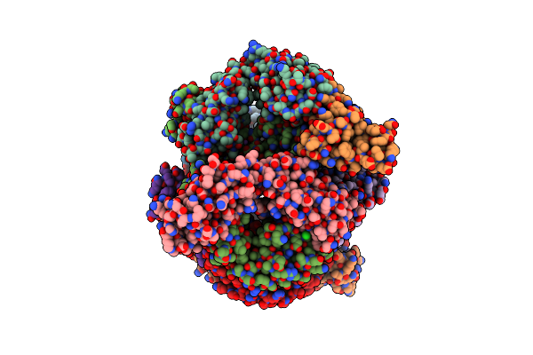
Crystal Structure Of At1412Dm Fab Fragment
Organism: Homo sapiens
Method: X-RAY DIFFRACTION Resolution:2.50 Å Release Date: 2021-05-12 Classification: IMMUNE SYSTEM Ligands: CL |
Organism: Homo sapiens
Method: X-RAY DIFFRACTION
Release Date: 2021-05-12
Ligands: CL
 |
Crystal Structure Of At1412Dm Fab Fragment In Complex With Cd9 Large Extracellular Loop
Organism: Homo sapiens
Method: X-RAY DIFFRACTION Resolution:2.20 Å Release Date: 2021-05-12 Classification: IMMUNE SYSTEM Ligands: CL, K, NA |
Organism: Homo sapiens
Method: X-RAY DIFFRACTION
Release Date: 2021-05-12
Ligands: CL, K, NA
 |
Soluble Epoxide Hydrolase In Complex With Talinolol
Organism: Homo sapiens
Method: X-RAY DIFFRACTION Resolution:2.00 Å Release Date: 2019-07-03 Classification: HYDROLASE Ligands: MG, G3Q |
Organism: Homo sapiens
Method: X-RAY DIFFRACTION
Release Date: 2019-07-03
Ligands: MG, G3Q
 |
Soluble Epoxide Hydrolase In Complex With 1-(4-((4-(Tert-Butyl)Morpholin-2-Yl)Methoxy)Phenyl)-3-Cyclohexylurea
Organism: Homo sapiens
Method: X-RAY DIFFRACTION Resolution:2.16 Å Release Date: 2019-07-03 Classification: HYDROLASE Ligands: MG, G3T |
Organism: Homo sapiens
Method: X-RAY DIFFRACTION
Release Date: 2019-07-03
Ligands: MG, G3T
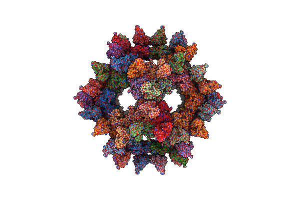 |
Structural Basis Of Human Parechovirus Neutralization By Human Monoclonal Antibodies
Organism: Human parechovirus 1 (strain harris), Homo sapiens
Method: ELECTRON MICROSCOPY Resolution:20.00 Å Release Date: 2015-07-22 Classification: VIRUS |
Organism: Human parechovirus 1 (strain harris), Homo sapiens
Method: ELECTRON MICROSCOPY
Release Date: 2015-07-22
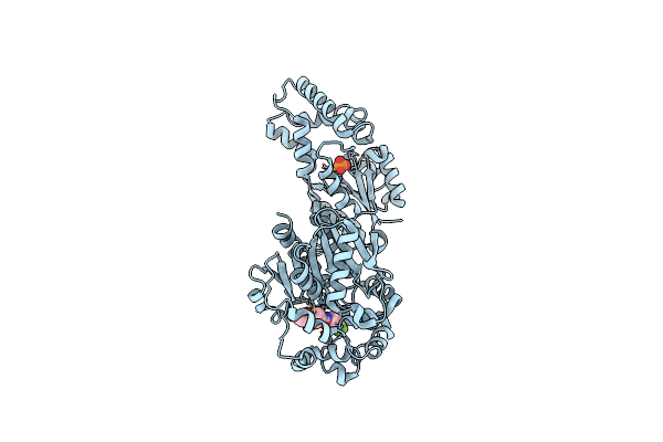 |
Crystal Structure Of Human Soluble Epoxide Hydrolase Complexed With 1-(1-Isobutyrylpiperidin-4-Yl)-3-(4-(Trifluoromethyl)Phenyl)Urea
Organism: Homo sapiens
Method: X-RAY DIFFRACTION Resolution:2.94 Å Release Date: 2014-09-24 Classification: HYDROLASE/HYDROLASE INHIBITOR Ligands: PO4, MG, 2RU |
Organism: Homo sapiens
Method: X-RAY DIFFRACTION
Release Date: 2014-09-24
Ligands: PO4, MG, 2RU
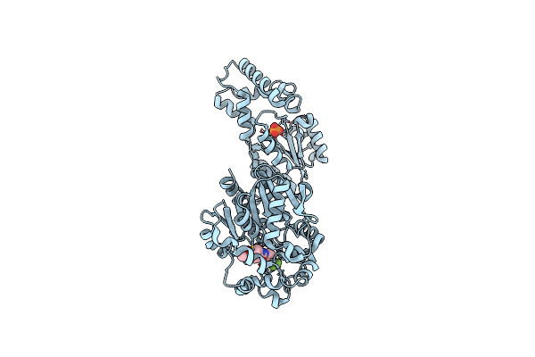 |
Crystal Structure Of Human Soluble Epoxide Hydrolase Complexed With 1-(1-Propanoylpiperidin-4-Yl)-3-[4-(Trifluoromethoxy)Phenyl]Urea
Organism: Homo sapiens
Method: X-RAY DIFFRACTION Resolution:2.92 Å Release Date: 2014-09-24 Classification: HYDROLASE/HYDROLASE INHIBITOR Ligands: PO4, MG, 2RV |
Organism: Homo sapiens
Method: X-RAY DIFFRACTION
Release Date: 2014-09-24
Ligands: PO4, MG, 2RV
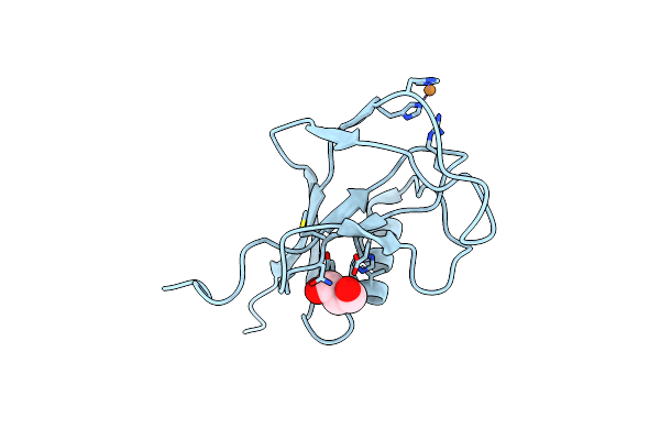 |
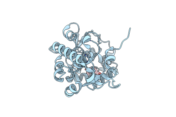
Crystal Structure Of The N-Terminal, Growth Factor-Like Domain Of The Amyloid Precursor Protein Bound To Copper
Organism: Homo sapiens
Method: X-RAY DIFFRACTION Resolution:1.75 Å Release Date: 2014-07-23 Classification: METAL BINDING PROTEIN Ligands: CU, GOL |
Organism: Homo sapiens
Method: X-RAY DIFFRACTION
Release Date: 2014-07-23
Ligands: CU, GOL
 |
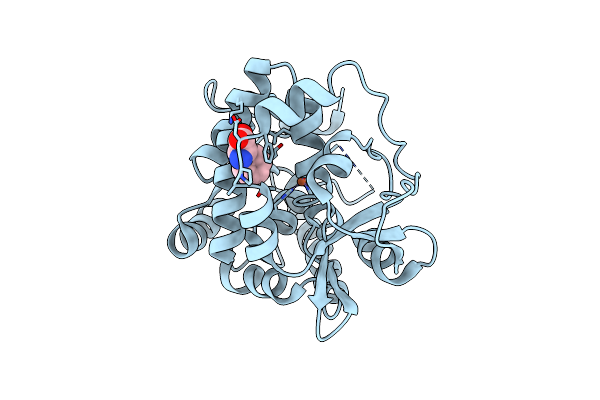
Crystal Structure Of Phenylalanine Hydroxylase S203P Mutant From Chromobacterium Violaceum
Organism: Chromobacterium violaceum
Method: X-RAY DIFFRACTION Resolution:1.55 Å Release Date: 2013-08-07 Classification: OXIDOREDUCTASE Ligands: PHE, CO |
Organism: Chromobacterium violaceum
Method: X-RAY DIFFRACTION
Release Date: 2013-08-07
Ligands: PHE, CO
 |
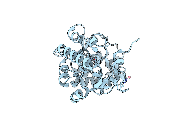
Iron And Phenylalanine Bound Crystal Structure Of Phenylalanine Hydroxylase From Chromobacterium Violaceum
Organism: Chromobacterium violaceum
Method: X-RAY DIFFRACTION Resolution:2.13 Å Release Date: 2013-08-07 Classification: OXIDOREDUCTASE Ligands: FE, PHE |
Organism: Chromobacterium violaceum
Method: X-RAY DIFFRACTION
Release Date: 2013-08-07
Ligands: FE, PHE
 |
Crystallographic Structure Of Phenylalanine Hydroxylase From Chromobacterium Violaceum Y155A Mutation
Organism: Chromobacterium violaceum
Method: X-RAY DIFFRACTION Resolution:1.35 Å Release Date: 2013-05-08 Classification: OXIDOREDUCTASE Ligands: CO |
Organism: Chromobacterium violaceum
Method: X-RAY DIFFRACTION
Release Date: 2013-05-08
Ligands: CO
 |
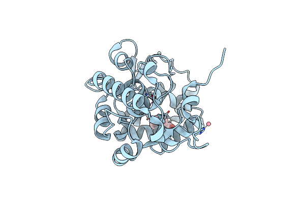
Crystallographic Structure Of Phenylalanine Hydroxylase From Chromobacterium Violaceum F258A Mutation
Organism: Chromobacterium violaceum
Method: X-RAY DIFFRACTION Resolution:1.49 Å Release Date: 2013-05-08 Classification: OXIDOREDUCTASE Ligands: CO |
Organism: Chromobacterium violaceum
Method: X-RAY DIFFRACTION
Release Date: 2013-05-08
Ligands: CO
 |
Crystallographic Structure Of Phenylalanine Hydroxylase From Chromobacterium Violaceum Cocrystallized With Phenylalanine In A Site Distal To The Active Site
Organism: Chromobacterium violaceum
Method: X-RAY DIFFRACTION Resolution:1.35 Å Release Date: 2012-09-05 Classification: OXIDOREDUCTASE Ligands: PHE, CO |
Organism: Chromobacterium violaceum
Method: X-RAY DIFFRACTION
Release Date: 2012-09-05
Ligands: PHE, CO
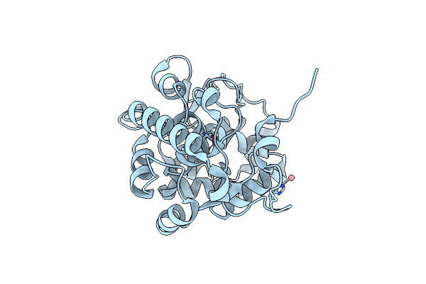 |
Crystal Structure Of Phenylalanine Hydroxylase From Chromobacterium Violaceum Bound To Cobalt
Organism: Chromobacterium violaceum
Method: X-RAY DIFFRACTION Resolution:1.50 Å Release Date: 2012-09-05 Classification: OXIDOREDUCTASE Ligands: CO |
Organism: Chromobacterium violaceum
Method: X-RAY DIFFRACTION
Release Date: 2012-09-05
Ligands: CO