

Search Count: 22
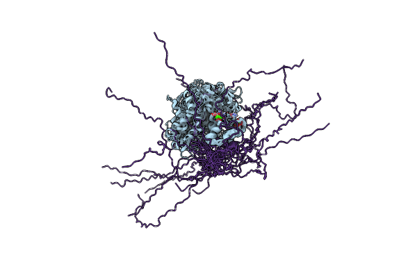 |
Solution Structure Of The Complex Of Trpv5(655-725) With A Calmodulin E32Q/E68Q Double Mutant
Organism: Homo sapiens
Method: SOLUTION NMR Release Date: 2018-04-25 Classification: MEMBRANE PROTEIN Ligands: CA |
Organism: Homo sapiens
Method: SOLUTION NMR
Release Date: 2018-04-25
Ligands: CA
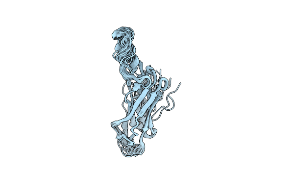 |
The Solution Structure Of Ca2+ Binding Domain 2B Of The Third Isoform Of The Na+/Ca2+ Exchanger
Organism: Mus musculus
Method: SOLUTION NMR Release Date: 2012-08-01 Classification: METAL BINDING PROTEIN |
Organism: Mus musculus
Method: SOLUTION NMR
Release Date: 2012-08-01
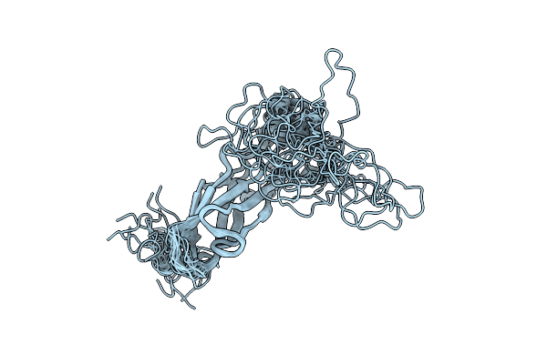 |
Apo-Form Of The Second Ca2+ Binding Domain Of Ncx1.4
Organism: Canis familiaris
Method: SOLUTION NMR Release Date: 2009-08-18 Classification: METAL BINDING PROTEIN |
Organism: Canis familiaris
Method: SOLUTION NMR
Release Date: 2009-08-18
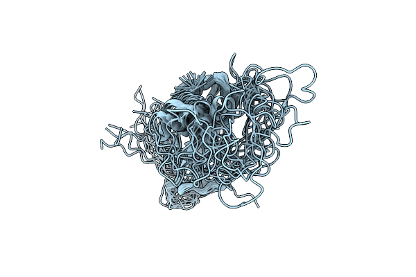 |
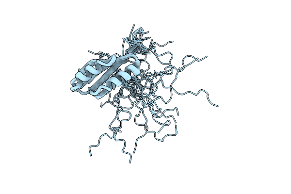
Second Ca2+ Binding Domain Of Ncx1.3
Organism: Canis familiaris
Method: SOLUTION NMR Release Date: 2009-08-18 Classification: METAL BINDING PROTEIN |
Organism: Canis familiaris
Method: SOLUTION NMR
Release Date: 2009-08-18
 |
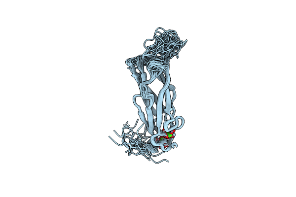
Solution Structure Of The E. Coli Tat Proofreading Chaperone Protein Napd
Organism: Escherichia coli k12
Method: SOLUTION NMR Release Date: 2007-08-21 Classification: CHAPERONE |
Organism: Escherichia coli k12
Method: SOLUTION NMR
Release Date: 2007-08-21
 |
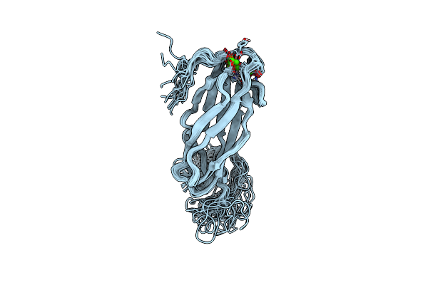
First Ca2+ Binding Domain Of The Na,Ca-Exchanger (Ncx1)
Organism: Canis lupus familiaris
Method: SOLUTION NMR Release Date: 2006-04-18 Classification: METAL TRANSPORT REGULATOR Ligands: CA |
Organism: Canis lupus familiaris
Method: SOLUTION NMR
Release Date: 2006-04-18
Ligands: CA
 |
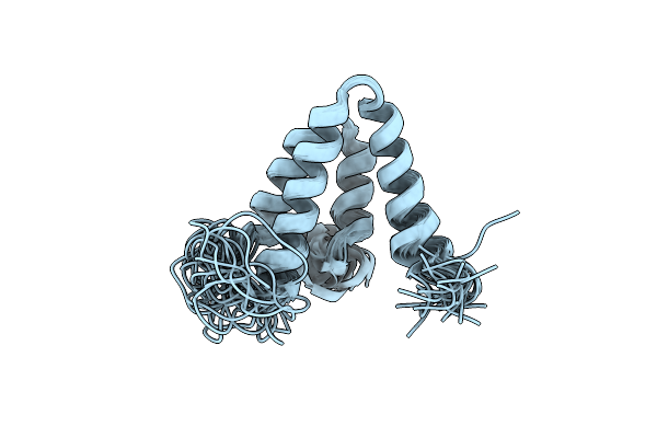
Second Ca2+ Binding Domain Of The Na,Ca-Exchanger (Ncx1)
Organism: Canis lupus familiaris
Method: SOLUTION NMR Release Date: 2006-04-18 Classification: METAL TRANSPORT REGULATOR Ligands: CA |
Organism: Canis lupus familiaris
Method: SOLUTION NMR
Release Date: 2006-04-18
Ligands: CA
 |
Solution Structure Of Free Pah2 Domain Of Msin3B
Organism: Mus musculus
Method: SOLUTION NMR Release Date: 2006-03-28 Classification: TRANSCRIPTION REPRESSOR |
Organism: Mus musculus
Method: SOLUTION NMR
Release Date: 2006-03-28
 |
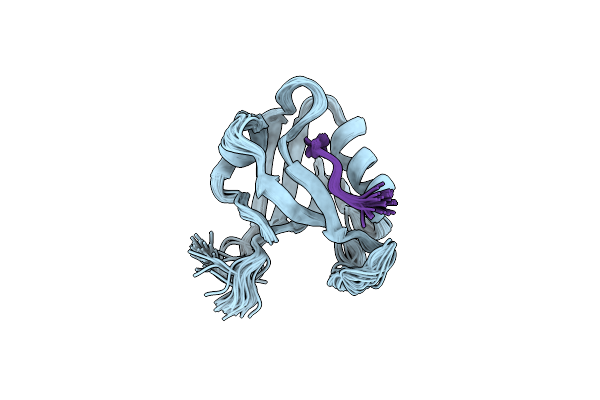
Solution Structure Of The Second Pdz Domain Of The Human Neuronal Adaptor X11Alpha
Organism: Homo sapiens
Method: SOLUTION NMR Release Date: 2005-11-22 Classification: PROTEIN TRANSPORT |
Organism: Homo sapiens
Method: SOLUTION NMR
Release Date: 2005-11-22
 |
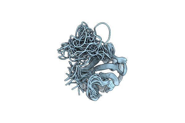
Pdz2 From Ptp-Bl In Complex With The C-Terminal Ligand From The Apc Protein
Organism: Mus musculus
Method: SOLUTION NMR Release Date: 2005-11-01 Classification: Hydrolase/Signaling Protein |
Organism: Mus musculus
Method: SOLUTION NMR
Release Date: 2005-11-01
 |
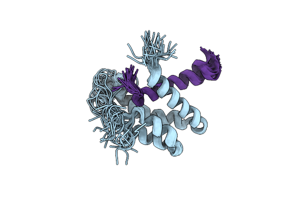
The Alternatively Spliced Pdz2 Domain Of Ptp-Bl
|
Organism: Mus musculus
Method: SOLUTION NMR
Release Date: 2004-01-27
 |
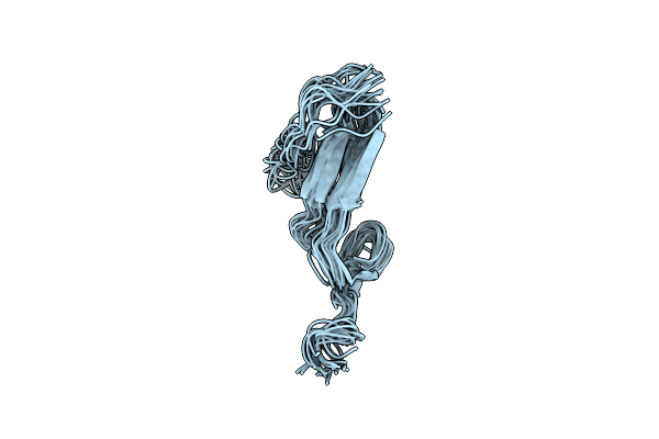
Extended Sid Of Mad1 Bound To The Pah2 Domain Of Msin3B
|
Organism: Mus musculus
Method: SOLUTION NMR
Release Date: 2004-01-20
 |
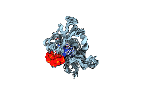
Solution Structure And Dynamics Of The Egf/Tgf-Alpha Chimera T1E
Organism: Homo sapiens
Method: SOLUTION NMR Release Date: 2003-10-07 Classification: HORMONE/GROWTH FACTOR |
Organism: Homo sapiens
Method: SOLUTION NMR
Release Date: 2003-10-07
 |
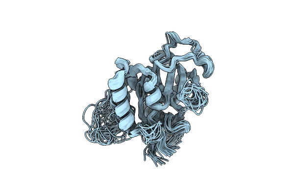
Atpase
Organism: Rattus norvegicus
Method: SOLUTION NMR Release Date: 2003-06-10 Classification: HYDROLASE Ligands: ATP |
Organism: Rattus norvegicus
Method: SOLUTION NMR
Release Date: 2003-06-10
Ligands: ATP
 |
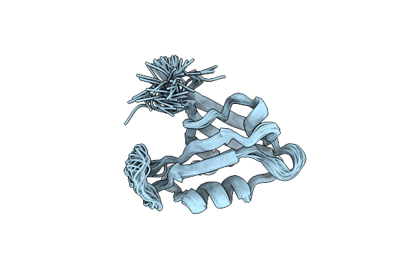
Atpase
|
Organism: Rattus norvegicus
Method: SOLUTION NMR
Release Date: 2003-06-03
 |
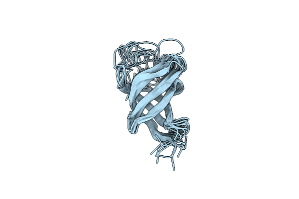
Second Pdz Domain (Pdz2) Of Ptp-Bl
|
Organism: Mus musculus
Method: SOLUTION NMR
Release Date: 2002-03-08
 |
Solution Structure Of The Single-Stranded Dna-Binding Cold Shock Domain (Csd) Of Human Y-Box Protein 1 (Yb1) Determined By Nmr (10 Lowest Energy Structures)
Organism: Homo sapiens
Method: SOLUTION NMR Release Date: 2002-02-21 Classification: TRANSLATION FACTOR |
Organism: Homo sapiens
Method: SOLUTION NMR
Release Date: 2002-02-21
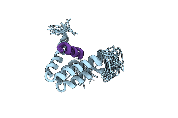 |
Structure Of The Complex Of The Mad1-Sin3B Interaction Domains
Organism: Mus musculus, Homo sapiens
Method: SOLUTION NMR Release Date: 2000-11-20 Classification: EUKARYOTIC TRANSCRIPTIONAL REGULATION |
Organism: Mus musculus, Homo sapiens
Method: SOLUTION NMR
Release Date: 2000-11-20
 |
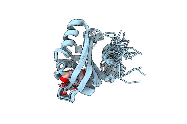
Solution Structure Of The Apo Eh1 Domain Of Mouse Epidermal Growth Factor Receptor Substrate 15, Eps15
|
Organism: Mus musculus
Method: SOLUTION NMR
Release Date: 2000-01-23
 |
Photoactive Yellow Protein, Dark State (Unbleached), Solution Structure, Nmr, 26 Structures
Organism: Halorhodospira halophila
Method: SOLUTION NMR Release Date: 1998-05-27 Classification: PHOTORECEPTOR Ligands: HC4 |
Organism: Halorhodospira halophila
Method: SOLUTION NMR
Release Date: 1998-05-27
Ligands: HC4