

Search Count: 30
 |
Crystal Structure Of The Hvm1 In Complex With Oxovanadium, Succinic Acid And Helvamide.
Organism: Aspergillus rugulosus
Method: X-RAY DIFFRACTION Resolution:1.96 Å Release Date: 2026-09-30 Classification: OXIDOREDUCTASE Ligands: SIN, VVO, A1E05 |
Organism: Aspergillus rugulosus
Method: X-RAY DIFFRACTION
Release Date: 2026-09-30
Ligands: SIN, VVO, A1E05
 |
X-Ray Structure Of Lysozyme Treated With V(V)-Lactate Complex (Structure C)
Organism: Gallus gallus
Method: X-RAY DIFFRACTION Resolution:1.52 Å Release Date: 2026-09-30 Classification: HYDROLASE Ligands: A1J62, A1J6T, VVO |
Organism: Gallus gallus
Method: X-RAY DIFFRACTION
Release Date: 2026-09-30
Ligands: A1J62, A1J6T, VVO
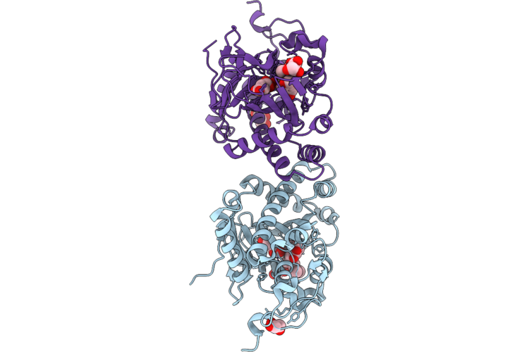 |
Crystal Structure Of Deoxypodophyllotoxin Synthase (Dps) Complexed With Vanadyl(Iv)-Oxo, Succinate And (-)-Hydroxy-Yatein
Organism: Sinopodophyllum hexandrum
Method: X-RAY DIFFRACTION Resolution:2.00 Å Release Date: 2026-06-03 Classification: OXIDOREDUCTASE Ligands: VVO, A1C3S, SIN, EDO, GOL, CL |
Organism: Sinopodophyllum hexandrum
Method: X-RAY DIFFRACTION
Release Date: 2026-06-03
Ligands: VVO, A1C3S, SIN, EDO, GOL, CL
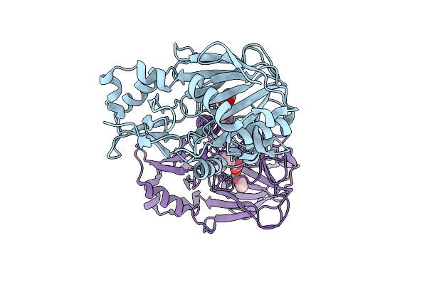 |
Crystal Structure Of Rv0097 G204F Variant With Vanadyl And Cada Bound
Organism: Mycobacterium tuberculosis
Method: X-RAY DIFFRACTION Resolution:1.55 Å Release Date: 2026-03-11 Classification: OXIDOREDUCTASE Ligands: ACT, VVO, VY9 |
Organism: Mycobacterium tuberculosis
Method: X-RAY DIFFRACTION
Release Date: 2026-03-11
Ligands: ACT, VVO, VY9
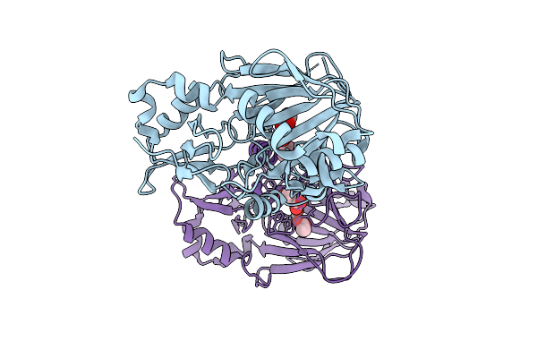 |
Crystal Structure Of Rv0097 G204F Variant With Vanadyl And Caba Bound
Organism: Mycobacterium tuberculosis
Method: X-RAY DIFFRACTION Resolution:1.70 Å Release Date: 2026-03-11 Classification: OXIDOREDUCTASE Ligands: ACT, VVO, 7UC |
Organism: Mycobacterium tuberculosis
Method: X-RAY DIFFRACTION
Release Date: 2026-03-11
Ligands: ACT, VVO, 7UC
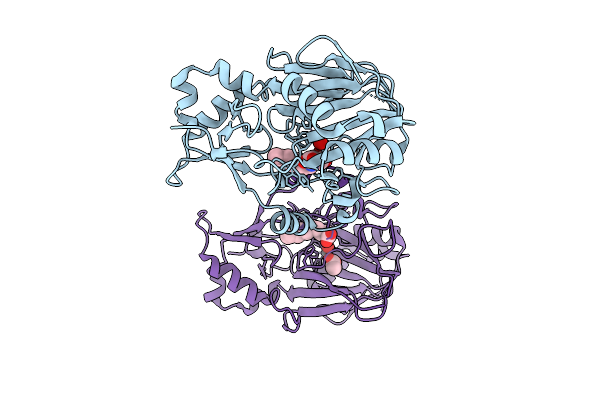 |
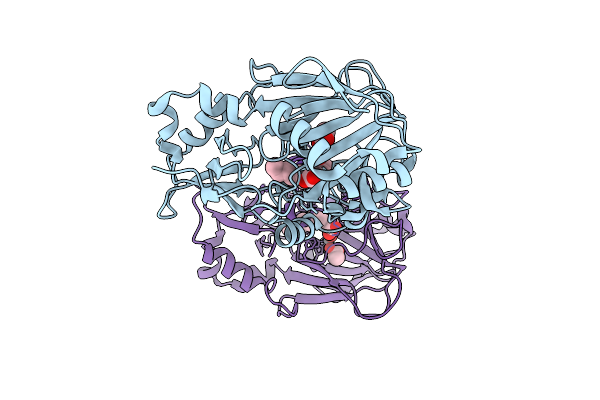
Crystal Structure Of Rv0097 F102W Variant With Vanadyl And Cada Bound
Organism: Mycobacterium tuberculosis
Method: X-RAY DIFFRACTION Resolution:2.00 Å Release Date: 2026-03-11 Classification: OXIDOREDUCTASE Ligands: VY9, VVO, ACT |
Organism: Mycobacterium tuberculosis
Method: X-RAY DIFFRACTION
Release Date: 2026-03-11
Ligands: VY9, VVO, ACT
 |
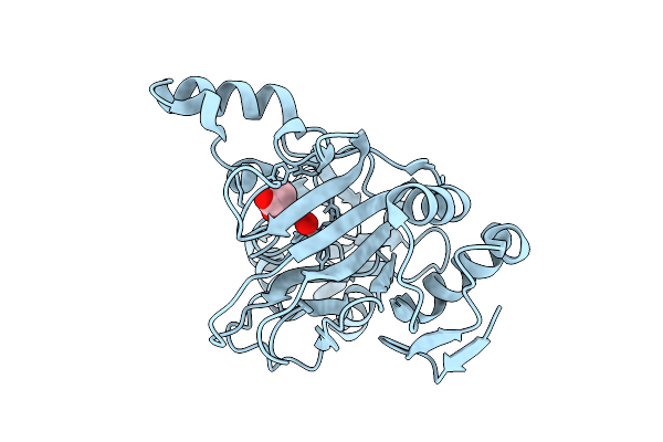
Crystal Structure Of Rv0097 F102Y Variant With Vanadyl And Cada Bound
Organism: Mycobacterium tuberculosis
Method: X-RAY DIFFRACTION Resolution:1.46 Å Release Date: 2026-03-11 Classification: OXIDOREDUCTASE Ligands: ACT, VY9, VVO |
Organism: Mycobacterium tuberculosis
Method: X-RAY DIFFRACTION
Release Date: 2026-03-11
Ligands: ACT, VY9, VVO
 |
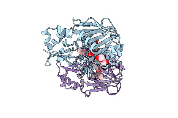
Crystal Structure Of Rv0097 F102A Variant With Vanadyl Bound
Organism: Mycobacterium tuberculosis
Method: X-RAY DIFFRACTION Resolution:1.82 Å Release Date: 2026-03-11 Classification: OXIDOREDUCTASE Ligands: ACT, VVO |
Organism: Mycobacterium tuberculosis
Method: X-RAY DIFFRACTION
Release Date: 2026-03-11
Ligands: ACT, VVO
 |
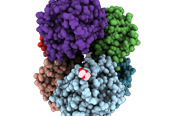
Crystal Structure Of Rv0097 With Vanadyl And Cada Bound
Organism: Mycobacterium tuberculosis
Method: X-RAY DIFFRACTION Resolution:1.36 Å Release Date: 2026-03-11 Classification: OXIDOREDUCTASE Ligands: ACT, VY9, VVO, PEG |
Organism: Mycobacterium tuberculosis
Method: X-RAY DIFFRACTION
Release Date: 2026-03-11
Ligands: ACT, VY9, VVO, PEG
 |
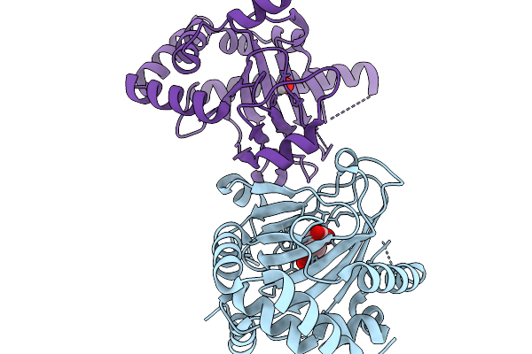
Hala With Lysine, Succinate, Chloride, And Vanadium(Iv)-Oxo At Ph 4.2
Organism: Actinoplanes teichomyceticus
Method: X-RAY DIFFRACTION Resolution:1.93 Å Release Date: 2025-11-19 Classification: BIOSYNTHETIC PROTEIN Ligands: LYS, SIN, PGE, VVO, CL, FLC, V |
Organism: Actinoplanes teichomyceticus
Method: X-RAY DIFFRACTION
Release Date: 2025-11-19
Ligands: LYS, SIN, PGE, VVO, CL, FLC, V
 |
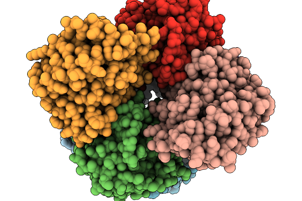
Hydrox With Succinate And Vanadium(Iv)-Oxo
Organism: Streptomyces roseifaciens
Method: X-RAY DIFFRACTION Resolution:1.90 Å Release Date: 2025-11-19 Classification: BIOSYNTHETIC PROTEIN Ligands: VVO, SIN |
Organism: Streptomyces roseifaciens
Method: X-RAY DIFFRACTION
Release Date: 2025-11-19
Ligands: VVO, SIN
 |
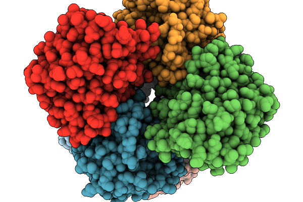
Hala With Lysine, Succinate, Chloride, And Vanadium(Iv)-Oxo At Ph 7
Organism: Actinoplanes teichomyceticus
Method: X-RAY DIFFRACTION Resolution:1.67 Å Release Date: 2025-11-19 Classification: BIOSYNTHETIC PROTEIN Ligands: LYS, SIN, VVO, CL |
Organism: Actinoplanes teichomyceticus
Method: X-RAY DIFFRACTION
Release Date: 2025-11-19
Ligands: LYS, SIN, VVO, CL
 |
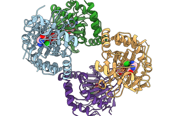
Hala I151N With Lysine, Succinate, Chloride, And Vanadium(Iv)-Oxo
Organism: Actinoplanes teichomyceticus
Method: X-RAY DIFFRACTION Resolution:2.17 Å Release Date: 2025-11-19 Classification: BIOSYNTHETIC PROTEIN Ligands: LYS, SIN, VVO, CL, V |
Organism: Actinoplanes teichomyceticus
Method: X-RAY DIFFRACTION
Release Date: 2025-11-19
Ligands: LYS, SIN, VVO, CL, V
 |
Hala N224V With Lysine, Succinate, Chloride, And Vanadium(Iv)-Oxo
Organism: Actinoplanes teichomyceticus
Method: X-RAY DIFFRACTION Resolution:2.40 Å Release Date: 2025-11-19 Classification: BIOSYNTHETIC PROTEIN Ligands: LYS, SIN, VVO, CL, V |
Organism: Actinoplanes teichomyceticus
Method: X-RAY DIFFRACTION
Release Date: 2025-11-19
Ligands: LYS, SIN, VVO, CL, V
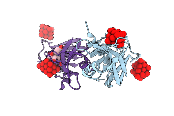 |
Rnase A In Complex With Uridine Vanadate And Decavanadates
Organism: Bos taurus
Method: X-RAY DIFFRACTION Resolution:1.83 Å Release Date: 2025-09-24 Classification: HYDROLASE Ligands: DVT, URI, VVO |
Organism: Bos taurus
Method: X-RAY DIFFRACTION
Release Date: 2025-09-24
Ligands: DVT, URI, VVO
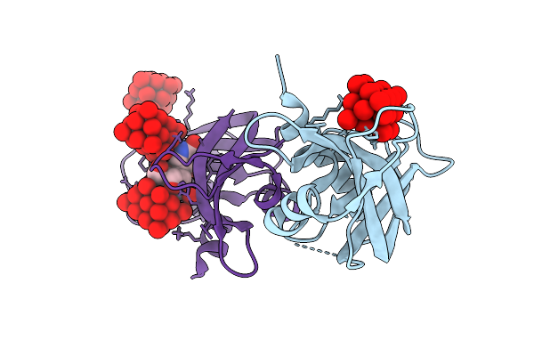 |
Rnase A In Complex With Pseudouridine Vanadate And Decavanadates
Organism: Bos taurus
Method: X-RAY DIFFRACTION Resolution:1.70 Å Release Date: 2025-09-24 Classification: HYDROLASE Ligands: DVT, FJF, VVO |
Organism: Bos taurus
Method: X-RAY DIFFRACTION
Release Date: 2025-09-24
Ligands: DVT, FJF, VVO
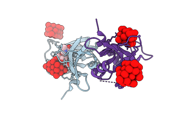 |
Rnase A In Complex With N1-Methylpseudouridine Vanadate And Decavanadates
Organism: Bos taurus
Method: X-RAY DIFFRACTION Resolution:1.71 Å Release Date: 2025-09-24 Classification: HYDROLASE Ligands: 8IZ, DVT, VVO |
Organism: Bos taurus
Method: X-RAY DIFFRACTION
Release Date: 2025-09-24
Ligands: 8IZ, DVT, VVO
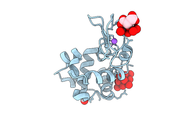 |
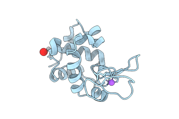
X-Ray Structure Of Decavanadate/Lysozyme Adduct Obtained When The Protein Is Treated With Cs2[V(V)2O4(Mal)2]2H2O (Structure A)
Organism: Gallus gallus
Method: X-RAY DIFFRACTION Resolution:1.46 Å Release Date: 2025-08-13 Classification: HYDROLASE Ligands: CL, NA, DVT, A1JFD, VVO |
Organism: Gallus gallus
Method: X-RAY DIFFRACTION
Release Date: 2025-08-13
Ligands: CL, NA, DVT, A1JFD, VVO
 |
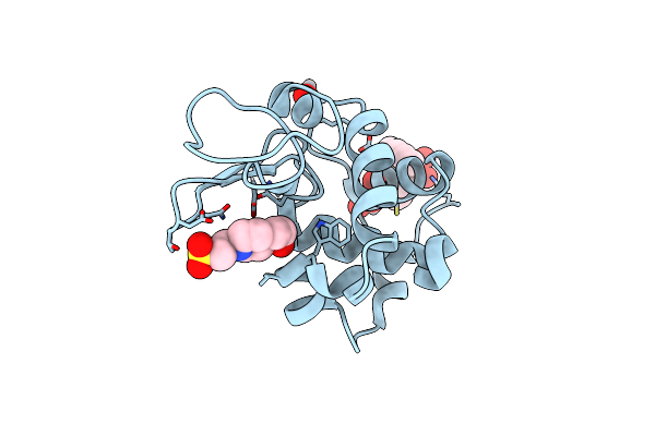
X-Ray Structure Of Lysozyme When Is Treated With Cs2[V(V)2O4(Mal)2]2H2O At 310K
Organism: Gallus gallus
Method: X-RAY DIFFRACTION Resolution:2.09 Å Release Date: 2025-08-13 Classification: HYDROLASE Ligands: CL, NA, VVO |
Organism: Gallus gallus
Method: X-RAY DIFFRACTION
Release Date: 2025-08-13
Ligands: CL, NA, VVO
 |
X-Ray Structure Of Lysozyme Obtained Upon Reaction With [Vivo(8-Hq)2] In Sodium Formate
Organism: Gallus gallus
Method: X-RAY DIFFRACTION Resolution:1.21 Å Release Date: 2025-02-12 Classification: HYDROLASE Ligands: EPE, A1H23, VVO, VVB |
Organism: Gallus gallus
Method: X-RAY DIFFRACTION
Release Date: 2025-02-12
Ligands: EPE, A1H23, VVO, VVB