

Search Count: 37
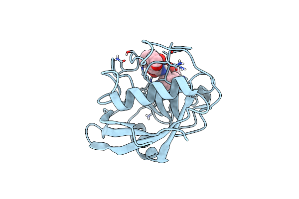 |
Structure Of Cyclophilin D Peptidyl-Prolyl Isomerase Domain Bound To Macrocyclic Inhibitor Jombt
Organism: Homo sapiens
Method: X-RAY DIFFRACTION Resolution:1.75 Å Release Date: 2022-08-24 Classification: ISOMERASE Ligands: I3Q |
Organism: Homo sapiens
Method: X-RAY DIFFRACTION
Release Date: 2022-08-24
Ligands: I3Q
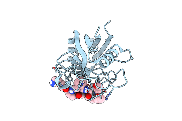 |
Structure Of Cyclophilin D Peptidyl-Prolyl Isomerase Domain Bound To Macrocyclic Inhibitor A26
Organism: Homo sapiens
Method: X-RAY DIFFRACTION Resolution:1.06 Å Release Date: 2022-08-24 Classification: ISOMERASE Ligands: I4F |
Organism: Homo sapiens
Method: X-RAY DIFFRACTION
Release Date: 2022-08-24
Ligands: I4F
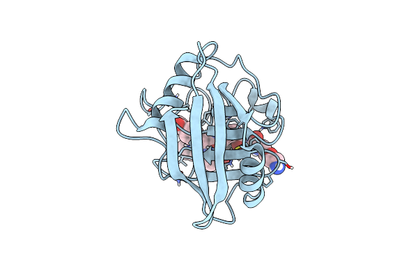 |
Structure Of Cyclophilin D Peptidyl-Prolyl Isomerase Domain Bound To Macrocyclic Inhibitor B1
Organism: Homo sapiens
Method: X-RAY DIFFRACTION Resolution:1.21 Å Release Date: 2022-08-24 Classification: ISOMERASE/ISOMERASE inhibitor Ligands: I44 |
Organism: Homo sapiens
Method: X-RAY DIFFRACTION
Release Date: 2022-08-24
Ligands: I44
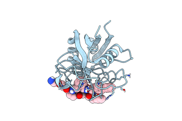 |
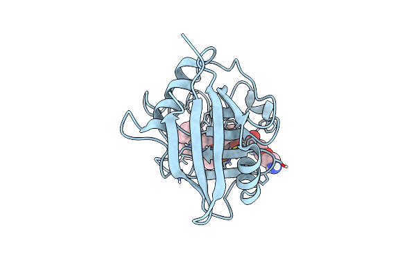
Structure Of Cyclophilin D Peptidyl-Prolyl Isomerase Domain Bound To Macrocyclic Inhibitor B2
Organism: Homo sapiens
Method: X-RAY DIFFRACTION Resolution:1.46 Å Release Date: 2022-08-24 Classification: ISOMERASE/ISOMERASE inhibitor Ligands: I3X |
Organism: Homo sapiens
Method: X-RAY DIFFRACTION
Release Date: 2022-08-24
Ligands: I3X
 |
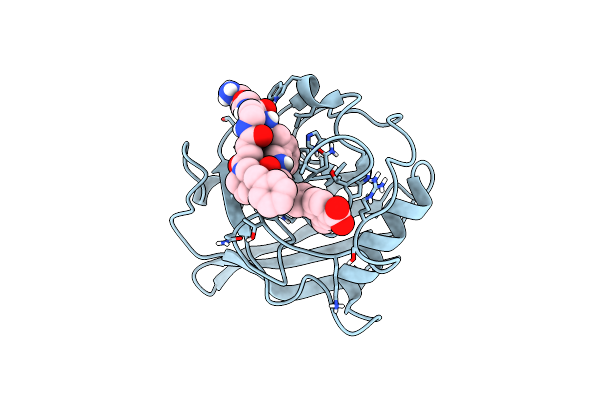
Structure Of Cyclophilin D Peptidyl-Prolyl Isomerase Domain Bound To Macrocyclic Inhibitor B3
Organism: Homo sapiens
Method: X-RAY DIFFRACTION Resolution:1.52 Å Release Date: 2022-08-24 Classification: ISOMERASE Ligands: I5J |
Organism: Homo sapiens
Method: X-RAY DIFFRACTION
Release Date: 2022-08-24
Ligands: I5J
 |
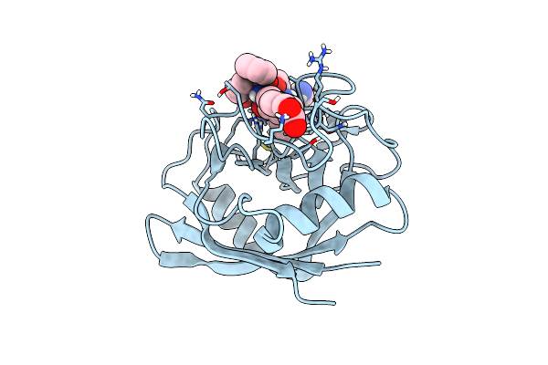
Structure Of Cyclophilin D Peptidyl-Prolyl Isomerase Domain Bound To Macrocyclic Inhibitor B21
Organism: Homo sapiens
Method: X-RAY DIFFRACTION Resolution:0.97 Å Release Date: 2022-08-24 Classification: ISOMERASE Ligands: I56 |
Organism: Homo sapiens
Method: X-RAY DIFFRACTION
Release Date: 2022-08-24
Ligands: I56
 |
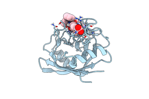
Structure Of Cyclophilin D Peptidyl-Prolyl Isomerase Domain Bound To Macrocyclic Inhibitor B23
Organism: Homo sapiens
Method: X-RAY DIFFRACTION Resolution:1.30 Å Release Date: 2022-08-24 Classification: ISOMERASE Ligands: I5E |
Organism: Homo sapiens
Method: X-RAY DIFFRACTION
Release Date: 2022-08-24
Ligands: I5E
 |
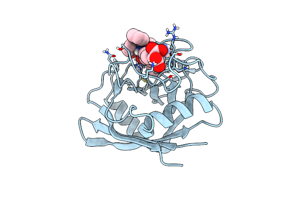
Structure Of Cyclophilin D Peptidyl-Prolyl Isomerase Domain Bound To Macrocyclic Inhibitor B25
Organism: Homo sapiens
Method: X-RAY DIFFRACTION Resolution:1.57 Å Release Date: 2022-08-24 Classification: ISOMERASE Ligands: I4Z |
Organism: Homo sapiens
Method: X-RAY DIFFRACTION
Release Date: 2022-08-24
Ligands: I4Z
 |
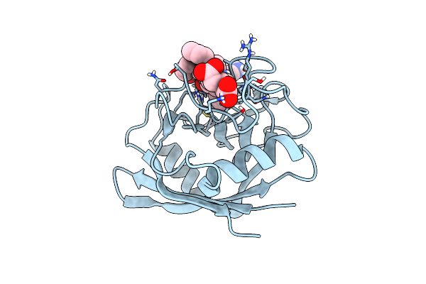
Structure Of Cyclophilin D Peptidyl-Prolyl Isomerase Domain Bound To Macrocyclic Inhibitor B52
Organism: Homo sapiens
Method: X-RAY DIFFRACTION Resolution:1.16 Å Release Date: 2022-08-24 Classification: ISOMERASE Ligands: I4V |
Organism: Homo sapiens
Method: X-RAY DIFFRACTION
Release Date: 2022-08-24
Ligands: I4V
 |
Structure Of Cyclophilin D Peptidyl-Prolyl Isomerase Domain Bound To Macrocyclic Inhibitor B53
Organism: Homo sapiens
Method: X-RAY DIFFRACTION Resolution:1.10 Å Release Date: 2022-08-24 Classification: ISOMERASE/ISOMERASE INHIBITOR Ligands: I4R |
Organism: Homo sapiens
Method: X-RAY DIFFRACTION
Release Date: 2022-08-24
Ligands: I4R
 |
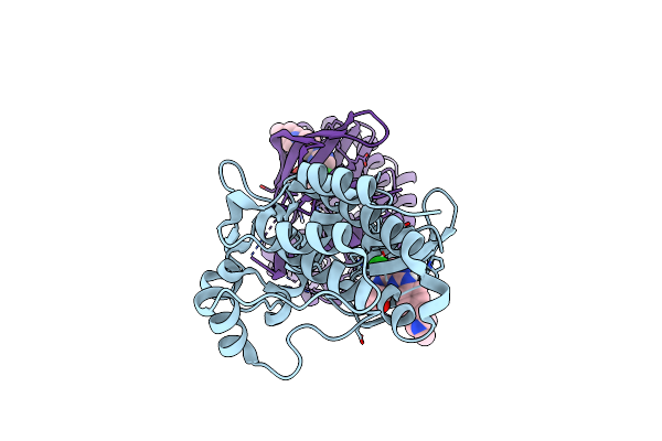
Fab Fragment Bound To The Cter Peptide Of Ly6G6D
Organism: Homo sapiens
Method: X-RAY DIFFRACTION Resolution:2.20 Å Release Date: 2022-04-13 Classification: SIGNALING PROTEIN Ligands: EPE, GOL |
Organism: Homo sapiens
Method: X-RAY DIFFRACTION
Release Date: 2022-04-13
Ligands: EPE, GOL
 |
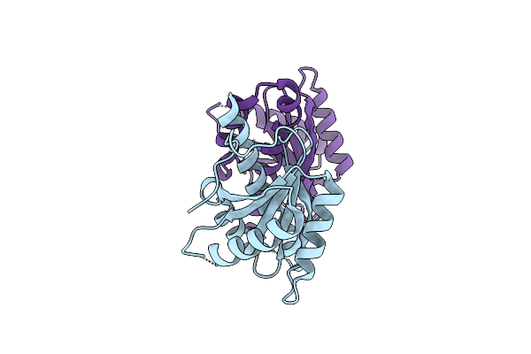
Structure Of Ack1 Kinase In Complex With A Selective Inhibitor
Organism: Homo sapiens
Method: X-RAY DIFFRACTION Resolution:1.79 Å Release Date: 2022-02-09 Classification: TRANSFERASE/INHIBITOR Ligands: WTP, CL |
Organism: Homo sapiens
Method: X-RAY DIFFRACTION
Release Date: 2022-02-09
Ligands: WTP, CL
 |
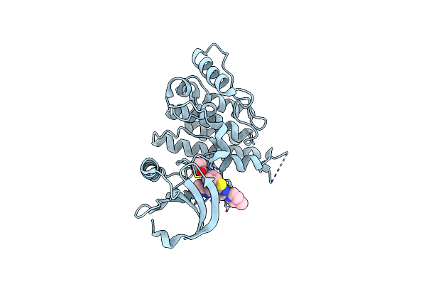
Structure-Guided Studies Of The Holliday Junction Resolvase Ruvx Provide Novel Insights Into Atp-Stimulated Cleavage Of Branched Dna And Rna Substrates
Organism: Mycobacterium tuberculosis h37rv
Method: X-RAY DIFFRACTION Resolution:1.93 Å Release Date: 2021-05-26 Classification: DNA BINDING PROTEIN |
Organism: Mycobacterium tuberculosis h37rv
Method: X-RAY DIFFRACTION
Release Date: 2021-05-26
 |
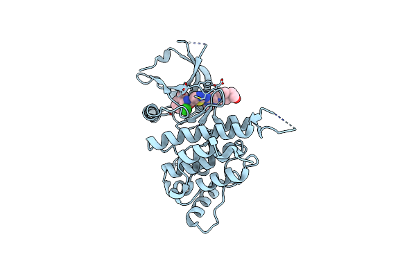
Ddr1 Bound To Vx-680
Organism: Homo sapiens
Method: X-RAY DIFFRACTION Resolution:2.23 Å Release Date: 2018-12-05 Classification: TRANSFERASE/TRANSFERASE INHIBITOR Ligands: VX6 |
Organism: Homo sapiens
Method: X-RAY DIFFRACTION
Release Date: 2018-12-05
Ligands: VX6
 |
Ddr1 Bound To Dasatinib
Organism: Homo sapiens
Method: X-RAY DIFFRACTION Resolution:2.61 Å Release Date: 2018-12-05 Classification: TRANSFERASE/TRANSFERASE INHIBITOR Ligands: 1N1 |
Organism: Homo sapiens
Method: X-RAY DIFFRACTION
Release Date: 2018-12-05
Ligands: 1N1
 |
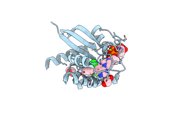
Co-Crystal Structure Of Kras (G12C) Covalently Bound With Quinazoline Based Inhibitor Jbi484
Organism: Homo sapiens
Method: X-RAY DIFFRACTION Resolution:1.70 Å Release Date: 2018-04-18 Classification: TRANSFERASE Ligands: MG, GDP, 94C, EDO |
Organism: Homo sapiens
Method: X-RAY DIFFRACTION
Release Date: 2018-04-18
Ligands: MG, GDP, 94C, EDO
 |
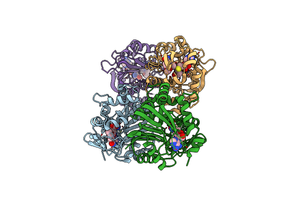
Co-Crystal Structure Of Kras (G12C) Covalently Bound With Quinazoline Based Inhibitor Jbi739
Organism: Homo sapiens
Method: X-RAY DIFFRACTION Resolution:1.69 Å Release Date: 2018-04-18 Classification: TRANSFERASE Ligands: MG, GDP, 94F, EDO |
Organism: Homo sapiens
Method: X-RAY DIFFRACTION
Release Date: 2018-04-18
Ligands: MG, GDP, 94F, EDO
 |
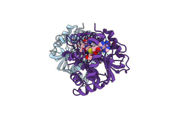
Co-Crystal Structure Of Human Nicotinamide N-Methyltransferase (Nnmt) With Small Molecule Analog Of Nicotinamide
Organism: Homo sapiens
Method: X-RAY DIFFRACTION Resolution:2.49 Å Release Date: 2018-03-21 Classification: TRANSFERASE Ligands: SAH, 8WO |
Organism: Homo sapiens
Method: X-RAY DIFFRACTION
Release Date: 2018-03-21
Ligands: SAH, 8WO
 |
Co-Crystal Structure Of Mouse Nicotinamide N-Methyltransferase (Nnmt) With Small Molecule Analog Of Nicotinamide
Organism: Mus musculus
Method: X-RAY DIFFRACTION Resolution:1.99 Å Release Date: 2018-03-21 Classification: TRANSFERASE Ligands: SAH, 8WO |
Organism: Mus musculus
Method: X-RAY DIFFRACTION
Release Date: 2018-03-21
Ligands: SAH, 8WO
 |
Crystal Structure Of Mouse Nicotinamide N-Methyltransferase (Nnmt) Bound With End Product, 1-Methyl Nicotinamide (Mna)
Organism: Mus musculus
Method: X-RAY DIFFRACTION Resolution:1.88 Å Release Date: 2017-08-02 Classification: TRANSFERASE Ligands: SAH, 8GC, GOL |
Organism: Mus musculus
Method: X-RAY DIFFRACTION
Release Date: 2017-08-02
Ligands: SAH, 8GC, GOL