

Search Count: 220
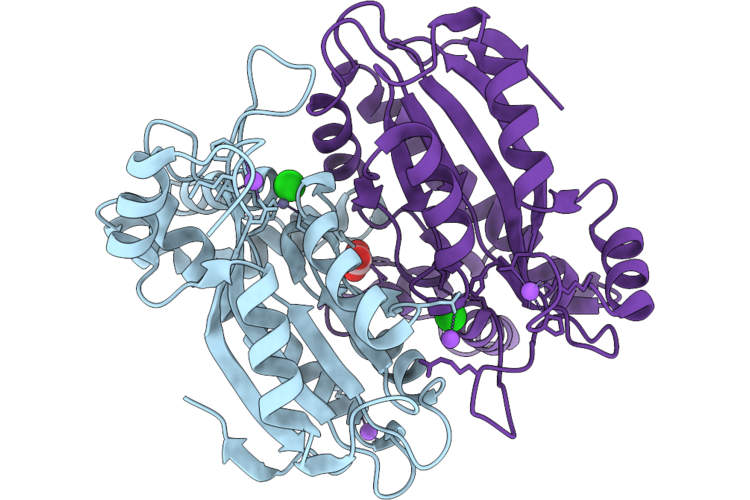 |
Crystal Structure Of C-Terminal Domain Of Theta Type Carbonic Anhydrase 3 From Marine Diatom Phaeodactylum Tricornutum
Organism: Phaeodactylum tricornutum
Method: X-RAY DIFFRACTION Resolution:1.70 Å Release Date: 2026-07-29 Classification: PHOTOSYNTHESIS Ligands: ZN, BCT, NA, CL |
Organism: Phaeodactylum tricornutum
Method: X-RAY DIFFRACTION
Release Date: 2026-07-29
Ligands: ZN, BCT, NA, CL
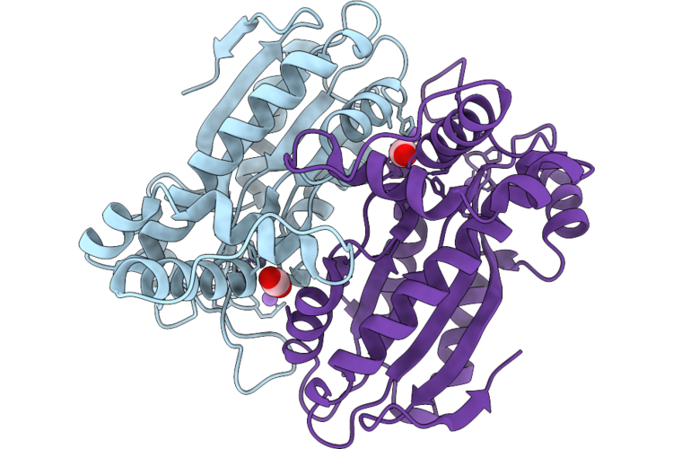 |
Crystal Structure Of C-Terminal Domain Of Theta Type Carbonic Anhydrase 3 From Marine Diatom Phaeodactylum Tricornutum
Organism: Phaeodactylum tricornutum
Method: X-RAY DIFFRACTION Resolution:1.50 Å Release Date: 2026-07-29 Classification: PHOTOSYNTHESIS Ligands: ZN, CO2, NA |
Organism: Phaeodactylum tricornutum
Method: X-RAY DIFFRACTION
Release Date: 2026-07-29
Ligands: ZN, CO2, NA
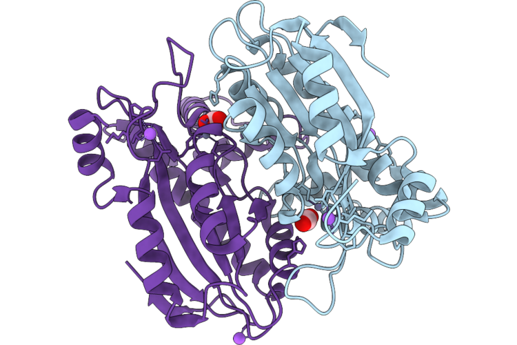 |
Crystal Structure Of C-Terminal Domain Of Theta Type Carbonic Anhydrase 3 (A88D Mutant) From Marine Diatom Phaeodactylum Tricornutum
Organism: Phaeodactylum tricornutum
Method: X-RAY DIFFRACTION Resolution:1.64 Å Release Date: 2026-07-29 Classification: PHOTOSYNTHESIS Ligands: CO2, NA, ZN, BCT |
Organism: Phaeodactylum tricornutum
Method: X-RAY DIFFRACTION
Release Date: 2026-07-29
Ligands: CO2, NA, ZN, BCT
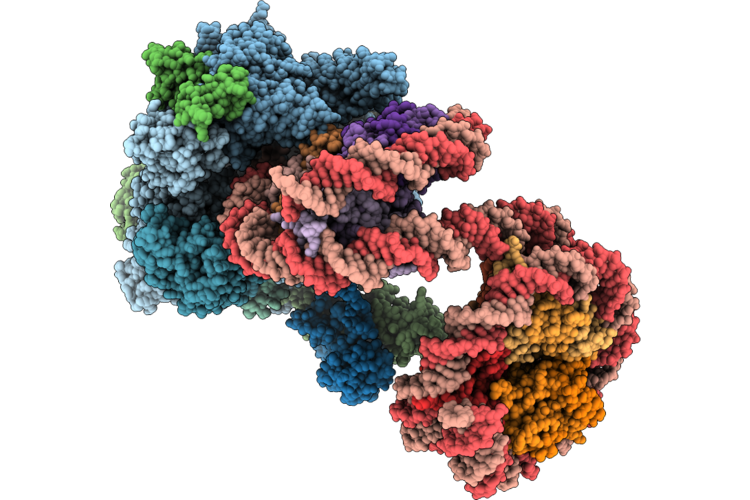 |
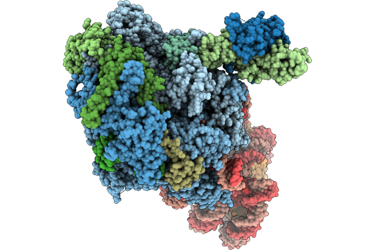
Rna Polymerase Ii Elongation Complex Stalled At Shl(-0.5) In The Hexasome Of The Overlapping Dinucleosome
Organism: Homo sapiens, Synthetic construct, Komagataella phaffii gs115
Method: ELECTRON MICROSCOPY Resolution:3.74 Å Release Date: 2026-07-01 Classification: TRANSCRIPTION Ligands: ZN, MG |
Organism: Homo sapiens, Synthetic construct, Komagataella phaffii gs115
Method: ELECTRON MICROSCOPY
Release Date: 2026-07-01
Ligands: ZN, MG
 |
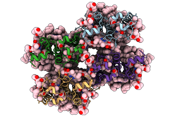
Rna Polymerase Ii Elongation Complex Stalled At Shl(-6) In The Hexasome Of The Overlapping Dinucleosome
Organism: Homo sapiens, Synthetic construct, Komagataella phaffii gs115
Method: ELECTRON MICROSCOPY Resolution:6.74 Å Release Date: 2026-07-01 Classification: TRANSCRIPTION Ligands: ZN, MG |
Organism: Homo sapiens, Synthetic construct, Komagataella phaffii gs115
Method: ELECTRON MICROSCOPY
Release Date: 2026-07-01
Ligands: ZN, MG
 |
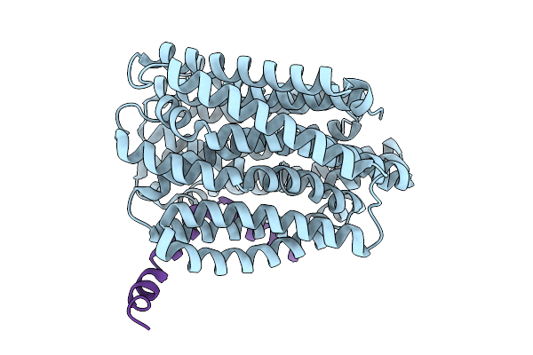
Cryo-Em Structure Of Violaxanthin-Chlorophyll-A-Binding Protein With Red Shifted Chl A (Rvcp) From Trachydiscus Minutus At 2.4 Angstrom
Organism: Trachydiscus minutus
Method: ELECTRON MICROSCOPY Resolution:2.42 Å Release Date: 2025-12-31 Classification: PHOTOSYNTHESIS Ligands: CLA, A1L1F, XAT |
Organism: Trachydiscus minutus
Method: ELECTRON MICROSCOPY
Release Date: 2025-12-31
Ligands: CLA, A1L1F, XAT
 |
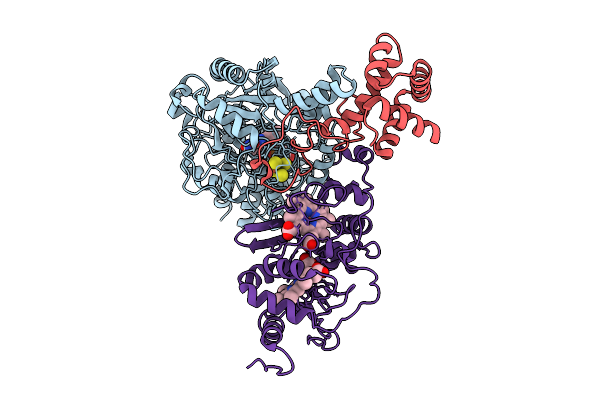
Jm Complex - E. Coli Murj, Levivirus M Lysis Protein Lysm (Sglm)
Organism: Escherichia coli, Enterobacteria phage m
Method: ELECTRON MICROSCOPY Release Date: 2025-09-10 Classification: VIRAL PROTEIN |
Organism: Escherichia coli, Enterobacteria phage m
Method: ELECTRON MICROSCOPY
Release Date: 2025-09-10
 |
Cryo-Em Structure Of Fructose Dehydrogenase Variant From Gluconobacter Japonicus Truncating Heme 1C And C-Terminal Hydrophobic Regions
Organism: Gluconobacter japonicus
Method: ELECTRON MICROSCOPY Release Date: 2025-08-06 Classification: OXIDOREDUCTASE Ligands: FAD, F3S, HEC |
Organism: Gluconobacter japonicus
Method: ELECTRON MICROSCOPY
Release Date: 2025-08-06
Ligands: FAD, F3S, HEC
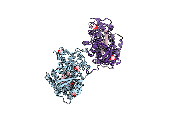 |
X-Ray Structure Of Cytochrome P450 Olet From Lacicoccus Alkaliphilus In Complex With Icosanoic Acid
Organism: Lacicoccus alkaliphilus dsm 16010
Method: X-RAY DIFFRACTION Resolution:2.44 Å Release Date: 2025-08-06 Classification: OXIDOREDUCTASE Ligands: HEM, DCR, GOL |
Organism: Lacicoccus alkaliphilus dsm 16010
Method: X-RAY DIFFRACTION
Release Date: 2025-08-06
Ligands: HEM, DCR, GOL
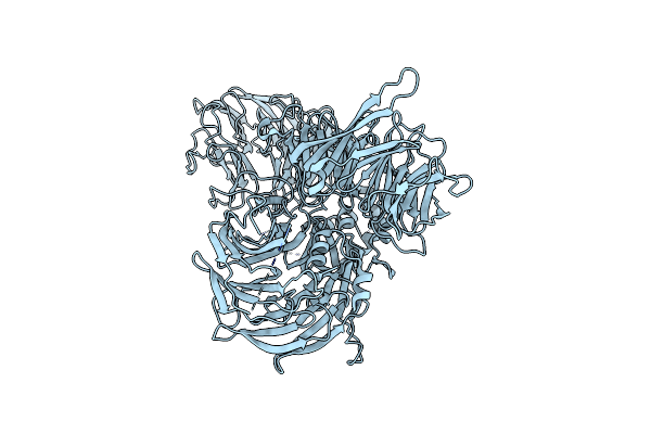 |
Hbx Fused Ddb1 4M Mutant
Organism: Hepatitis b virus, Homo sapiens
Method: ELECTRON MICROSCOPY Release Date: 2025-06-04 Classification: VIRAL PROTEIN |
Organism: Hepatitis b virus, Homo sapiens
Method: ELECTRON MICROSCOPY
Release Date: 2025-06-04
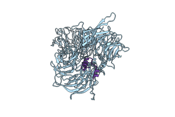 |
Hbx Complexed With Ddb1
Organism: Homo sapiens, Hepatitis b virus
Method: ELECTRON MICROSCOPY Release Date: 2025-06-04 Classification: VIRAL PROTEIN |
Organism: Homo sapiens, Hepatitis b virus
Method: ELECTRON MICROSCOPY
Release Date: 2025-06-04
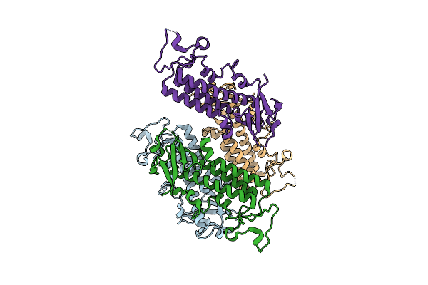 |
X-Ray Structure Of Fbb18 From Chlamydomonas Reinhardtii.
Organism: Chlamydomonas reinhardtii
Method: X-RAY DIFFRACTION Resolution:2.30 Å Release Date: 2025-04-02 Classification: MOTOR PROTEIN |
Organism: Chlamydomonas reinhardtii
Method: X-RAY DIFFRACTION
Release Date: 2025-04-02
 |
Neutron Structure Of Cellulase Cel6A From Phanerochaete Chrysosporium At Room Temperature
Organism: Phanerodontia chrysosporium
Method: X-RAY DIFFRACTION, NEUTRON DIFFRACTION Resolution:1.36 Å, 1.86 Å Release Date: 2025-03-12 Classification: HYDROLASE |
Organism: Phanerodontia chrysosporium
Method: X-RAY DIFFRACTION, NEUTRON DIFFRACTION
Release Date: 2025-03-12
 |
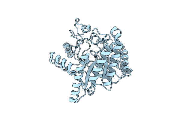
Neutron Structure Of Cellulase Cel6A From Phanerochaete Chrysosporium At Room Temperature, Enzyme-Product Complex
Organism: Phanerodontia chrysosporium
Method: X-RAY DIFFRACTION, NEUTRON DIFFRACTION Resolution:1.40 Å, 1.8 Å Release Date: 2025-03-12 Classification: HYDROLASE Ligands: BGC, NA |
Organism: Phanerodontia chrysosporium
Method: X-RAY DIFFRACTION, NEUTRON DIFFRACTION
Release Date: 2025-03-12
Ligands: BGC, NA
 |
Neutron Structure Of Cellulase Cel6A From Phanerochaete Chrysosporium At Room Temperature, Low-D2O-Solvent
Organism: Phanerodontia chrysosporium
Method: X-RAY DIFFRACTION, NEUTRON DIFFRACTION Resolution:1.40 Å, 2.15 Å Release Date: 2025-03-12 Classification: HYDROLASE |
Organism: Phanerodontia chrysosporium
Method: X-RAY DIFFRACTION, NEUTRON DIFFRACTION
Release Date: 2025-03-12
 |
Neutron Structure Of Cellulase Cel6A From Phanerochaete Chrysosporium At Room Temperature, Enzyme-Product Complex, H2O Solvent
Organism: Phanerodontia chrysosporium
Method: X-RAY DIFFRACTION, NEUTRON DIFFRACTION Resolution:1.80 Å, 2.59 Å Release Date: 2025-03-12 Classification: HYDROLASE Ligands: BGC, NA |
Organism: Phanerodontia chrysosporium
Method: X-RAY DIFFRACTION, NEUTRON DIFFRACTION
Release Date: 2025-03-12
Ligands: BGC, NA
 |
High-Resolution X-Ray Structure Of Cellulase Cel6A From Phanerochaete Chrysosporium At Cryogenic Temperature
Organism: Phanerodontia chrysosporium
Method: X-RAY DIFFRACTION Resolution:0.80 Å Release Date: 2025-03-12 Classification: HYDROLASE Ligands: MPD, ACT |
Organism: Phanerodontia chrysosporium
Method: X-RAY DIFFRACTION
Release Date: 2025-03-12
Ligands: MPD, ACT
 |
High-Resolution X-Ray Structure Of Cellulase Cel6A From Phanerochaete Chrysosporium At Cryogenic Temperature, Enzyme-Product Complex
Organism: Phanerodontia chrysosporium
Method: X-RAY DIFFRACTION Resolution:0.85 Å Release Date: 2025-03-12 Classification: HYDROLASE Ligands: BGC, NA, CL, PEG |
Organism: Phanerodontia chrysosporium
Method: X-RAY DIFFRACTION
Release Date: 2025-03-12
Ligands: BGC, NA, CL, PEG
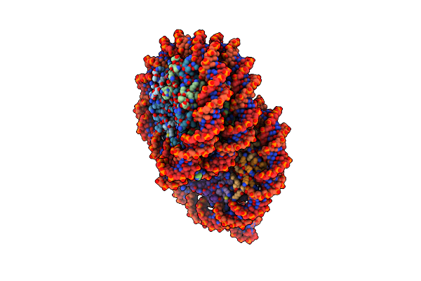 |
Cryo-Em Structure Of The Overlapping Di-Nucleosome (Closed Form)
Organism: Homo sapiens, Synthetic construct
Method: ELECTRON MICROSCOPY Release Date: 2025-01-29 Classification: GENE REGULATION/DNA |
Organism: Homo sapiens, Synthetic construct
Method: ELECTRON MICROSCOPY
Release Date: 2025-01-29
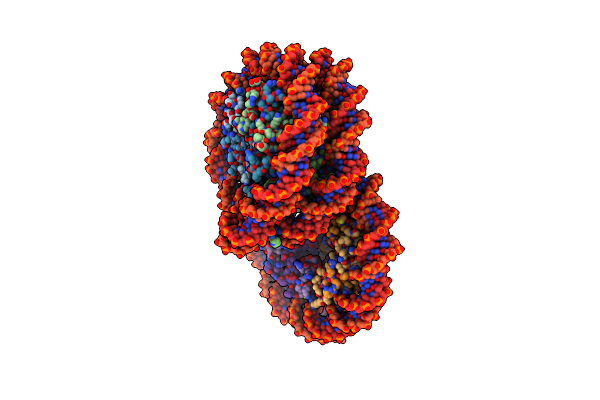 |
Cryo-Em Structure Of The Overlapping Di-Nucleosome (Intermediate Form2)
Organism: Homo sapiens, Synthetic construct
Method: ELECTRON MICROSCOPY Release Date: 2025-01-29 Classification: GENE REGULATION/DNA |
Organism: Homo sapiens, Synthetic construct
Method: ELECTRON MICROSCOPY
Release Date: 2025-01-29