
Search Count: 35
All
Selected
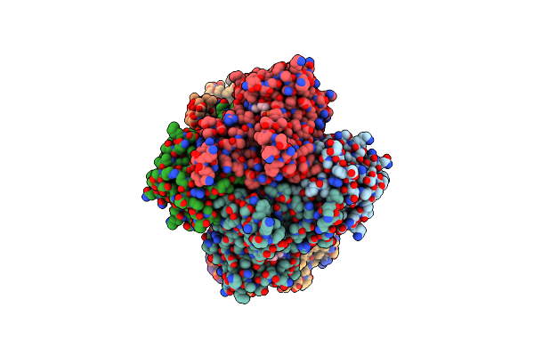 |
Structure Of Human Mitochondrial Rnase P In Complex With Mitochondrial Pre-Trna-His(5,Ser)
Organism: Homo sapiens
Method: ELECTRON MICROSCOPY Release Date: 2024-06-12 Classification: RNA BINDING PROTEIN Ligands: NAD, ZN, SAH, MG |
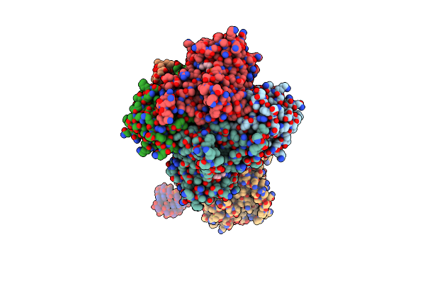 |
Structure Of Human Mitochondrial Rnase Z In Complex With Mitochondrial Pre-Trna-His(0,Ser)
Organism: Homo sapiens
Method: ELECTRON MICROSCOPY Release Date: 2024-06-12 Classification: RNA BINDING PROTEIN Ligands: NAD, ZN, SAH |
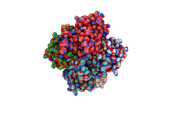 |
Structure Of Human Mitochondrial Cca-Adding Enzyme In Complex With Mitochondrial Pre-Trna-Ile
Organism: Homo sapiens
Method: ELECTRON MICROSCOPY Release Date: 2024-06-12 Classification: RNA BINDING PROTEIN Ligands: NAD, CDP, SAH |
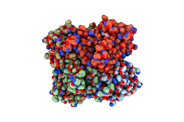 |
Structure Of Human Mitochondrial Mrpp1-Mrpp2 In Complex With Mitochondrial Pre-Trna-Ile
Organism: Homo sapiens
Method: ELECTRON MICROSCOPY Release Date: 2024-06-12 Classification: RNA BINDING PROTEIN Ligands: NAD, SAH |
 |
Organism: Homo sapiens
Method: X-RAY DIFFRACTION Resolution:1.65 Å Release Date: 2023-10-25 Classification: RNA BINDING PROTEIN |
 |
Organism: Homo sapiens, Synthetic construct
Method: X-RAY DIFFRACTION Resolution:2.80 Å Release Date: 2023-10-25 Classification: RNA BINDING PROTEIN |
 |
Organism: Homo sapiens
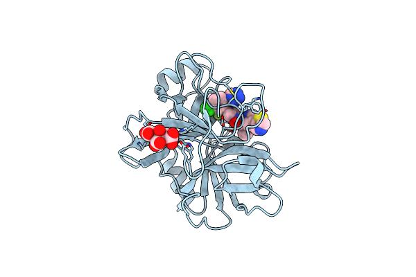
Method: X-RAY DIFFRACTION Resolution:1.80 Å Release Date: 2022-08-03 Classification: Blood Clotting, Hydrolase Ligands: OQ6 |
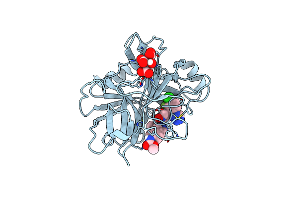 |
Organism: Homo sapiens
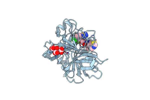
Method: X-RAY DIFFRACTION Resolution:1.80 Å Release Date: 2022-08-03 Classification: BLOOD CLOTTING, Hydrolase Ligands: OQI, CIT |
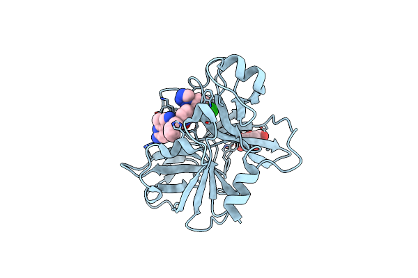 |
Organism: Homo sapiens
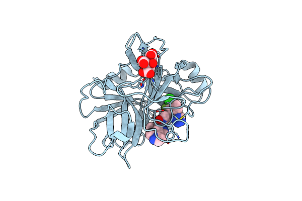
Method: X-RAY DIFFRACTION Resolution:1.47 Å Release Date: 2022-08-03 Classification: BLOOD CLOTTING, hydrolase Ligands: OQO, CIT |
 |
Organism: Homo sapiens
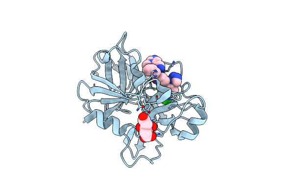
Method: X-RAY DIFFRACTION Resolution:1.63 Å Release Date: 2022-08-03 Classification: BLOOD CLOTTING, Hydrolase Ligands: OR0, CIT |
 |
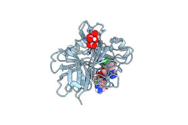
Organism: Homo sapiens
Method: X-RAY DIFFRACTION Resolution:1.59 Å Release Date: 2022-08-03 Classification: BLOOD CLOTTING, Hydrolase Ligands: CIT, ORF |
 |
Organism: Homo sapiens
Method: X-RAY DIFFRACTION Resolution:1.70 Å Release Date: 2022-08-03 Classification: BLOOD CLOTTING, Hydrolase Ligands: CIT, ORU |
 |
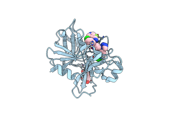
Organism: Homo sapiens
Method: X-RAY DIFFRACTION Resolution:1.68 Å Release Date: 2022-08-03 Classification: BLOOD CLOTTING, Hydrolase Ligands: CIT, OSX |
 |
Organism: Homo sapiens
Method: X-RAY DIFFRACTION Resolution:1.51 Å Release Date: 2022-08-03 Classification: BLOOD CLOTTING, Hydrolase Ligands: CIT, OT7 |
 |
Organism: Homo sapiens
Method: X-RAY DIFFRACTION Resolution:1.52 Å Release Date: 2022-08-03 Classification: BLOOD CLOTTING, Hydrolase Ligands: CIT, OTL |
 |
Organism: Homo sapiens
Method: X-RAY DIFFRACTION Resolution:1.73 Å Release Date: 2022-08-03 Classification: BLOOD CLOTTING, Hydrolase Ligands: CIT, OTX |
 |
Organism: Escherichia coli k-12, Synthetic construct
Method: X-RAY DIFFRACTION Resolution:2.29 Å Release Date: 2022-06-01 Classification: RNA BINDING PROTEIN Ligands: 6D6 |
 |
Organism: Escherichia coli k-12, Synthetic construct
Method: X-RAY DIFFRACTION Resolution:1.59 Å Release Date: 2022-06-01 Classification: RNA BINDING PROTEIN Ligands: 6D6 |
 |
Organism: Escherichia coli k-12
Method: X-RAY DIFFRACTION Resolution:2.10 Å Release Date: 2022-05-25 Classification: RNA BINDING PROTEIN Ligands: 0Y0 |
 |
Organism: Pseudomonas aeruginosa pao1
Method: X-RAY DIFFRACTION Resolution:1.27 Å Release Date: 2021-04-07 Classification: TOXIN |