

Search Count: 19
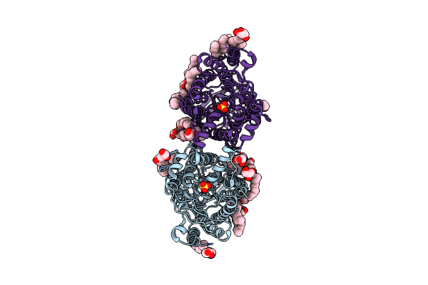 |
Yeee(Tsua)-Yeed(Tsub) Complex For Thiosulfate Uptake
Organism: Spirochaeta thermophila dsm 6578, Corynebacterium pollutisoli
Method: X-RAY DIFFRACTION Resolution:3.34 Å Release Date: 2024-03-27 Classification: TRANSPORT PROTEIN Ligands: THJ, OLC |
Organism: Spirochaeta thermophila dsm 6578, Corynebacterium pollutisoli
Method: X-RAY DIFFRACTION
Release Date: 2024-03-27
Ligands: THJ, OLC
 |
Yeee(Tsua)-Yeed(Tsub) Complex For Thiosulfate Uptake
Organism: Spirochaeta thermophila dsm 6578, Corynebacterium pollutisoli
Method: X-RAY DIFFRACTION Resolution:2.60 Å Release Date: 2024-03-27 Classification: TRANSPORT PROTEIN Ligands: OLC |
Organism: Spirochaeta thermophila dsm 6578, Corynebacterium pollutisoli
Method: X-RAY DIFFRACTION
Release Date: 2024-03-27
Ligands: OLC
 |
Crystal Structure Of Cgl1 From Crassostrea Gigas, Mannobiose-Bound Form (Cgl1/Man(Alpha)1-2Man)
Organism: Crassostrea gigas
Method: X-RAY DIFFRACTION Resolution:1.00 Å Release Date: 2023-10-25 Classification: SUGAR BINDING PROTEIN Ligands: ACY, MAN, CAC |
Organism: Crassostrea gigas
Method: X-RAY DIFFRACTION
Release Date: 2023-10-25
Ligands: ACY, MAN, CAC
 |
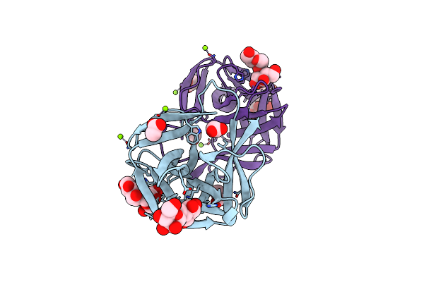
Crystal Structure Of Cgl1 From Crassostrea Gigas, Mannotriose-Bound Form (Cgl1/Man(Alpha)1-2Man(Alpha)1-2Man)
Organism: Crassostrea gigas
Method: X-RAY DIFFRACTION Resolution:0.97 Å Release Date: 2023-10-25 Classification: SUGAR BINDING PROTEIN Ligands: ACY, MG, CAC, MAN |
Organism: Crassostrea gigas
Method: X-RAY DIFFRACTION
Release Date: 2023-10-25
Ligands: ACY, MG, CAC, MAN
 |
Crystal Structure Of Cgl1 From Crassostrea Gigas, Mannotetraose-Bound Form (Cgl1/Man(Alpha)1-2Man(Alpha)1-2Man(Alpha)1-6Man)
Organism: Crassostrea gigas
Method: X-RAY DIFFRACTION Resolution:1.30 Å Release Date: 2023-10-25 Classification: SUGAR BINDING PROTEIN Ligands: ACY, MG |
Organism: Crassostrea gigas
Method: X-RAY DIFFRACTION
Release Date: 2023-10-25
Ligands: ACY, MG
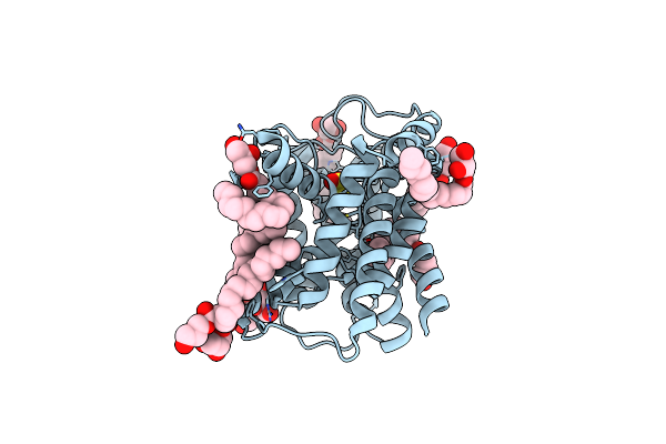 |
Crystal Structure Of Thiosulfate Transporter Yeee From Spirochaeta Thermophila
Organism: Spirochaeta thermophila
Method: X-RAY DIFFRACTION Resolution:2.52 Å Release Date: 2020-09-02 Classification: TRANSPORT PROTEIN Ligands: THJ, OLC |
Organism: Spirochaeta thermophila
Method: X-RAY DIFFRACTION
Release Date: 2020-09-02
Ligands: THJ, OLC
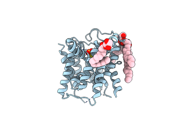 |
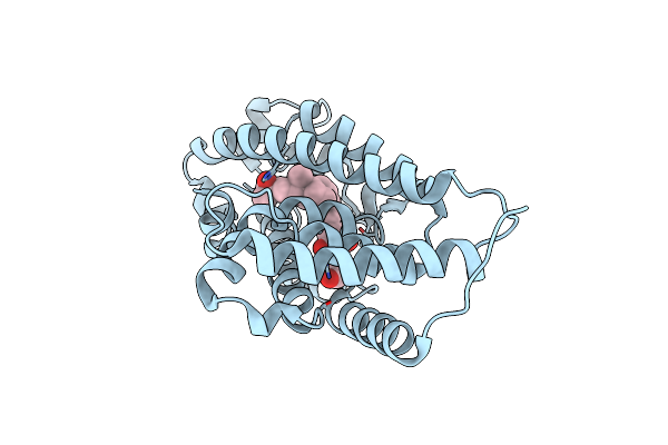
Crystal Structure Of Thiosulfate Transporter Yeee Inactive Mutant - C91A
Organism: Spirochaeta thermophila
Method: X-RAY DIFFRACTION Resolution:2.60 Å Release Date: 2020-09-02 Classification: TRANSPORT PROTEIN Ligands: OLC, THJ |
Organism: Spirochaeta thermophila
Method: X-RAY DIFFRACTION
Release Date: 2020-09-02
Ligands: OLC, THJ
 |
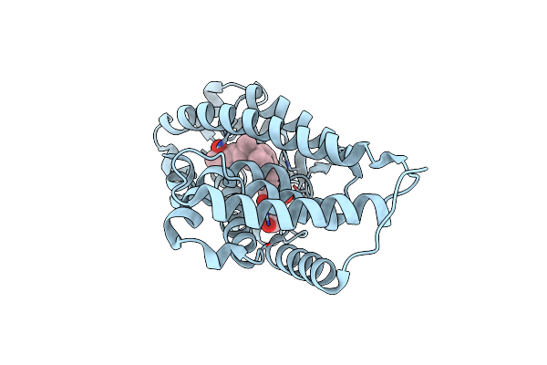
Crystal Structure Of The Human Vitamin D Receptor Ligand Binding Domain Complexed With (1R,2R,3R)-5-[(E)-2-{(1R,3As,7Ar)-1-[(R)-6-Hydroxy-6-Methylheptan-2-Yl]-7A-Methyl-2,3,3A,6,7,7A-Hexahydro-1H-Inden-4-Yl}Vinyl]-2-(3-Hydroxypropyl)Cyclohex-4-Ene-1,3-Diol
Organism: Homo sapiens
Method: X-RAY DIFFRACTION Resolution:2.00 Å Release Date: 2018-04-11 Classification: TRANSCRIPTION Ligands: 90L |
Organism: Homo sapiens
Method: X-RAY DIFFRACTION
Release Date: 2018-04-11
Ligands: 90L
 |
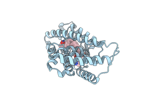
Crystal Structure Of The Human Vitamin D Receptor Ligand Binding Domain Complexed With (1R,2S,3R)-5-[(E)-2-{(1R,3As,7Ar)-1-[(R)-6-Hydroxy-6-Methylheptan-2-Yl]-7A-Methyl-2,3,3A,6,7,7A-Hexahydro-1H-Inden-4-Yl}Vinyl]-2-(3-Hydroxypropyl)Cyclohex-4-Ene-1,3-Diol
Organism: Homo sapiens
Method: X-RAY DIFFRACTION Resolution:2.00 Å Release Date: 2018-04-11 Classification: TRANSCRIPTION Ligands: 90O |
Organism: Homo sapiens
Method: X-RAY DIFFRACTION
Release Date: 2018-04-11
Ligands: 90O
 |
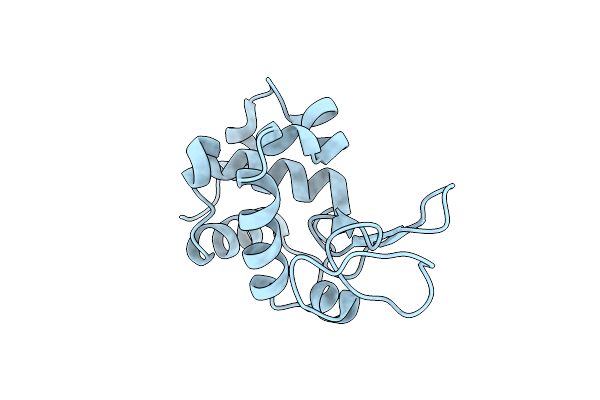
Crystal Structure Of The Human Vitamin D Receptor Ligand Binding Domain Complexed With 1-((((1S,2R,6R,Z)-2,6-Dihydroxy-4-((E)-2-((1R,3As,7Ar)-1-((R)-6-Hydroxy-6-Methylheptan-2-Yl)-7A-Methylhexahydro-1H-Inden-4(2H)-Ylidene)Ethylidene)-3-Methylenecyclohexyl)Oxy)Methyl)Cyclopropanecarbonitrile
Organism: Homo sapiens
Method: X-RAY DIFFRACTION Resolution:3.18 Å Release Date: 2014-12-31 Classification: TRANSCRIPTION Ligands: 3AJ |
Organism: Homo sapiens
Method: X-RAY DIFFRACTION
Release Date: 2014-12-31
Ligands: 3AJ
 |
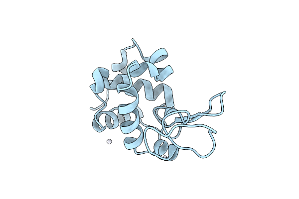
Spatiotemporal Development Of Soaked Protein Crystal; Native
Organism: Gallus gallus
Method: X-RAY DIFFRACTION Resolution:2.00 Å Release Date: 2014-07-30 Classification: HYDROLASE |
Organism: Gallus gallus
Method: X-RAY DIFFRACTION
Release Date: 2014-07-30
 |
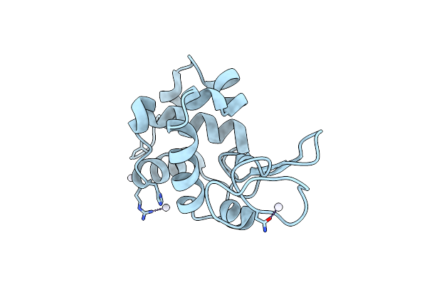
Spatiotemporal Development Of Soaked Protein Crystal; 750 Sec
Organism: Gallus gallus
Method: X-RAY DIFFRACTION Resolution:2.00 Å Release Date: 2014-07-30 Classification: HYDROLASE Ligands: PT4 |
Organism: Gallus gallus
Method: X-RAY DIFFRACTION
Release Date: 2014-07-30
Ligands: PT4
 |
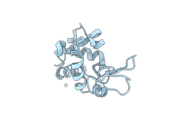
Spatiotemporal Development Of Soaked Protein Crystal; 2510 Sec
Organism: Gallus gallus
Method: X-RAY DIFFRACTION Resolution:2.00 Å Release Date: 2014-07-30 Classification: HYDROLASE Ligands: PT4 |
Organism: Gallus gallus
Method: X-RAY DIFFRACTION
Release Date: 2014-07-30
Ligands: PT4
 |
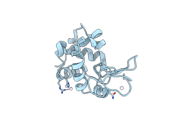
Spatiotemporal Development Of Soaked Protein Crystal; Derivative 250 Sec
Organism: Gallus gallus
Method: X-RAY DIFFRACTION Resolution:2.00 Å Release Date: 2014-07-30 Classification: HYDROLASE Ligands: PT4 |
Organism: Gallus gallus
Method: X-RAY DIFFRACTION
Release Date: 2014-07-30
Ligands: PT4
 |
Spatiotemporal Development Of Soaked Protein Crystal; Derivative 1080 Sec
Organism: Gallus gallus
Method: X-RAY DIFFRACTION Resolution:2.00 Å Release Date: 2014-07-30 Classification: HYDROLASE Ligands: PT4 |
Organism: Gallus gallus
Method: X-RAY DIFFRACTION
Release Date: 2014-07-30
Ligands: PT4
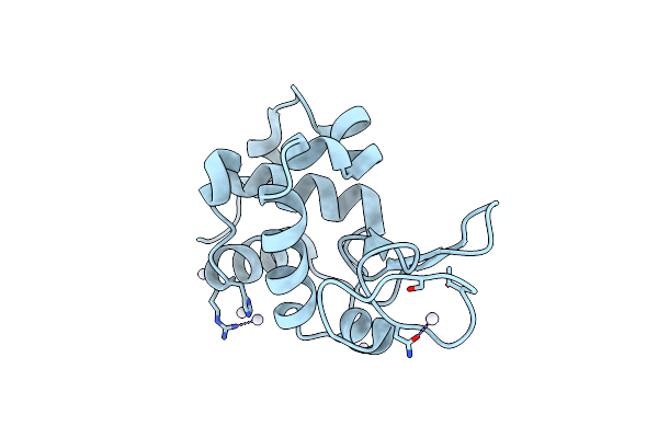 |
Spatiotemporal Development Of Soaked Protein Crystal; Derivative 1580 Sec
Organism: Gallus gallus
Method: X-RAY DIFFRACTION Resolution:2.00 Å Release Date: 2014-07-30 Classification: HYDROLASE Ligands: PT4 |
Organism: Gallus gallus
Method: X-RAY DIFFRACTION
Release Date: 2014-07-30
Ligands: PT4
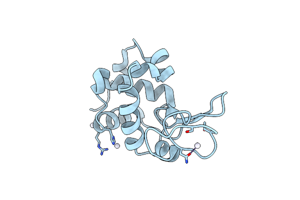 |
Spatiotemporal Development Of Soaked Protein Crystal; Derivative 3610 Sec
Organism: Gallus gallus
Method: X-RAY DIFFRACTION Resolution:2.00 Å Release Date: 2014-07-30 Classification: HYDROLASE Ligands: PT4 |
Organism: Gallus gallus
Method: X-RAY DIFFRACTION
Release Date: 2014-07-30
Ligands: PT4
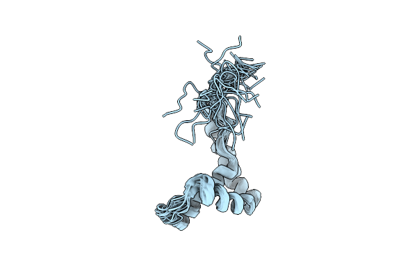 |
Solution Structure Of Oxidized Human Hmgb1 A Box
Organism: Homo sapiens
Method: SOLUTION NMR Release Date: 2014-03-05 Classification: DNA BINDING PROTEIN |
Organism: Homo sapiens
Method: SOLUTION NMR
Release Date: 2014-03-05
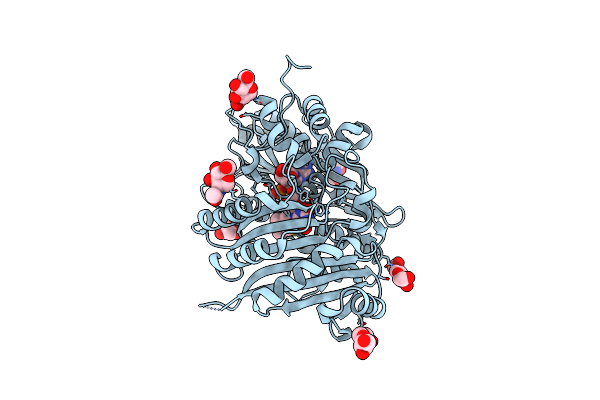 |
Crystal Structure Of Tetrahydrocannabinolic Acid Synthase From Cannabis Sativa
Organism: Cannabis sativa
Method: X-RAY DIFFRACTION Resolution:2.75 Å Release Date: 2012-07-25 Classification: OXIDOREDUCTASE Ligands: NAG, FAD |
Organism: Cannabis sativa
Method: X-RAY DIFFRACTION
Release Date: 2012-07-25
Ligands: NAG, FAD