

Search Count: 24
 |
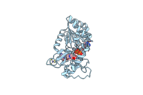
Crystal Structure Of Human Sirt5 In Complex With Succinylated Prx1 Fragment
Organism: Homo sapiens
Method: X-RAY DIFFRACTION Resolution:1.45 Å Release Date: 2024-10-09 Classification: TRANSFERASE Ligands: ZN, DMS |
Organism: Homo sapiens
Method: X-RAY DIFFRACTION
Release Date: 2024-10-09
Ligands: ZN, DMS
 |
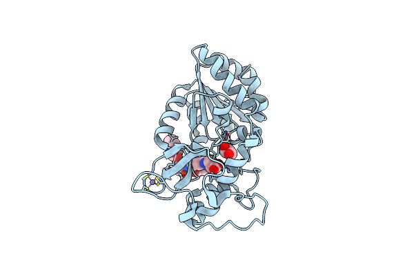
Crystal Structure Of Human Sirt5 In Complex With Succprx1 And Adp Ribose
Organism: Homo sapiens
Method: X-RAY DIFFRACTION Resolution:1.83 Å Release Date: 2024-10-09 Classification: TRANSFERASE Ligands: ADP, ZN |
Organism: Homo sapiens
Method: X-RAY DIFFRACTION
Release Date: 2024-10-09
Ligands: ADP, ZN
 |
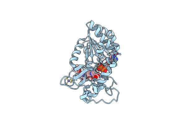
Crystal Structure Of Human Q140L-Sirt5 In Complex With Succinylated Prx1 Fragment
Organism: Homo sapiens
Method: X-RAY DIFFRACTION Resolution:1.81 Å Release Date: 2024-10-09 Classification: TRANSFERASE Ligands: ZN, GOL, CL |
Organism: Homo sapiens
Method: X-RAY DIFFRACTION
Release Date: 2024-10-09
Ligands: ZN, GOL, CL
 |
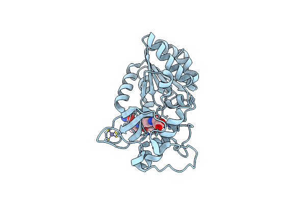
Crystal Structure Of Human Q140L-Sirt5 In Complex With Succinylated Prx1 Fragment And Adp Ribose
Organism: Homo sapiens
Method: X-RAY DIFFRACTION Resolution:1.96 Å Release Date: 2024-10-09 Classification: TRANSFERASE Ligands: ZN, ADP |
Organism: Homo sapiens
Method: X-RAY DIFFRACTION
Release Date: 2024-10-09
Ligands: ZN, ADP
 |
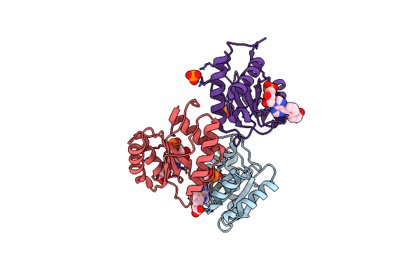
Crystal Structure Of Human N141V-Sirt5 In Complex With Succinylated Prx1 Fragment
Organism: Homo sapiens
Method: X-RAY DIFFRACTION Resolution:2.41 Å Release Date: 2024-10-09 Classification: TRANSFERASE Ligands: ZN, CL |
Organism: Homo sapiens
Method: X-RAY DIFFRACTION
Release Date: 2024-10-09
Ligands: ZN, CL
 |
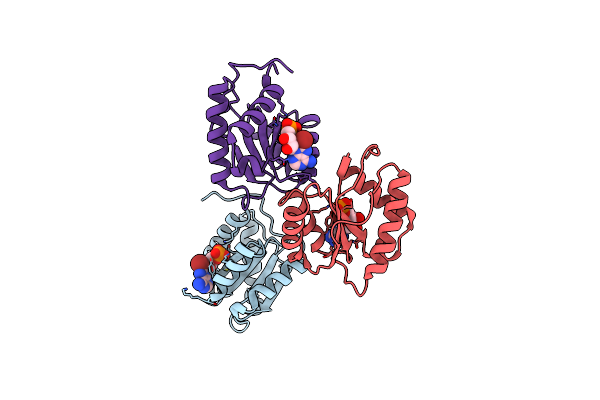
Crystal Structure Of Ago2 Mid Domain In Complex With 6-(3-(2-Carboxyethyl)Phenyl)Purine Riboside Monophosphate
Organism: Homo sapiens
Method: X-RAY DIFFRACTION Resolution:1.70 Å Release Date: 2020-11-25 Classification: RNA BINDING PROTEIN Ligands: K2R, PO4 |
Organism: Homo sapiens
Method: X-RAY DIFFRACTION
Release Date: 2020-11-25
Ligands: K2R, PO4
 |
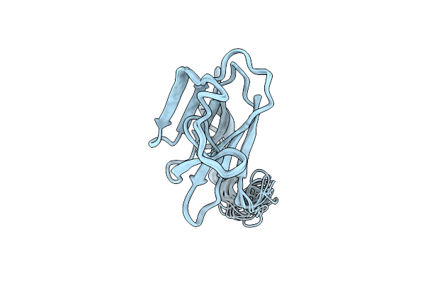
Crystal Structure Of Ago2 Mid Domain In Complex With 8-Br-Adenosin-5'-Monophosphate
Organism: Homo sapiens
Method: X-RAY DIFFRACTION Resolution:2.00 Å Release Date: 2020-11-25 Classification: RNA BINDING PROTEIN Ligands: 8BR |
Organism: Homo sapiens
Method: X-RAY DIFFRACTION
Release Date: 2020-11-25
Ligands: 8BR
 |
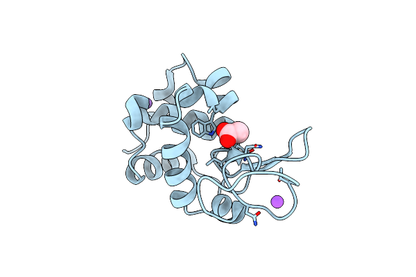
Structure Of The Plasmodium 6-Cysteine S48/45 Domain
Organism: Plasmodium falciparum 3d7
Method: SOLUTION NMR Release Date: 2012-04-18 Classification: MEMBRANE PROTEIN |
Organism: Plasmodium falciparum 3d7
Method: SOLUTION NMR
Release Date: 2012-04-18
 |
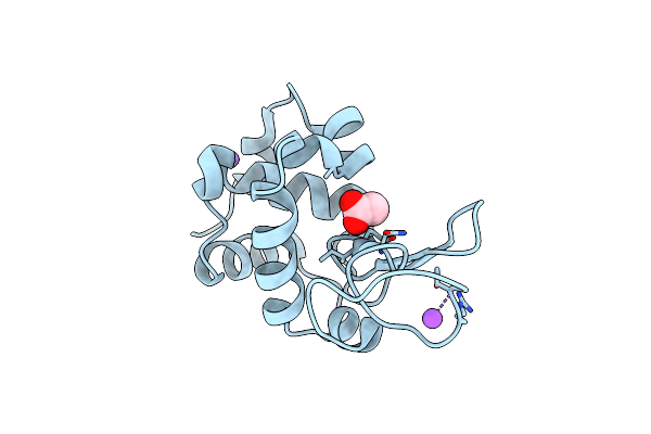
Crystal Structure Of Tetragonal Hen Egg White Lysozyme At 84.2% Relative Humidity
Organism: Gallus gallus
Method: X-RAY DIFFRACTION Resolution:2.10 Å Release Date: 2011-03-30 Classification: HYDROLASE Ligands: ACT, NA, CL |
Organism: Gallus gallus
Method: X-RAY DIFFRACTION
Release Date: 2011-03-30
Ligands: ACT, NA, CL
 |
Crystal Structure Of Tetragonal Hen Egg White Lysozyme At 71.9% Relative Humidity
Organism: Gallus gallus
Method: X-RAY DIFFRACTION Resolution:2.10 Å Release Date: 2011-03-30 Classification: HYDROLASE Ligands: ACT, NA, CL |
Organism: Gallus gallus
Method: X-RAY DIFFRACTION
Release Date: 2011-03-30
Ligands: ACT, NA, CL
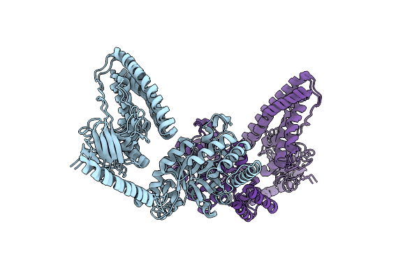 |
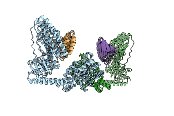
Solution Structure Of The H189Q Mutant Of The Enzyme I Dimer Using Residual Dipolar Couplings And Small Angle X-Ray Scattering
Organism: Escherichia coli
Method: SOLUTION NMR, SOLUTION SCATTERING Release Date: 2011-01-12 Classification: TRANSFERASE |
Organism: Escherichia coli
Method: SOLUTION NMR, SOLUTION SCATTERING
Release Date: 2011-01-12
 |
Solution Structure Of The Enzyme I Dimer Complexed With Hpr Using Residual Dipolar Couplings And Small Angle X-Ray Scattering
Organism: Escherichia coli
Method: SOLUTION NMR, SOLUTION SCATTERING Release Date: 2010-09-22 Classification: TRANSFERASE |
Organism: Escherichia coli
Method: SOLUTION NMR, SOLUTION SCATTERING
Release Date: 2010-09-22
 |
Solution Structure Of The Enzyme I Dimer Using Residual Dipolar Couplings And Small Angle X-Ray Scattering
Organism: Escherichia coli
Method: SOLUTION NMR, SOLUTION SCATTERING Release Date: 2010-09-15 Classification: TRANSFERASE |
Organism: Escherichia coli
Method: SOLUTION NMR, SOLUTION SCATTERING
Release Date: 2010-09-15
 |
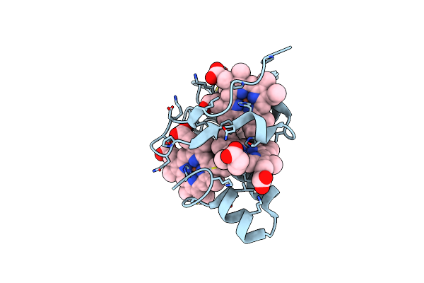
The Y66L Mutant Of Tetraheme Cytochrome C3 From Desulfovibrio Vulgaris Miyazaki F
Organism: Desulfovibrio vulgaris str. 'miyazaki f'
Method: X-RAY DIFFRACTION Resolution:1.60 Å Release Date: 2008-06-17 Classification: ELECTRON TRANSPORT Ligands: HEC |
Organism: Desulfovibrio vulgaris str. 'miyazaki f'
Method: X-RAY DIFFRACTION
Release Date: 2008-06-17
Ligands: HEC
 |
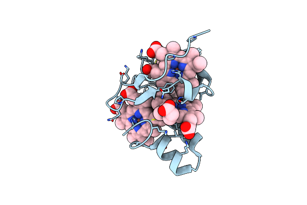
The F20M Mutant Of Tetraheme Cytochrome C3 From Desulfovibrio Vulgaris Miyazaki F
Organism: Desulfovibrio vulgaris str. 'miyazaki f'
Method: X-RAY DIFFRACTION Resolution:1.33 Å Release Date: 2008-05-06 Classification: ELECTRON TRANSPORT Ligands: HEC |
Organism: Desulfovibrio vulgaris str. 'miyazaki f'
Method: X-RAY DIFFRACTION
Release Date: 2008-05-06
Ligands: HEC
 |
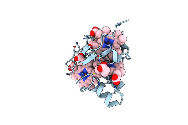
The Y65A Mutant Of Tetraheme Cytochrome C3 From Desulfovibrio Vulgaris Miyazaki F
Organism: Desulfovibrio vulgaris str. 'miyazaki f'
Method: X-RAY DIFFRACTION Resolution:1.00 Å Release Date: 2008-05-06 Classification: ELECTRON TRANSPORT Ligands: HEC |
Organism: Desulfovibrio vulgaris str. 'miyazaki f'
Method: X-RAY DIFFRACTION
Release Date: 2008-05-06
Ligands: HEC
 |
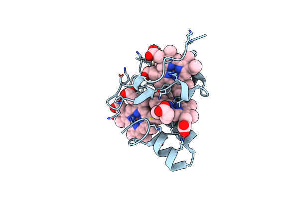
The H25M Mutant Of Tetraheme Cytochrome C3 From Desulfovibrio Vulgaris Miyazaki F
Organism: Desulfovibrio vulgaris str. 'miyazaki f'
Method: X-RAY DIFFRACTION Resolution:1.50 Å Release Date: 2008-04-29 Classification: ELECTRON TRANSPORT Ligands: HEC |
Organism: Desulfovibrio vulgaris str. 'miyazaki f'
Method: X-RAY DIFFRACTION
Release Date: 2008-04-29
Ligands: HEC
 |
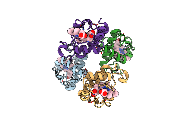
The T24V Mutant Of Tetraheme Cytochrome C3 From Desulfovibrio Vulgaris Miyazaki F
Organism: Desulfovibrio vulgaris str. 'miyazaki f'
Method: X-RAY DIFFRACTION Resolution:1.00 Å Release Date: 2006-11-28 Classification: ELECTRON TRANSPORT Ligands: HEM |
Organism: Desulfovibrio vulgaris str. 'miyazaki f'
Method: X-RAY DIFFRACTION
Release Date: 2006-11-28
Ligands: HEM
 |
Crystal Structure Of Oxidized Cytochrome C6A From Arabidopsis Thaliana
Organism: Arabidopsis thaliana
Method: X-RAY DIFFRACTION Resolution:1.50 Å Release Date: 2006-07-04 Classification: ELECTRON TRANSPORT Ligands: ZN, HEC |
Organism: Arabidopsis thaliana
Method: X-RAY DIFFRACTION
Release Date: 2006-07-04
Ligands: ZN, HEC
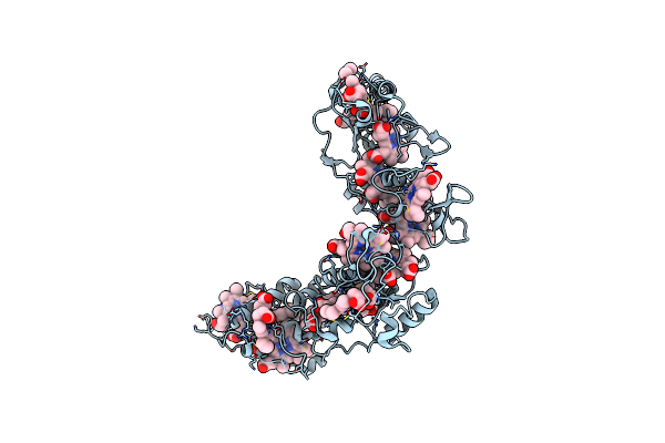 |
Crystal Structure Of High-Molecular Weight Cytochrome C From Desulfovibrio Vulgaris (Hildenborough)
Organism: Desulfovibrio vulgaris
Method: X-RAY DIFFRACTION Resolution:2.00 Å Release Date: 2006-06-06 Classification: ELECTRON TRANSPORT Ligands: HEC |
Organism: Desulfovibrio vulgaris
Method: X-RAY DIFFRACTION
Release Date: 2006-06-06
Ligands: HEC