

Search Count: 153
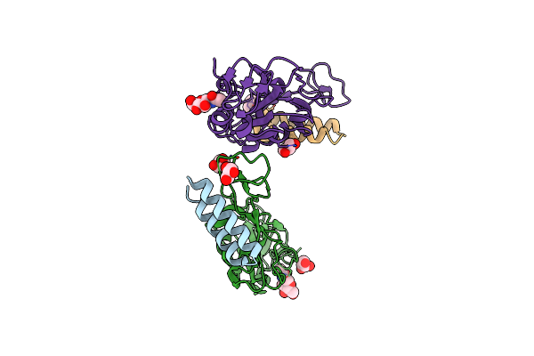 |
Crystal Structure Of The Receptor Binding Domain Of Sars-Cov-2 Spike Protein In Complex With Ce9
Organism: Severe acute respiratory syndrome coronavirus 2, Synthetic construct
Method: X-RAY DIFFRACTION Resolution:2.30 Å Release Date: 2025-01-15 Classification: VIRAL PROTEIN/INHIBITOR Ligands: NAG, GOL |
Organism: Severe acute respiratory syndrome coronavirus 2, Synthetic construct
Method: X-RAY DIFFRACTION
Release Date: 2025-01-15
Ligands: NAG, GOL
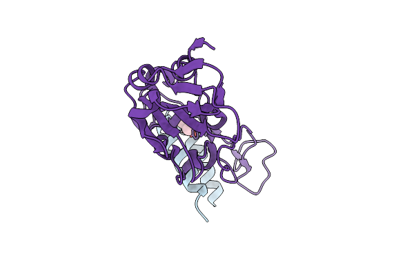 |
Crystal Structure Of The Receptor Binding Domain Of Sars-Cov-2 Omicron Ba.2 Variant Spike Protein In Complex With Ce149
Organism: Severe acute respiratory syndrome coronavirus 2, Synthetic construct
Method: X-RAY DIFFRACTION Resolution:1.90 Å Release Date: 2025-01-15 Classification: VIRAL PROTEIN/INHIBITOR Ligands: GOL |
Organism: Severe acute respiratory syndrome coronavirus 2, Synthetic construct
Method: X-RAY DIFFRACTION
Release Date: 2025-01-15
Ligands: GOL
 |
Crystal Structure Of The Receptor Binding Domain Of Sars-Cov-2 Alpha Variant Spike Protein In Complex With Ce59
Organism: Severe acute respiratory syndrome coronavirus 2, Synthetic construct
Method: X-RAY DIFFRACTION Resolution:2.30 Å Release Date: 2025-01-15 Classification: VIRAL PROTEIN/INHIBITOR Ligands: NAG |
Organism: Severe acute respiratory syndrome coronavirus 2, Synthetic construct
Method: X-RAY DIFFRACTION
Release Date: 2025-01-15
Ligands: NAG
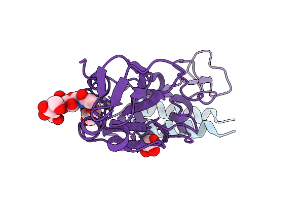 |
Crystal Structure Of The Receptor Binding Domain Of Sars-Cov-2 Delta Variant Spike Protein In Complex With Ce59
Organism: Severe acute respiratory syndrome coronavirus 2, Synthetic construct
Method: X-RAY DIFFRACTION Resolution:1.90 Å Release Date: 2025-01-15 Classification: VIRAL PROTEIN/INHIBITOR Ligands: GOL |
Organism: Severe acute respiratory syndrome coronavirus 2, Synthetic construct
Method: X-RAY DIFFRACTION
Release Date: 2025-01-15
Ligands: GOL
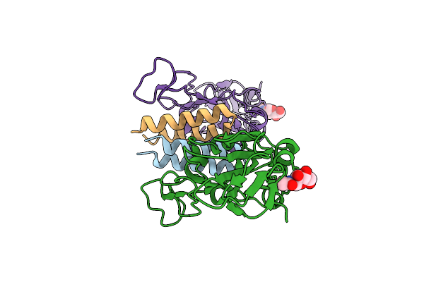 |
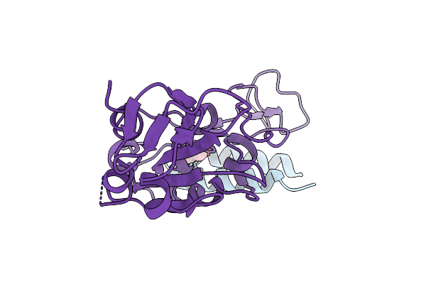
Crystal Structure Of The Receptor Binding Domain Of Sars-Cov-2 Alpha Variant Spike Protein In Complex With Ce41
Organism: Severe acute respiratory syndrome coronavirus 2, Synthetic construct
Method: X-RAY DIFFRACTION Resolution:2.50 Å Release Date: 2025-01-15 Classification: VIRAL PROTEIN/INHIBITOR Ligands: NAG |
Organism: Severe acute respiratory syndrome coronavirus 2, Synthetic construct
Method: X-RAY DIFFRACTION
Release Date: 2025-01-15
Ligands: NAG
 |
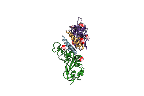
Crystal Structure Of The Receptor Binding Domain Of Sars-Cov-2 Omicron Ba.2 Variant Spike Protein In Complex With Cespiace
Organism: Severe acute respiratory syndrome coronavirus 2, Synthetic construct
Method: X-RAY DIFFRACTION Resolution:1.70 Å Release Date: 2025-01-15 Classification: VIRAL PROTEIN/INHIBITOR Ligands: GOL |
Organism: Severe acute respiratory syndrome coronavirus 2, Synthetic construct
Method: X-RAY DIFFRACTION
Release Date: 2025-01-15
Ligands: GOL
 |
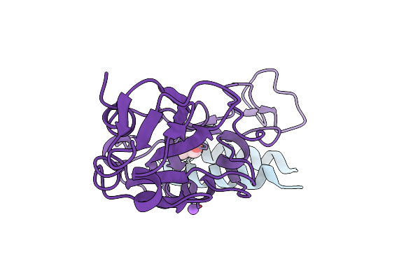
Crystal Structure Of The Receptor Binding Domain Of Sars-Cov-2 Omicron Ba.5 Variant Spike Protein In Complex With Cespiace
Organism: Severe acute respiratory syndrome coronavirus 2, Synthetic construct
Method: X-RAY DIFFRACTION Resolution:2.20 Å Release Date: 2025-01-15 Classification: VIRAL PROTEIN/INHIBITOR Ligands: GOL |
Organism: Severe acute respiratory syndrome coronavirus 2, Synthetic construct
Method: X-RAY DIFFRACTION
Release Date: 2025-01-15
Ligands: GOL
 |
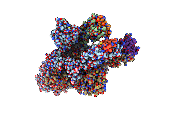
Crystal Structure Of The Receptor Binding Domain Of Sars-Cov-2 Omicron Xbb.1.5 Variant Spike Protein In Complex With Cespiace
Organism: Severe acute respiratory syndrome coronavirus 2, Synthetic construct
Method: X-RAY DIFFRACTION Resolution:2.00 Å Release Date: 2025-01-15 Classification: VIRAL PROTEIN/INHIBITOR Ligands: GOL, NA |
Organism: Severe acute respiratory syndrome coronavirus 2, Synthetic construct
Method: X-RAY DIFFRACTION
Release Date: 2025-01-15
Ligands: GOL, NA
 |
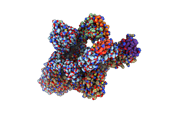
Cryo-Em Structure Of Sars-Cov-2 Spike Ectodomain (Hexapro, Omicron Ba.2 Variant) In Complex With Cespiace
Organism: Severe acute respiratory syndrome coronavirus 2, Synthetic construct
Method: ELECTRON MICROSCOPY Release Date: 2025-01-15 Classification: VIRAL PROTEIN/INHIBITOR Ligands: NAG |
Organism: Severe acute respiratory syndrome coronavirus 2, Synthetic construct
Method: ELECTRON MICROSCOPY
Release Date: 2025-01-15
Ligands: NAG
 |
Cryo-Em Structure Of Sars-Cov-2 Spike Ectodomain (Hexapro, Omicron Ba.5 Variant) In Complex With Cespiace, Class 1
Organism: Severe acute respiratory syndrome coronavirus 2, Synthetic construct
Method: ELECTRON MICROSCOPY Release Date: 2025-01-15 Classification: VIRAL PROTEIN/INHIBITOR Ligands: NAG |
Organism: Severe acute respiratory syndrome coronavirus 2, Synthetic construct
Method: ELECTRON MICROSCOPY
Release Date: 2025-01-15
Ligands: NAG
 |
Cryo-Em Structure Of Sars-Cov-2 Spike Ectodomain (Hexapro, Omicron Ba.5 Variant) In Complex With Cespiace, Class 2
Organism: Severe acute respiratory syndrome coronavirus 2, Synthetic construct
Method: ELECTRON MICROSCOPY Release Date: 2025-01-15 Classification: VIRAL PROTEIN/INHIBITOR Ligands: NAG |
Organism: Severe acute respiratory syndrome coronavirus 2, Synthetic construct
Method: ELECTRON MICROSCOPY
Release Date: 2025-01-15
Ligands: NAG
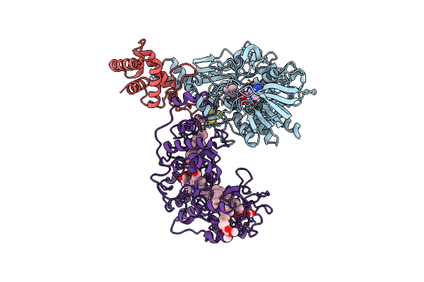 |
Cryo-Em Structure Of Membrane-Bound Fructose Dehydrogenase From Gluconobacter Japonicus Variant-H1147A
Organism: Gluconobacter japonicus
Method: ELECTRON MICROSCOPY Release Date: 2024-05-22 Classification: OXIDOREDUCTASE Ligands: FAD, F3S, HEC, U10 |
Organism: Gluconobacter japonicus
Method: ELECTRON MICROSCOPY
Release Date: 2024-05-22
Ligands: FAD, F3S, HEC, U10
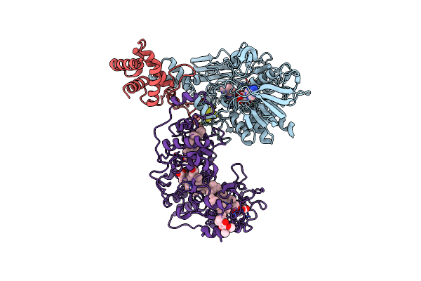 |
Cryo-Em Structure Of Membrane-Bound Fructose Dehydrogenase From Gluconobacter Japonicus Variant-N1146A
Organism: Gluconobacter japonicus
Method: ELECTRON MICROSCOPY Release Date: 2024-05-22 Classification: OXIDOREDUCTASE Ligands: FAD, F3S, HEC, U10 |
Organism: Gluconobacter japonicus
Method: ELECTRON MICROSCOPY
Release Date: 2024-05-22
Ligands: FAD, F3S, HEC, U10
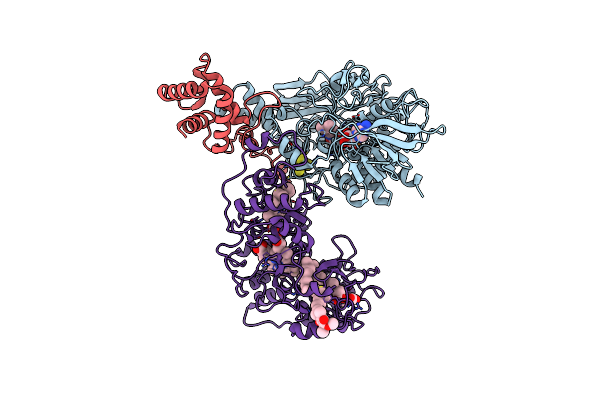 |
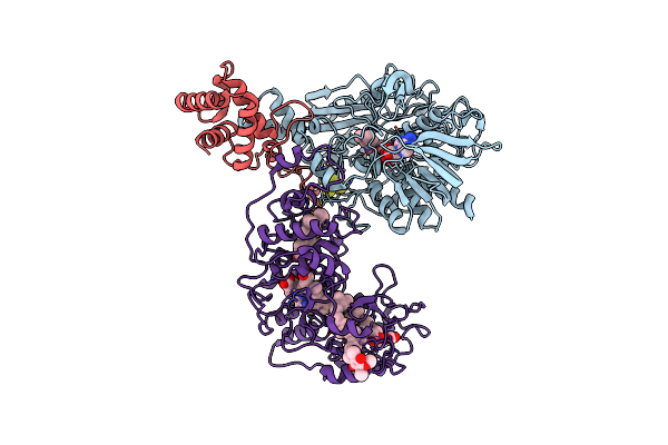
Cryo-Em Structure Of Membrane-Bound Fructose Dehydrogenase From Gluconobacter Japonicus Variant-N1146Q
Organism: Gluconobacter japonicus
Method: ELECTRON MICROSCOPY Release Date: 2024-05-22 Classification: OXIDOREDUCTASE Ligands: FAD, F3S, HEC, U10 |
Organism: Gluconobacter japonicus
Method: ELECTRON MICROSCOPY
Release Date: 2024-05-22
Ligands: FAD, F3S, HEC, U10
 |
Cryo-Em Structure Of Membrane-Bound Fructose Dehydrogenase From Gluconobacter Japonicus Variant-N1190A
Organism: Gluconobacter japonicus
Method: ELECTRON MICROSCOPY Release Date: 2024-05-22 Classification: OXIDOREDUCTASE Ligands: FAD, F3S, HEC, U10 |
Organism: Gluconobacter japonicus
Method: ELECTRON MICROSCOPY
Release Date: 2024-05-22
Ligands: FAD, F3S, HEC, U10
 |
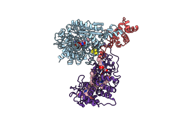
Cryo-Em Structure Of Na-Dithionite Reduced Membrane-Bound Fructose Dehydrogenase From Gluconobacter Japonicus
Organism: Gluconobacter japonicus
Method: ELECTRON MICROSCOPY Release Date: 2023-10-25 Classification: OXIDOREDUCTASE Ligands: FAD, F3S, HEC, U10 |
Organism: Gluconobacter japonicus
Method: ELECTRON MICROSCOPY
Release Date: 2023-10-25
Ligands: FAD, F3S, HEC, U10
 |
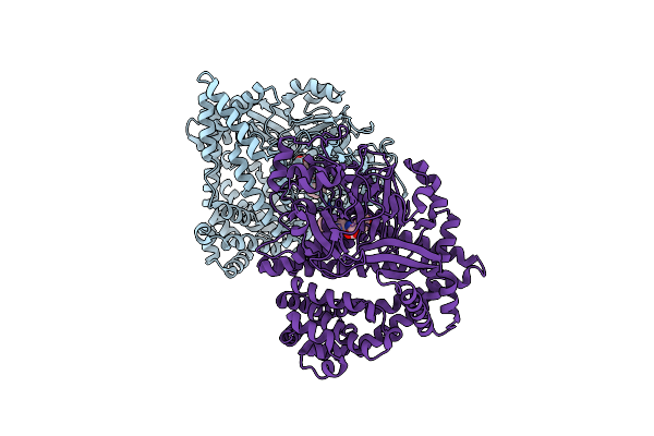
Cryo-Em Structure Of K-Ferricyanide Oxidized Membrane-Bound Fructose Dehydrogenase From Gluconobacter Japonicus
Organism: Gluconobacter japonicus
Method: ELECTRON MICROSCOPY Release Date: 2023-10-25 Classification: OXIDOREDUCTASE Ligands: FAD, F3S, HEC, U10 |
Organism: Gluconobacter japonicus
Method: ELECTRON MICROSCOPY
Release Date: 2023-10-25
Ligands: FAD, F3S, HEC, U10
 |
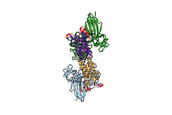
Small Dipeptide Analogues Developed By Co-Crystal Structure Of Stenotrophomonas Maltophilia Dipeptidyl Peptidase 7
Organism: Stenotrophomonas maltophilia (strain r551-3)
Method: X-RAY DIFFRACTION Resolution:2.59 Å Release Date: 2023-09-06 Classification: HYDROLASE Ligands: ALC, TYR |
Organism: Stenotrophomonas maltophilia (strain r551-3)
Method: X-RAY DIFFRACTION
Release Date: 2023-09-06
Ligands: ALC, TYR
 |
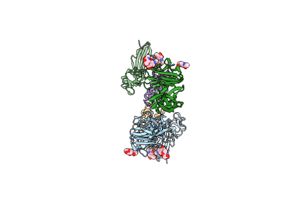
Ectodomain Of Full-Length Wild-Type Kit-Scf Dimers
Organism: Homo sapiens
Method: ELECTRON MICROSCOPY Release Date: 2023-03-29 Classification: TRANSFERASE Ligands: NAG |
Organism: Homo sapiens
Method: ELECTRON MICROSCOPY
Release Date: 2023-03-29
Ligands: NAG
 |
Ectodomain Of Full-Length Kit(Dupa502,Y503)-Scf Dimers
Organism: Homo sapiens
Method: ELECTRON MICROSCOPY Release Date: 2023-03-29 Classification: TRANSFERASE Ligands: NAG |
Organism: Homo sapiens
Method: ELECTRON MICROSCOPY
Release Date: 2023-03-29
Ligands: NAG