
Search Count: 268
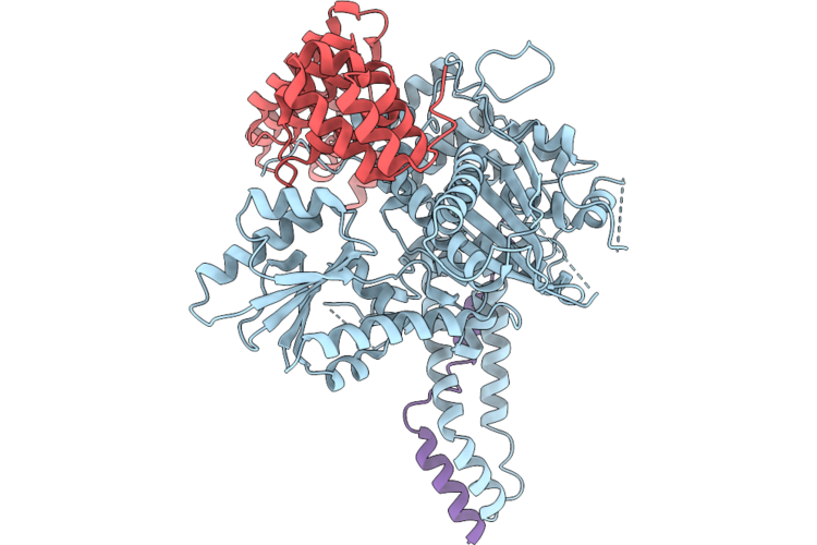 |
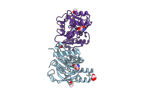
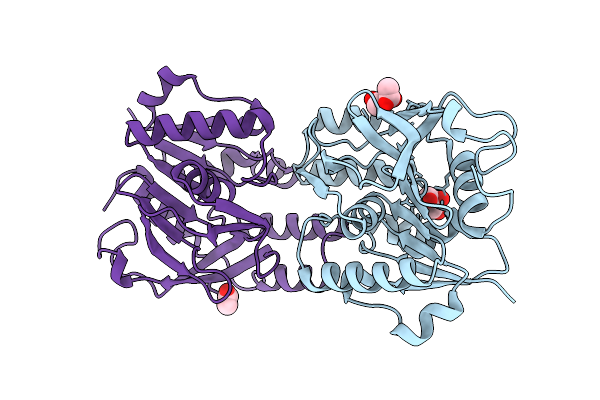
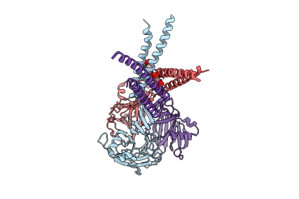
Revised Structure Of A Complex Between The Snare Nyv1 And The Hops Vps33-Vps16 Subcomplex
Organism: Thermochaetoides thermophila dsm 1495
Method: X-RAY DIFFRACTION Resolution:3.05 Å Release Date: 2026-06-03 Classification: TRANSPORT PROTEIN |
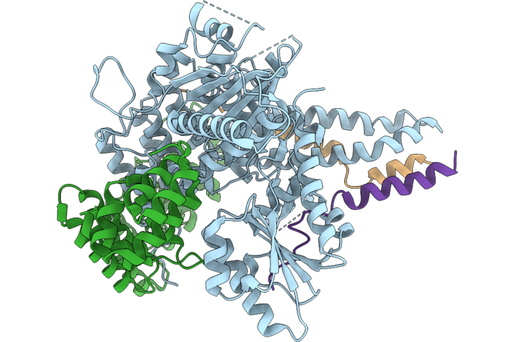 |
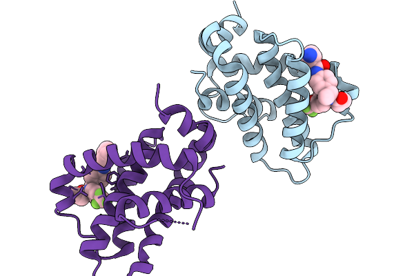
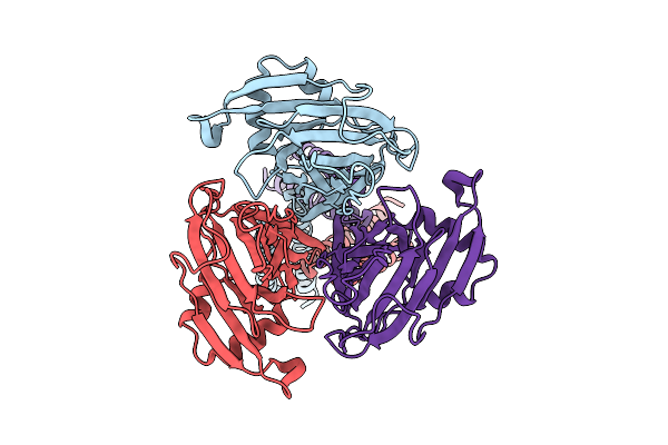
Crystal Structure Of Hops Subunits Vps33 And Vps16 In Complex With The Nyv1 And Vam3 Snare Motifs
Organism: Thermochaetoides thermophila dsm 1495
Method: X-RAY DIFFRACTION Resolution:3.19 Å Release Date: 2026-06-03 Classification: TRANSPORT PROTEIN |
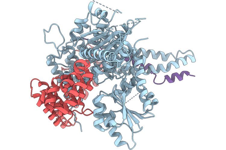 |
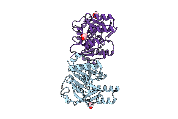
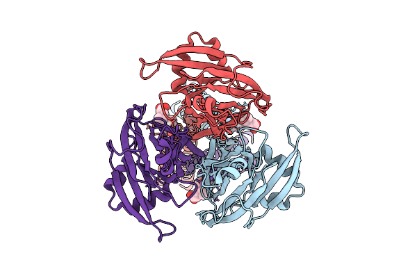
Crystal Structure Of Hops Subunits Vps33 And Vps16 In Complex With The Nyv1 Snare Motif
Organism: Thermochaetoides thermophila dsm 1495
Method: X-RAY DIFFRACTION Resolution:2.90 Å Release Date: 2026-06-03 Classification: TRANSPORT PROTEIN |
 |
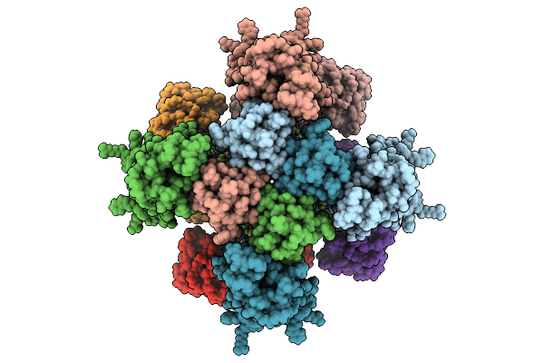
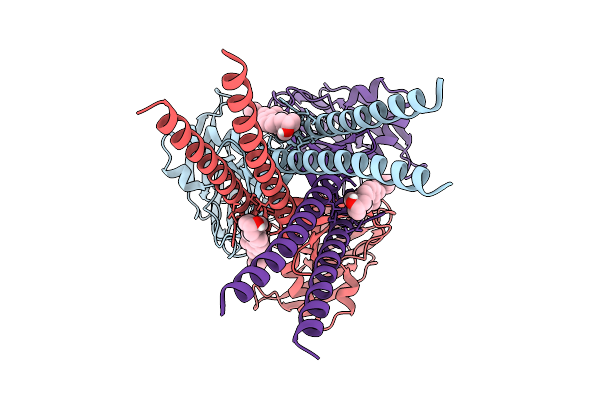
Organism: Streptomyces mobaraensis
Method: ELECTRON MICROSCOPY Resolution:2.01 Å Release Date: 2026-04-22 Classification: BIOSYNTHETIC PROTEIN Ligands: ZN, GTP |
 |
Crystal Structure Of 1L-Myo-Inositol 1-Phosphate Synthase 1 From Oryza Sativa
Organism: Oryza sativa
Method: X-RAY DIFFRACTION Resolution:1.79 Å Release Date: 2026-02-04 Classification: ISOMERASE Ligands: KPG, NAI, EDO, K |
 |
Organism: Homo sapiens
Method: ELECTRON MICROSCOPY Release Date: 2026-01-28 Classification: MEMBRANE PROTEIN Ligands: PIO, A1LVR |
 |
Organism: Homo sapiens
Method: ELECTRON MICROSCOPY Release Date: 2026-01-28 Classification: MEMBRANE PROTEIN Ligands: PIO, A1LWZ |
 |
Organism: Homo sapiens
Method: ELECTRON MICROSCOPY Release Date: 2026-01-21 Classification: MEMBRANE PROTEIN Ligands: A1LVR |
 |
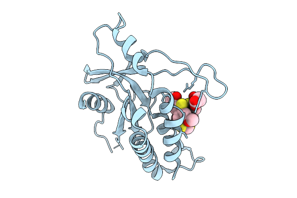
Structure Of Bfl1 In Complex With A Covalent Inhibitor, Alternative Series, Cmpd25
Organism: Homo sapiens
Method: X-RAY DIFFRACTION Resolution:1.43 Å Release Date: 2026-01-14 Classification: APOPTOSIS Ligands: A1JL2 |
 |
Organism: Arabidopsis thaliana
Method: X-RAY DIFFRACTION Resolution:1.74 Å Release Date: 2025-10-29 Classification: HYDROLASE Ligands: GLU, ACT, GOL |
 |
Organism: Arabidopsis thaliana
Method: X-RAY DIFFRACTION Resolution:1.90 Å Release Date: 2025-10-29 Classification: HYDROLASE Ligands: ACT, GOL |
 |
Organism: Arabidopsis thaliana
Method: X-RAY DIFFRACTION Resolution:2.04 Å Release Date: 2025-10-29 Classification: HYDROLASE Ligands: ACT, GOL |
 |
Gamma-Glutamyl Peptidase 1 From Arabidopsis Thaliana (H192N Gamma-Glu Intermediate)
Organism: Arabidopsis thaliana
Method: X-RAY DIFFRACTION Resolution:1.59 Å Release Date: 2025-10-29 Classification: HYDROLASE Ligands: GLU, ACT, GOL |
 |
Organism: Homo sapiens
Method: X-RAY DIFFRACTION Resolution:2.31 Å Release Date: 2025-09-24 Classification: SIGNALING PROTEIN Ligands: A1JA0 |
 |
Organism: Homo sapiens
Method: ELECTRON MICROSCOPY Resolution:3.43 Å Release Date: 2025-07-30 Classification: MEMBRANE PROTEIN |
 |
Organism: Homo sapiens
Method: ELECTRON MICROSCOPY Release Date: 2025-07-30 Classification: MEMBRANE PROTEIN Ligands: DXC |
 |
Organism: Homo sapiens
Method: ELECTRON MICROSCOPY Release Date: 2025-07-23 Classification: MEMBRANE PROTEIN Ligands: IZ8 |
 |
Organism: Homo sapiens
Method: ELECTRON MICROSCOPY Release Date: 2025-07-23 Classification: MEMBRANE PROTEIN Ligands: CLR |
 |
Organism: Homo sapiens
Method: ELECTRON MICROSCOPY Release Date: 2025-04-16 Classification: MEMBRANE PROTEIN |
 |
Organism: Homo sapiens
Method: ELECTRON MICROSCOPY Release Date: 2025-04-16 Classification: MEMBRANE PROTEIN Ligands: PIO |