

Search Count: 15
 |
Structure Of Human Nhe9
Organism: Homo sapiens
Method: ELECTRON MICROSCOPY Release Date: 2026-09-16 Classification: MEMBRANE PROTEIN |
Organism: Homo sapiens
Method: ELECTRON MICROSCOPY
Release Date: 2026-09-16
 |
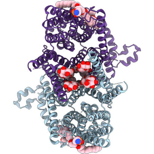
Structure Of Human Nhe9 (Core-Tm Domain)
Organism: Homo sapiens
Method: ELECTRON MICROSCOPY Release Date: 2026-09-16 Classification: MEMBRANE PROTEIN |
Organism: Homo sapiens
Method: ELECTRON MICROSCOPY
Release Date: 2026-09-16
 |
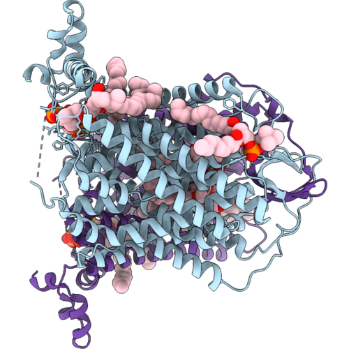
Structure Of Human Nhe6.0
Organism: Homo sapiens
Method: ELECTRON MICROSCOPY Release Date: 2026-09-16 Classification: MEMBRANE PROTEIN Ligands: LMT, 3PE |
Organism: Homo sapiens
Method: ELECTRON MICROSCOPY
Release Date: 2026-09-16
Ligands: LMT, 3PE
 |
Structure Of Human Nhe6.1 Bound To Pip2
Organism: Homo sapiens
Method: ELECTRON MICROSCOPY Release Date: 2026-09-16 Classification: MEMBRANE PROTEIN Ligands: LMT, PC1, PCF, A1JDN |
Organism: Homo sapiens
Method: ELECTRON MICROSCOPY
Release Date: 2026-09-16
Ligands: LMT, PC1, PCF, A1JDN
 |
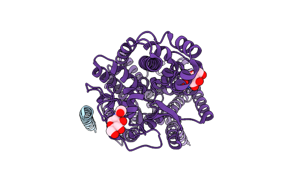
Structure Of Human Nhe6.1 Bound To Pip2 (Core-Tm Domain)
Organism: Homo sapiens
Method: ELECTRON MICROSCOPY Release Date: 2026-09-16 Classification: MEMBRANE PROTEIN |
Organism: Homo sapiens
Method: ELECTRON MICROSCOPY
Release Date: 2026-09-16
 |
Structure Of Human Sit1 (Focussed Map / Refinement)
Organism: Homo sapiens
Method: ELECTRON MICROSCOPY Release Date: 2024-06-12 Classification: MEMBRANE PROTEIN Ligands: NAG |
Organism: Homo sapiens
Method: ELECTRON MICROSCOPY
Release Date: 2024-06-12
Ligands: NAG
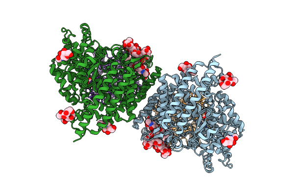 |
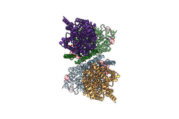
Structure Of Human Sit1:Ace2 Complex (Open Pd Conformation)
Organism: Homo sapiens
Method: ELECTRON MICROSCOPY Release Date: 2024-06-12 Classification: MEMBRANE PROTEIN Ligands: ZN, NDG, NAG |
Organism: Homo sapiens
Method: ELECTRON MICROSCOPY
Release Date: 2024-06-12
Ligands: ZN, NDG, NAG
 |
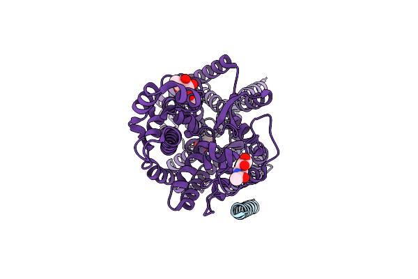
Structure Of Human Sit1:Ace2 Complex (Closed Pd Conformation)
Organism: Homo sapiens
Method: ELECTRON MICROSCOPY Release Date: 2024-06-12 Classification: MEMBRANE PROTEIN Ligands: ZN, NDG, NAG |
Organism: Homo sapiens
Method: ELECTRON MICROSCOPY
Release Date: 2024-06-12
Ligands: ZN, NDG, NAG
 |
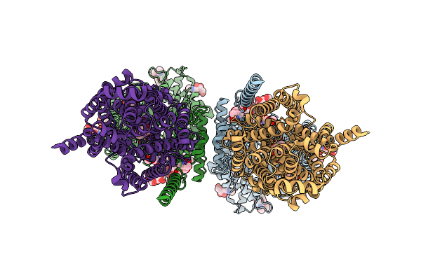
Structure Of Human Sit1 Bound To L-Pipecolate (Focussed Map / Refinement)
Organism: Homo sapiens
Method: ELECTRON MICROSCOPY Release Date: 2024-06-12 Classification: MEMBRANE PROTEIN Ligands: NAG, YCP, CL |
Organism: Homo sapiens
Method: ELECTRON MICROSCOPY
Release Date: 2024-06-12
Ligands: NAG, YCP, CL
 |
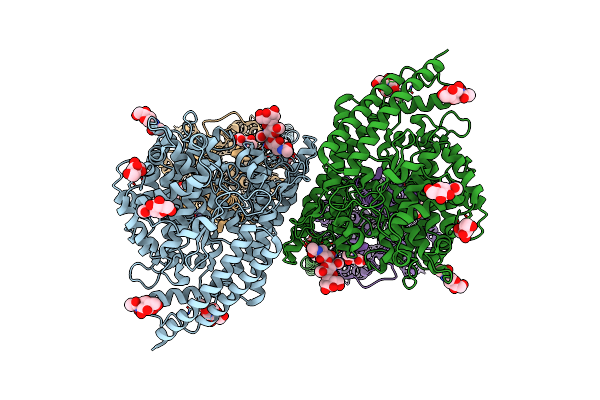
Structure Of Human Sit1:Ace2 Complex (Open Pd Conformation) Bound To L-Pipecolate
Organism: Homo sapiens
Method: ELECTRON MICROSCOPY Release Date: 2024-06-12 Classification: MEMBRANE PROTEIN Ligands: ZN, NDG, NAG, YCP, CL |
Organism: Homo sapiens
Method: ELECTRON MICROSCOPY
Release Date: 2024-06-12
Ligands: ZN, NDG, NAG, YCP, CL
 |
Structure Of Human Sit1:Ace2 Complex (Closed Pd Conformation) Bound To L-Pipecolate
Organism: Homo sapiens
Method: ELECTRON MICROSCOPY Release Date: 2024-06-12 Classification: MEMBRANE PROTEIN Ligands: ZN, NDG, NAG, YCP |
Organism: Homo sapiens
Method: ELECTRON MICROSCOPY
Release Date: 2024-06-12
Ligands: ZN, NDG, NAG, YCP
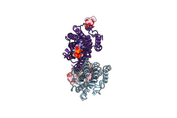 |
Crystal Structure Of Human Elovl Fatty Acid Elongase 7 (Elovl7)
Organism: Homo sapiens
Method: X-RAY DIFFRACTION Resolution:2.05 Å Release Date: 2020-05-13 Classification: TRANSFERASE Ligands: OFN, 37X, CL |
Organism: Homo sapiens
Method: X-RAY DIFFRACTION
Release Date: 2020-05-13
Ligands: OFN, 37X, CL
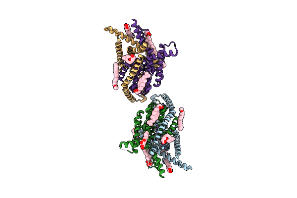 |
Crystal Structure Of The Human Two Pore Domain Potassium Ion Channel Task-1 (K2P3.1) In A Closed Conformation
Organism: Homo sapiens
Method: X-RAY DIFFRACTION Resolution:3.00 Å Release Date: 2019-08-07 Classification: MEMBRANE PROTEIN Ligands: K, Y01, DMU, PC1 |
Organism: Homo sapiens
Method: X-RAY DIFFRACTION
Release Date: 2019-08-07
Ligands: K, Y01, DMU, PC1
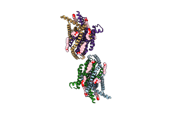 |
Crystal Structure Of The Human Two Pore Domain Potassium Ion Channel Task-1 (K2P3.1) In A Closed Conformation With A Bound Inhibitor Bay 1000493
Organism: Homo sapiens
Method: X-RAY DIFFRACTION Resolution:2.90 Å Release Date: 2019-08-07 Classification: MEMBRANE PROTEIN Ligands: K, Y01, DMU, PC1, KKQ |
Organism: Homo sapiens
Method: X-RAY DIFFRACTION
Release Date: 2019-08-07
Ligands: K, Y01, DMU, PC1, KKQ
 |
Crystal Structure Of The Human Two Pore Domain Potassium Ion Channel Task-1 (K2P3.1) In A Closed Conformation With A Bound Inhibitor Bay 2341237
Organism: Homo sapiens
Method: X-RAY DIFFRACTION Resolution:3.10 Å Release Date: 2019-08-07 Classification: MEMBRANE PROTEIN Ligands: K, Y01, KKZ, PC1 |
Organism: Homo sapiens
Method: X-RAY DIFFRACTION
Release Date: 2019-08-07
Ligands: K, Y01, KKZ, PC1