

Search Count: 45
 |
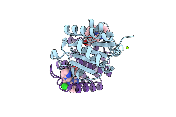
Crystal Structure Of Dnph1 Bound By Compound 2.
Organism: Homo sapiens
Method: X-RAY DIFFRACTION Resolution:1.60 Å Release Date: 2025-11-12 Classification: HYDROLASE Ligands: A1IZR |
Organism: Homo sapiens
Method: X-RAY DIFFRACTION
Release Date: 2025-11-12
Ligands: A1IZR
 |
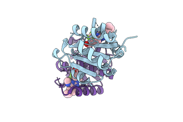
Crystal Structure Of Dnph1 Bound By Compound 39.
Organism: Homo sapiens
Method: X-RAY DIFFRACTION Resolution:1.22 Å Release Date: 2025-11-12 Classification: HYDROLASE Ligands: MG, A1IZW, CL |
Organism: Homo sapiens
Method: X-RAY DIFFRACTION
Release Date: 2025-11-12
Ligands: MG, A1IZW, CL
 |
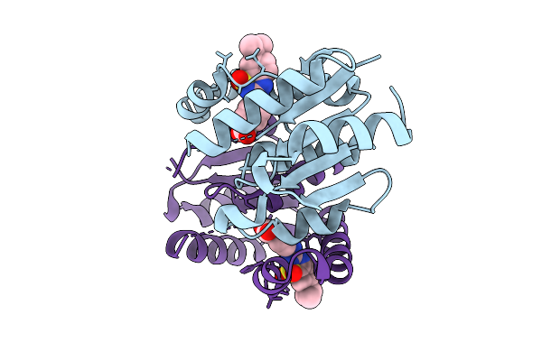
Crystal Structure Of Dnph1 Bound By Compound 31.
Organism: Homo sapiens
Method: X-RAY DIFFRACTION Resolution:1.21 Å Release Date: 2025-11-12 Classification: HYDROLASE Ligands: A1IZX |
Organism: Homo sapiens
Method: X-RAY DIFFRACTION
Release Date: 2025-11-12
Ligands: A1IZX
 |
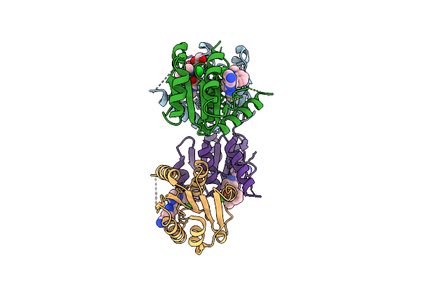
Crystal Structure Of Dnph1 Bound By Compound 5.
Organism: Homo sapiens
Method: X-RAY DIFFRACTION Resolution:1.49 Å Release Date: 2025-11-12 Classification: HYDROLASE Ligands: A1I0K |
Organism: Homo sapiens
Method: X-RAY DIFFRACTION
Release Date: 2025-11-12
Ligands: A1I0K
 |
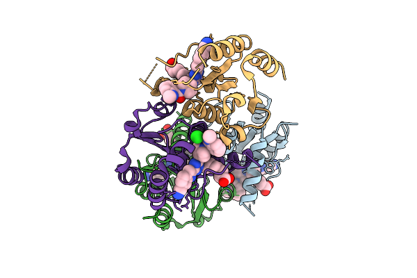
Crystal Structure Of Dnph1 Bound By Compound 2.
Organism: Homo sapiens
Method: X-RAY DIFFRACTION Resolution:1.72 Å Release Date: 2025-11-12 Classification: HYDROLASE Ligands: EOH, EDO, A1I1M, CL |
Organism: Homo sapiens
Method: X-RAY DIFFRACTION
Release Date: 2025-11-12
Ligands: EOH, EDO, A1I1M, CL
 |
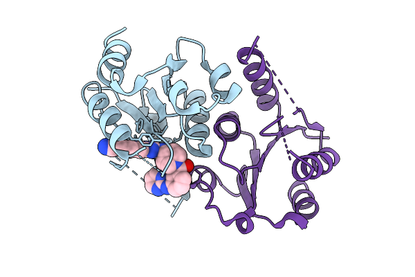
Crystal Structure Of Dnph1 Bound By Compound 3.
Organism: Homo sapiens
Method: X-RAY DIFFRACTION Resolution:2.21 Å Release Date: 2025-11-12 Classification: HYDROLASE Ligands: CL, A1I1U |
Organism: Homo sapiens
Method: X-RAY DIFFRACTION
Release Date: 2025-11-12
Ligands: CL, A1I1U
 |
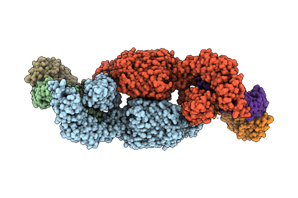
Crystal Structure Of Dnph1 Bound By Compound 1.
Organism: Homo sapiens
Method: X-RAY DIFFRACTION Resolution:2.07 Å Release Date: 2025-11-12 Classification: HYDROLASE Ligands: A1JFX |
Organism: Homo sapiens
Method: X-RAY DIFFRACTION
Release Date: 2025-11-12
Ligands: A1JFX
 |
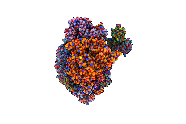
Fusobacterium Nucleatum Bama-Fab 9 Complex
Organism: Fusobacterium nucleatum, Synthetic construct
Method: ELECTRON MICROSCOPY Release Date: 2025-09-17 Classification: MEMBRANE PROTEIN |
Organism: Fusobacterium nucleatum, Synthetic construct
Method: ELECTRON MICROSCOPY
Release Date: 2025-09-17
 |
Structure Of Pol Ii-Tc-Ner-Stk19 Complex
Organism: Homo sapiens, Sus scrofa domesticus
Method: ELECTRON MICROSCOPY Release Date: 2024-11-13 Classification: TRANSCRIPTION Ligands: ZN, MG |
Organism: Homo sapiens, Sus scrofa domesticus
Method: ELECTRON MICROSCOPY
Release Date: 2024-11-13
Ligands: ZN, MG
 |
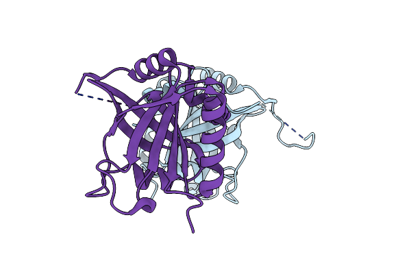
Crystal Structure Of The Spoc Domain Of Human Sharp (Spen) In Complex With Rna Polymerase Ii Ctd Heptapeptide Phosphorylated On Ser5
Organism: Homo sapiens
Method: X-RAY DIFFRACTION Resolution:1.55 Å Release Date: 2022-12-07 Classification: TRANSCRIPTION |
Organism: Homo sapiens
Method: X-RAY DIFFRACTION
Release Date: 2022-12-07
 |
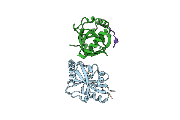
Crystal Structure Of The Spoc Domain Of Human Rbm15
Organism: Homo sapiens
Method: X-RAY DIFFRACTION Resolution:1.45 Å Release Date: 2022-12-07 Classification: TRANSCRIPTION |
Organism: Homo sapiens
Method: X-RAY DIFFRACTION
Release Date: 2022-12-07
 |
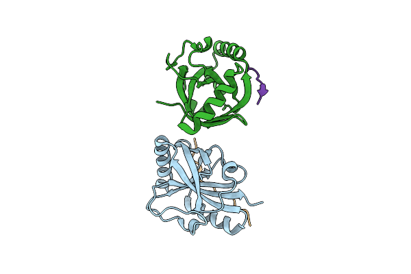
Crystal Structure Of The Spoc Domain Of Human Phf3 In Complex With Rna Polymerase Ii Ctd Diheptapeptide Phosphorylated On Ser2
Organism: Homo sapiens
Method: X-RAY DIFFRACTION Resolution:1.93 Å Release Date: 2020-07-15 Classification: PROTEIN BINDING |
Organism: Homo sapiens
Method: X-RAY DIFFRACTION
Release Date: 2020-07-15
 |
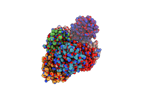
Crystal Structure Of The Spoc Domain Of Human Phf3 In Complex With Rna Polymerase Ii Ctd Diheptapeptide Phosphorylated On Ser2Ser7
Organism: Homo sapiens
Method: X-RAY DIFFRACTION Resolution:1.75 Å Release Date: 2020-07-15 Classification: TRANSCRIPTION |
Organism: Homo sapiens
Method: X-RAY DIFFRACTION
Release Date: 2020-07-15
 |
Crystal Structure Of Spoc Domain Of Human Phd-Finger Protein 3 (Phf3)
Organism: Homo sapiens
Method: X-RAY DIFFRACTION Resolution:2.59 Å Release Date: 2020-07-15 Classification: TRANSCRIPTION Ligands: GOL |
Organism: Homo sapiens
Method: X-RAY DIFFRACTION
Release Date: 2020-07-15
Ligands: GOL
 |
Crystal Structure Of The Spoc Domain Of Human Phf3 In Complex With Rna Polymerase Ii Ctd Diheptapeptide Phosphorylated On Ser2Ser5
Organism: Homo sapiens
Method: X-RAY DIFFRACTION Resolution:2.85 Å Release Date: 2020-07-15 Classification: TRANSCRIPTION |
Organism: Homo sapiens
Method: X-RAY DIFFRACTION
Release Date: 2020-07-15
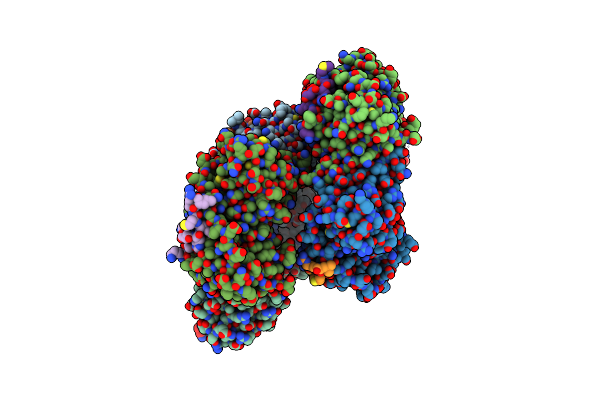 |
Crystal Structure Of Human Pcna In Complex With Parg (Poly(Adp-Ribose) Glycohydrolase) Peptide.
Organism: Homo sapiens
Method: X-RAY DIFFRACTION Resolution:2.58 Å Release Date: 2017-08-02 Classification: TRANSCRIPTION |
Organism: Homo sapiens
Method: X-RAY DIFFRACTION
Release Date: 2017-08-02
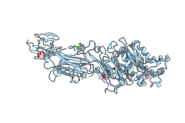 |
Crystal Structure Of Protein Arginine Deiminase 2 (0 Mm Ca2+)
Organism: Homo sapiens
Method: X-RAY DIFFRACTION Resolution:1.66 Å Release Date: 2015-02-04 Classification: HYDROLASE Ligands: MPD, ACT, CA |
Organism: Homo sapiens
Method: X-RAY DIFFRACTION
Release Date: 2015-02-04
Ligands: MPD, ACT, CA
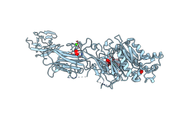 |
Crystal Structure Of Protein Arginine Deiminase 2 (50 Um Ca2+)
Organism: Homo sapiens
Method: X-RAY DIFFRACTION Resolution:1.89 Å Release Date: 2015-02-04 Classification: HYDROLASE Ligands: ACT, GOL, CA, MPD |
Organism: Homo sapiens
Method: X-RAY DIFFRACTION
Release Date: 2015-02-04
Ligands: ACT, GOL, CA, MPD
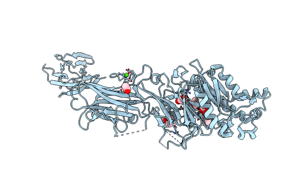 |
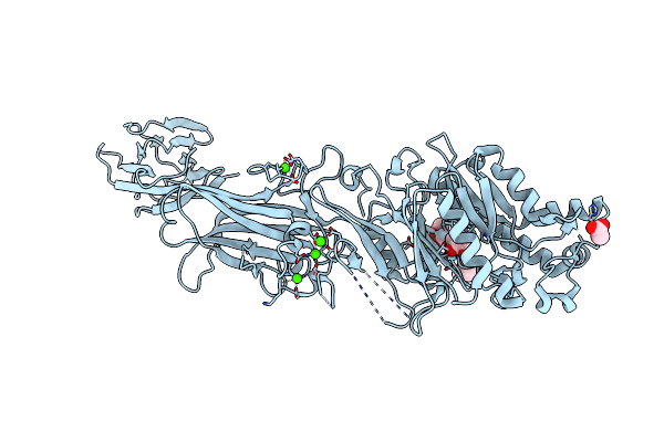
Crystal Structure Of Protein Arginine Deiminase 2 (100 Um Ca2+)
Organism: Homo sapiens
Method: X-RAY DIFFRACTION Resolution:1.97 Å Release Date: 2015-02-04 Classification: HYDROLASE Ligands: MRD, MPD, CA, GOL, ACT |
Organism: Homo sapiens
Method: X-RAY DIFFRACTION
Release Date: 2015-02-04
Ligands: MRD, MPD, CA, GOL, ACT
 |
Crystal Structure Of Protein Arginine Deiminase 2 (250 Um Ca2+)
Organism: Homo sapiens
Method: X-RAY DIFFRACTION Resolution:1.93 Å Release Date: 2015-02-04 Classification: HYDROLASE Ligands: MRD, MPD, ACT, CA |
Organism: Homo sapiens
Method: X-RAY DIFFRACTION
Release Date: 2015-02-04
Ligands: MRD, MPD, ACT, CA