

Search Count: 12
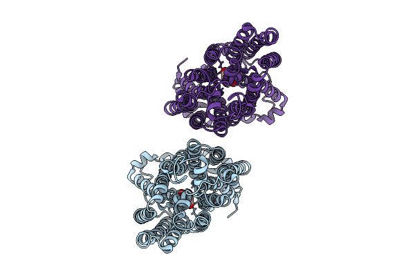 |
Cryo-Em Structure Of Pfht1 Bound To 2,5-Anhydro-D-Mannitol
Organism: Plasmodium falciparum, Aequorea victoria
Method: ELECTRON MICROSCOPY Release Date: 2026-04-15 Classification: TRANSPORT PROTEIN Ligands: A1IVP |
Organism: Plasmodium falciparum, Aequorea victoria
Method: ELECTRON MICROSCOPY
Release Date: 2026-04-15
Ligands: A1IVP
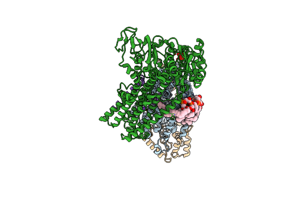 |
Cryo-Em Map Of The Wt Kdpfabc Complex In The E1-P Tight Conformation, Stabilised With The Inhibitor Orthovanadate
Organism: Escherichia coli
Method: ELECTRON MICROSCOPY Release Date: 2022-11-16 Classification: MEMBRANE PROTEIN Ligands: K, CDL, VO4 |
Organism: Escherichia coli
Method: ELECTRON MICROSCOPY
Release Date: 2022-11-16
Ligands: K, CDL, VO4
 |
Cryo-Em Map Of The Wt Kdpfabc Complex In The E1-P Tight Conformation, Under Turnover Conditions
Organism: Escherichia coli
Method: ELECTRON MICROSCOPY Release Date: 2022-11-16 Classification: MEMBRANE PROTEIN Ligands: K, CDL |
Organism: Escherichia coli
Method: ELECTRON MICROSCOPY
Release Date: 2022-11-16
Ligands: K, CDL
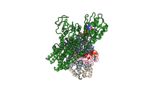 |
Cryo-Em Map Of The Wt Kdpfabc Complex In The E1_Atpearly Conformation, Under Turnover Conditions
Organism: Escherichia coli
Method: ELECTRON MICROSCOPY Release Date: 2022-11-16 Classification: MEMBRANE PROTEIN Ligands: K, CDL, ATP |
Organism: Escherichia coli
Method: ELECTRON MICROSCOPY
Release Date: 2022-11-16
Ligands: K, CDL, ATP
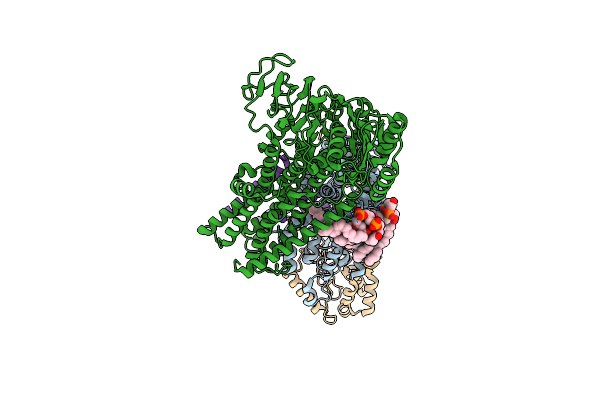 |
Cryo-Em Structure Of The Kdpfabc Complex In A Nucleotide-Free E1 Conformation Loaded With K+
Organism: Escherichia coli
Method: ELECTRON MICROSCOPY Release Date: 2022-11-16 Classification: MEMBRANE PROTEIN Ligands: K, CDL |
Organism: Escherichia coli
Method: ELECTRON MICROSCOPY
Release Date: 2022-11-16
Ligands: K, CDL
 |
Cryo-Em Structure Of The Kdpfabc Complex In A Nucleotide-Free E1 Conformation Loaded With K+
Organism: Escherichia coli
Method: ELECTRON MICROSCOPY Release Date: 2022-11-16 Classification: MEMBRANE PROTEIN Ligands: K, CDL |
Organism: Escherichia coli
Method: ELECTRON MICROSCOPY
Release Date: 2022-11-16
Ligands: K, CDL
 |
Cryo-Em Structure Of The Kdpfabc Complex In A Nucleotide-Free E1 Conformation Loaded With K+
Organism: Escherichia coli
Method: ELECTRON MICROSCOPY Release Date: 2022-11-16 Classification: MEMBRANE PROTEIN Ligands: K, CDL |
Organism: Escherichia coli
Method: ELECTRON MICROSCOPY
Release Date: 2022-11-16
Ligands: K, CDL
 |
Cryo-Em Map Of The Wt Kdpfabc Complex In The E1-P_Adp Conformation, Under Turnover Conditions
Organism: Escherichia coli
Method: ELECTRON MICROSCOPY Release Date: 2022-11-16 Classification: MEMBRANE PROTEIN Ligands: K, CDL, ADP |
Organism: Escherichia coli
Method: ELECTRON MICROSCOPY
Release Date: 2022-11-16
Ligands: K, CDL, ADP
 |
Cryo-Em Map Of The Unphosphorylated Kdpfabc Complex In The E2-P Conformation, Under Turnover Conditions
Organism: Escherichia coli k-12
Method: ELECTRON MICROSCOPY Release Date: 2022-11-16 Classification: MEMBRANE PROTEIN Ligands: K |
Organism: Escherichia coli k-12
Method: ELECTRON MICROSCOPY
Release Date: 2022-11-16
Ligands: K
 |
Cryo-Em Map Of The Unphosphorylated Kdpfabc Complex In The E1-P_Adp Conformation, Under Turnover Conditions
Organism: Escherichia coli
Method: ELECTRON MICROSCOPY Release Date: 2022-11-16 Classification: MEMBRANE PROTEIN Ligands: K, ADP, MG |
Organism: Escherichia coli
Method: ELECTRON MICROSCOPY
Release Date: 2022-11-16
Ligands: K, ADP, MG
 |
Cryo-Em Structure Of The Kdpfabc Complex In An E1-Atp Conformation Loaded With K+
Organism: Escherichia coli
Method: ELECTRON MICROSCOPY Release Date: 2021-07-28 Classification: MEMBRANE PROTEIN Ligands: K, CDL, ACP |
Organism: Escherichia coli
Method: ELECTRON MICROSCOPY
Release Date: 2021-07-28
Ligands: K, CDL, ACP
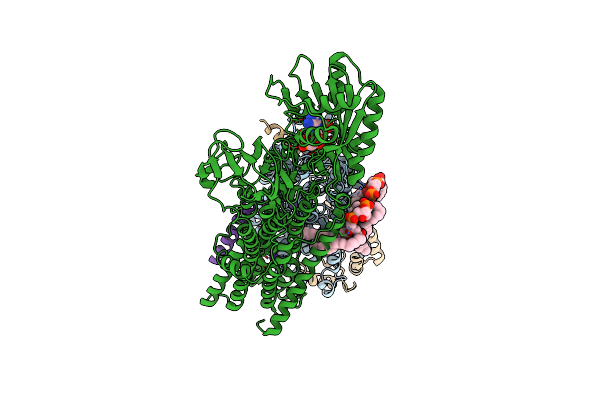 |
Rb-Loaded Cryo-Em Structure Of The E1-Atp Kdpfabc Complex.
Organism: Escherichia coli
Method: ELECTRON MICROSCOPY Release Date: 2021-07-28 Classification: MEMBRANE PROTEIN Ligands: RB, CDL, ACP |
Organism: Escherichia coli
Method: ELECTRON MICROSCOPY
Release Date: 2021-07-28
Ligands: RB, CDL, ACP