

Search Count: 18
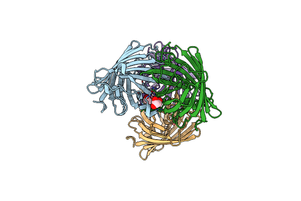 |
Structure Of The Fluorescent Protein Aausfp1 From Aequorea Cf. Australis At Ph 7.0
Organism: Aequorea australis
Method: X-RAY DIFFRACTION Resolution:2.47 Å Release Date: 2020-07-22 Classification: FLUORESCENT PROTEIN Ligands: GOL |
Organism: Aequorea australis
Method: X-RAY DIFFRACTION
Release Date: 2020-07-22
Ligands: GOL
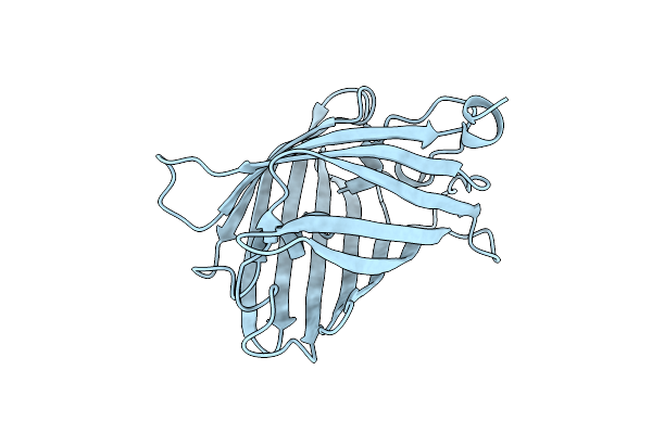 |
Structure Of The Fluorescent Protein Aausfp2 From Aequorea Cf. Australis At Ph 7.6
Organism: Aequorea australis
Method: X-RAY DIFFRACTION Resolution:2.06 Å Release Date: 2020-07-22 Classification: FLUORESCENT PROTEIN |
Organism: Aequorea australis
Method: X-RAY DIFFRACTION
Release Date: 2020-07-22
 |
Crystal Structure Of Human Phosphodiesterase 4D2 Catalytic Domain With Inhibitor Npd-1335
Organism: Homo sapiens
Method: X-RAY DIFFRACTION Resolution:1.99 Å Release Date: 2019-07-24 Classification: HYDROLASE Ligands: EDO, PEG, ZN, FFZ, MG, EPE, DMS |
Organism: Homo sapiens
Method: X-RAY DIFFRACTION
Release Date: 2019-07-24
Ligands: EDO, PEG, ZN, FFZ, MG, EPE, DMS
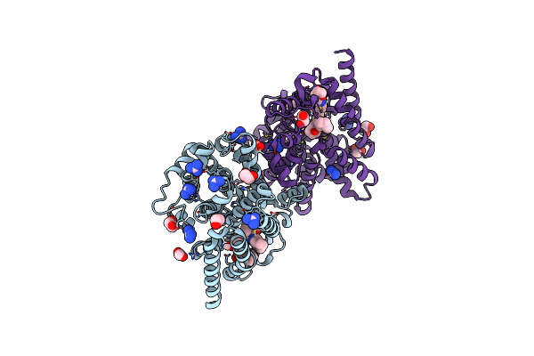 |
Crystal Structure Of T. Brucei Pde-B1 Catalytic Domain With Inhibitor Npd-053
Organism: Trypanosoma brucei
Method: X-RAY DIFFRACTION Resolution:1.90 Å Release Date: 2019-07-24 Classification: HYDROLASE Ligands: MG, ZN, GAI, EDO, FMT, JX2 |
Organism: Trypanosoma brucei
Method: X-RAY DIFFRACTION
Release Date: 2019-07-24
Ligands: MG, ZN, GAI, EDO, FMT, JX2
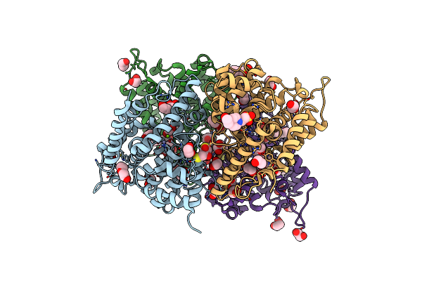 |
Crystal Structure Of Human Phosphodiesterase 4D2 Catalytic Domain With Inhibitor Npd-053
Organism: Homo sapiens
Method: X-RAY DIFFRACTION Resolution:2.08 Å Release Date: 2019-07-24 Classification: HYDROLASE Ligands: ZN, MG, EDO, PEG, DTT, EPE, JX2 |
Organism: Homo sapiens
Method: X-RAY DIFFRACTION
Release Date: 2019-07-24
Ligands: ZN, MG, EDO, PEG, DTT, EPE, JX2
 |
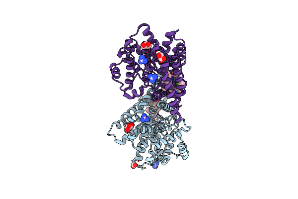
Crystal Structure Of T. Brucei Pde-B1 Catalytic Domain With Inhibitor Npd-1018
Organism: Trypanosoma brucei
Method: X-RAY DIFFRACTION Resolution:2.29 Å Release Date: 2019-07-24 Classification: HYDROLASE Ligands: ZN, MG, FMT, GAI, GOL, K3W |
Organism: Trypanosoma brucei
Method: X-RAY DIFFRACTION
Release Date: 2019-07-24
Ligands: ZN, MG, FMT, GAI, GOL, K3W
 |
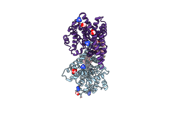
Crystal Structure Of T. Brucei Pde-B1 Catalytic Domain With Inhibitor Npd-1039
Organism: Trypanosoma brucei
Method: X-RAY DIFFRACTION Resolution:2.18 Å Release Date: 2019-07-24 Classification: HYDROLASE Ligands: ZN, MG, FMT, GAI, GOL, K1Q |
Organism: Trypanosoma brucei
Method: X-RAY DIFFRACTION
Release Date: 2019-07-24
Ligands: ZN, MG, FMT, GAI, GOL, K1Q
 |
Crystal Structure Of T. Brucei Pde-B1 Catalytic Domain With Inhibitor Npd-055
Organism: Trypanosoma brucei
Method: X-RAY DIFFRACTION Resolution:2.03 Å Release Date: 2019-07-24 Classification: HYDROLASE Ligands: ZN, MG, FMT, GOL, GAI, K3N |
Organism: Trypanosoma brucei
Method: X-RAY DIFFRACTION
Release Date: 2019-07-24
Ligands: ZN, MG, FMT, GOL, GAI, K3N
 |
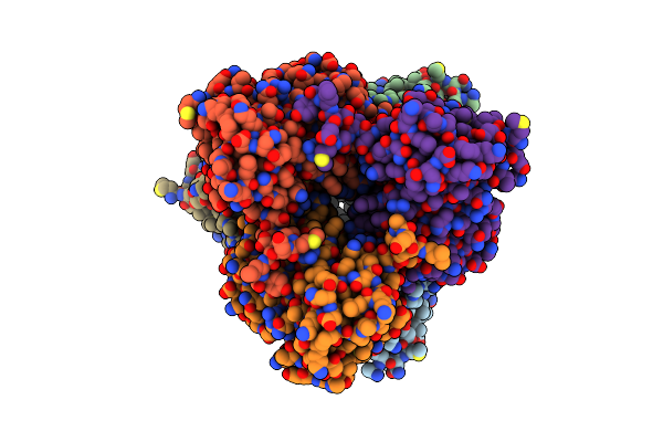
Crystal Structure Of T. Brucei Pde-B1 Catalytic Domain With Inhibitor Npd-1335
Organism: Trypanosoma brucei
Method: X-RAY DIFFRACTION Resolution:1.96 Å Release Date: 2019-07-10 Classification: HYDROLASE Ligands: MG, ZN, GAI, FFZ, GOL |
Organism: Trypanosoma brucei
Method: X-RAY DIFFRACTION
Release Date: 2019-07-10
Ligands: MG, ZN, GAI, FFZ, GOL
 |
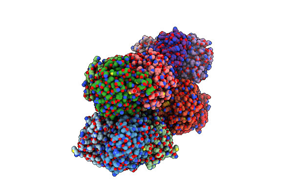
Structure Of The Yellow-Green Fluorescent Protein Mneongreen From Branchiostoma Lanceolatum At The Acidic Ph 4.5
Organism: Branchiostoma lanceolatum
Method: X-RAY DIFFRACTION Resolution:1.70 Å Release Date: 2016-12-21 Classification: FLUORESCENT PROTEIN Ligands: CL, FLC |
Organism: Branchiostoma lanceolatum
Method: X-RAY DIFFRACTION
Release Date: 2016-12-21
Ligands: CL, FLC
 |
Structure Of The Yellow Fluorescent Protein Lanyfp From Branchiostoma Lanceolatum At Ph 7.5
Organism: Branchiostoma lanceolatum
Method: X-RAY DIFFRACTION Resolution:2.05 Å Release Date: 2016-12-07 Classification: FLUORESCENT PROTEIN Ligands: CL |
Organism: Branchiostoma lanceolatum
Method: X-RAY DIFFRACTION
Release Date: 2016-12-07
Ligands: CL
 |
Structure Of The Yellow-Green Fluorescent Protein Mneongreen From Branchiostoma Lanceolatum At The Near Physiological Ph 8.0
Organism: Branchiostoma lanceolatum
Method: X-RAY DIFFRACTION Resolution:1.21 Å Release Date: 2016-12-07 Classification: FLUORESCENT PROTEIN Ligands: CL |
Organism: Branchiostoma lanceolatum
Method: X-RAY DIFFRACTION
Release Date: 2016-12-07
Ligands: CL
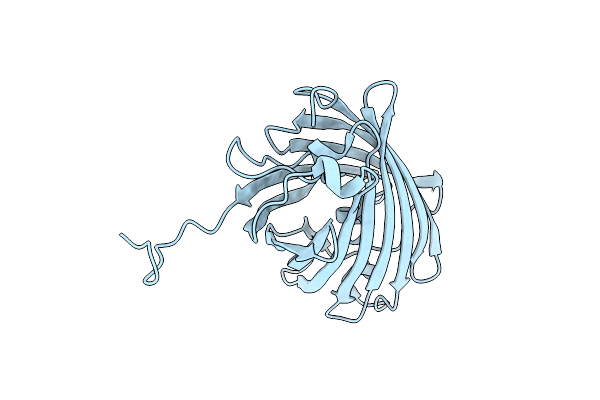 |
Crystal Structure Of Red Fluorescent Protein Neptune At Ph 7.0
Organism: Entacmaea quadricolor
Method: X-RAY DIFFRACTION Resolution:1.60 Å Release Date: 2009-12-15 Classification: FLUORESCENT PROTEIN |
Organism: Entacmaea quadricolor
Method: X-RAY DIFFRACTION
Release Date: 2009-12-15
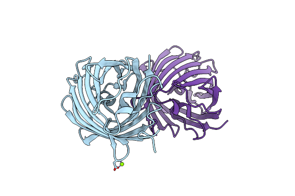 |
Crystal Structure Of Morange
Organism: Discosoma sp.
Method: X-RAY DIFFRACTION Resolution:1.08 Å Release Date: 2006-08-22 Classification: LUMINESCENT PROTEIN Ligands: MG |
Organism: Discosoma sp.
Method: X-RAY DIFFRACTION
Release Date: 2006-08-22
Ligands: MG
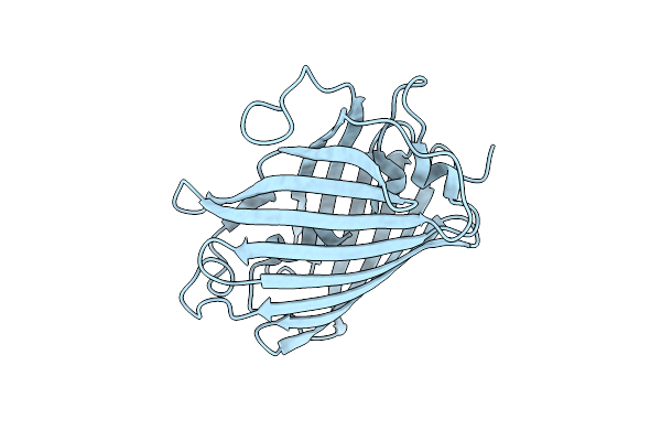 |
Crystal Structure Of Mstrawberry At Ph 9.5
Organism: Discosoma sp.
Method: X-RAY DIFFRACTION Resolution:1.21 Å Release Date: 2006-08-22 Classification: LUMINESCENT PROTEIN |
Organism: Discosoma sp.
Method: X-RAY DIFFRACTION
Release Date: 2006-08-22
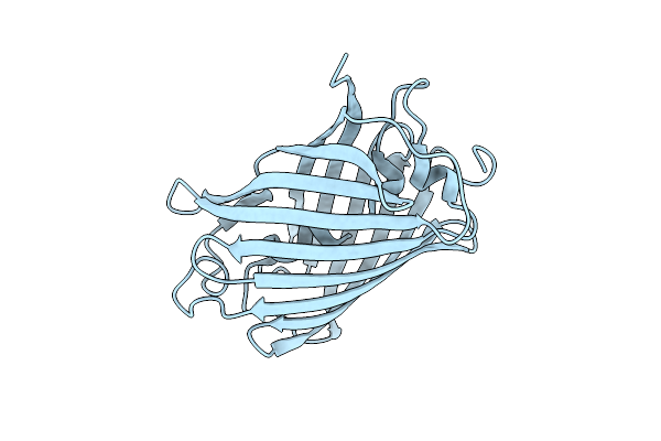 |
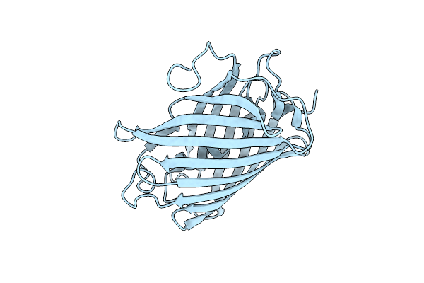
Crystal Structure Of Mcherry
Organism: Discosoma sp.
Method: X-RAY DIFFRACTION Resolution:1.36 Å Release Date: 2006-08-22 Classification: LUMINESCENT PROTEIN |
Organism: Discosoma sp.
Method: X-RAY DIFFRACTION
Release Date: 2006-08-22
 |
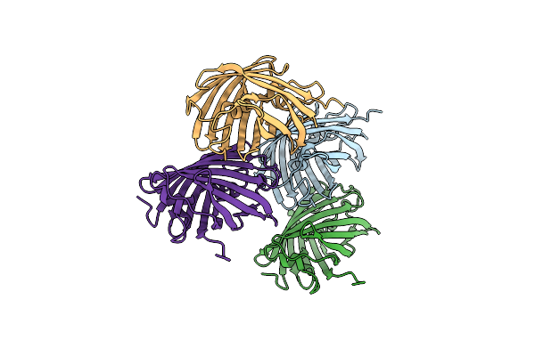
Crystal Structure Of Mstrawberry At Ph 10.5
Organism: Discosoma sp.
Method: X-RAY DIFFRACTION Resolution:1.60 Å Release Date: 2006-08-22 Classification: LUMINESCENT PROTEIN |
Organism: Discosoma sp.
Method: X-RAY DIFFRACTION
Release Date: 2006-08-22
 |
Crystal Structure Of A Redshifted Mutant (K83M) Of The Red Fluorescent Protein Drfp583/Dsred
Organism: Discosoma sp.
Method: X-RAY DIFFRACTION Resolution:2.00 Å Release Date: 2006-08-22 Classification: LUMINESCENT PROTEIN |
Organism: Discosoma sp.
Method: X-RAY DIFFRACTION
Release Date: 2006-08-22