

Search Count: 52
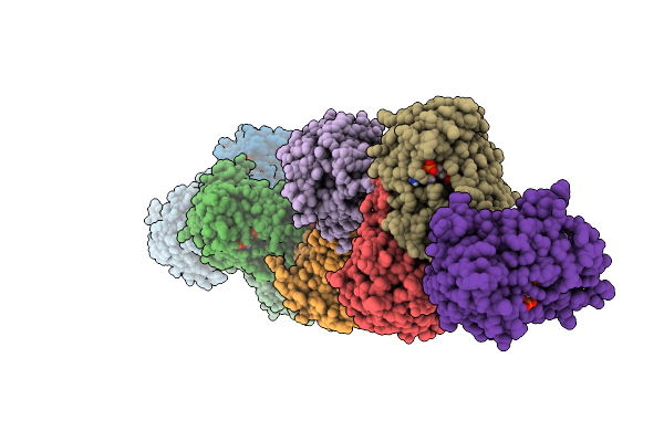 |
Crystal Structure Of 11Betahsd1 In Complex With Compound 1
Organism: Homo sapiens
Method: X-RAY DIFFRACTION Resolution:3.20 Å Release Date: 2025-10-08 Classification: OXIDOREDUCTASE Ligands: NAP, A1MBC |
Organism: Homo sapiens
Method: X-RAY DIFFRACTION
Release Date: 2025-10-08
Ligands: NAP, A1MBC
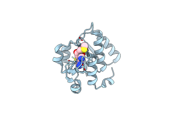 |
The X-Ray Structure Of N-Terminal Catalytic Domain Of Thermoplasma Acidophilum Trna Methyltransferase Trm56 (Ta0931) In Complex With 5'-Methylthioadenosine
Organism: Thermoplasma acidophilum dsm 1728
Method: X-RAY DIFFRACTION Resolution:1.99 Å Release Date: 2025-04-02 Classification: TRANSFERASE Ligands: MTA |
Organism: Thermoplasma acidophilum dsm 1728
Method: X-RAY DIFFRACTION
Release Date: 2025-04-02
Ligands: MTA
 |
The X-Ray Structure Of N-Terminal Catalytic Domain Of Thermoplasma Acidophilum Trna Methyltransferase Trm56 (Ta0931).
Organism: Thermoplasma acidophilum dsm 1728
Method: X-RAY DIFFRACTION Resolution:1.80 Å Release Date: 2024-11-27 Classification: TRANSFERASE |
Organism: Thermoplasma acidophilum dsm 1728
Method: X-RAY DIFFRACTION
Release Date: 2024-11-27
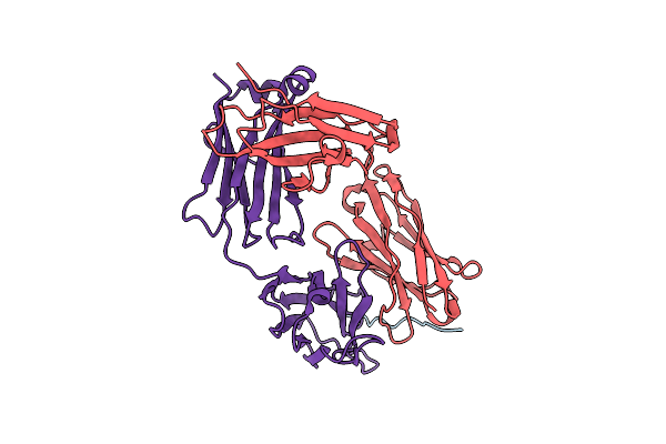 |
24B3 Antibody-Peptide Complex
Organism: Homo sapiens, Mus musculus
Method: X-RAY DIFFRACTION Resolution:2.60 Å Release Date: 2022-08-17 Classification: IMMUNE SYSTEM |
Organism: Homo sapiens, Mus musculus
Method: X-RAY DIFFRACTION
Release Date: 2022-08-17
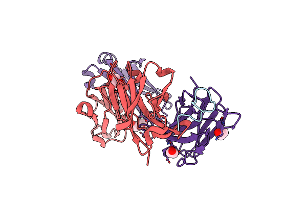 |
Fab-Amyloid Beta Fragment Complex
Organism: Homo sapiens, Mus musculus
Method: X-RAY DIFFRACTION Resolution:2.50 Å Release Date: 2022-01-05 Classification: IMMUNE SYSTEM Ligands: EDO, PEG |
Organism: Homo sapiens, Mus musculus
Method: X-RAY DIFFRACTION
Release Date: 2022-01-05
Ligands: EDO, PEG
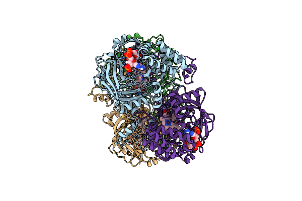 |
Electron Crystallographic Structure Of Catalase Using A Direct Electron Detector At 300 Kv
Organism: Bos taurus
Method: ELECTRON CRYSTALLOGRAPHY Resolution:3.20 Å Release Date: 2020-12-09 Classification: OXIDOREDUCTASE Ligands: HEM, NDP |
Organism: Bos taurus
Method: ELECTRON CRYSTALLOGRAPHY
Release Date: 2020-12-09
Ligands: HEM, NDP
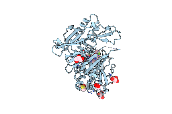 |
Structure Of Bace1 In Complex With N-(3-((4R,5R,6S)-2-Amino-6-(1,1-Difluoroethyl)-5-Fluoro-4-Methyl-5,6-Dihydro-4H-1,3-Oxazin-4-Yl)-4-Fluorophenyl)-5-(Fluoromethoxy)Pyrazine-2-Carboxamide
Organism: Homo sapiens
Method: X-RAY DIFFRACTION Resolution:2.20 Å Release Date: 2018-08-08 Classification: HYDROLASE/INHIBITOR Ligands: IOD, GOL, DMS, 0B5 |
Organism: Homo sapiens
Method: X-RAY DIFFRACTION
Release Date: 2018-08-08
Ligands: IOD, GOL, DMS, 0B5
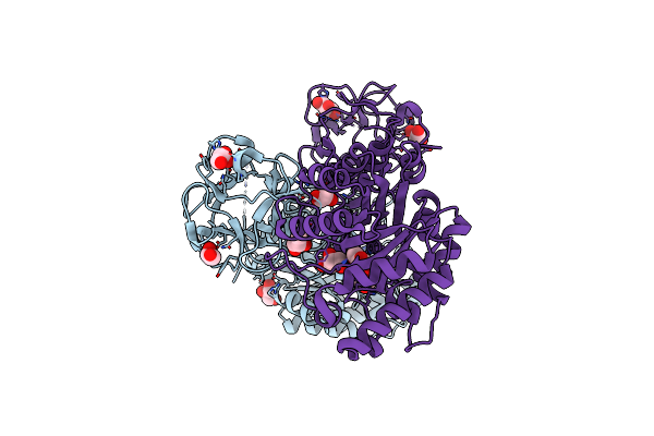 |
Crystal Structure Of Covalent Glycosyl-Enzyme Intermediate Of Xylanase Mutant (T82A, N127S, And E128H) From Streptomyces Olivaceoviridis E-86
Organism: Streptomyces olivaceoviridis
Method: X-RAY DIFFRACTION Resolution:1.80 Å Release Date: 2017-08-09 Classification: HYDROLASE Ligands: GOL |
Organism: Streptomyces olivaceoviridis
Method: X-RAY DIFFRACTION
Release Date: 2017-08-09
Ligands: GOL
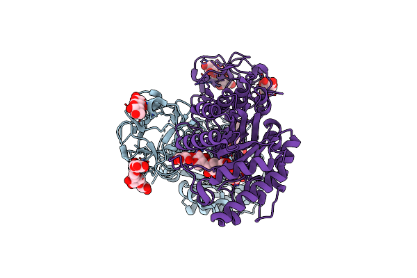 |
Crystal Structure Of Michaelis Complex Of Xylanase Mutant (T82A, N127S, And E128H) From Streptomyces Olivaceoviridis E-86
Organism: Streptomyces olivaceoviridis
Method: X-RAY DIFFRACTION Resolution:2.50 Å Release Date: 2017-08-09 Classification: HYDROLASE |
Organism: Streptomyces olivaceoviridis
Method: X-RAY DIFFRACTION
Release Date: 2017-08-09
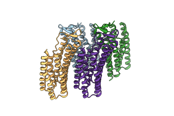 |
Crystal Structure Of Thermophilic Rhodopsin.
Organism: Thermus thermophilus jl-18
Method: X-RAY DIFFRACTION Resolution:2.80 Å Release Date: 2016-04-27 Classification: TRANSPORT PROTEIN |
Organism: Thermus thermophilus jl-18
Method: X-RAY DIFFRACTION
Release Date: 2016-04-27
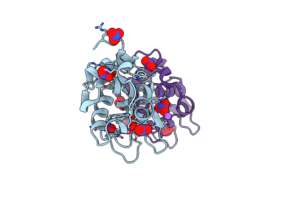 |
Monoclinic Lysozyme At 0.96 A Resolution
Organism: Gallus gallus
Method: X-RAY DIFFRACTION Resolution:0.96 Å Release Date: 2014-11-12 Classification: HYDROLASE Ligands: NO3, NA, EDO |
Organism: Gallus gallus
Method: X-RAY DIFFRACTION
Release Date: 2014-11-12
Ligands: NO3, NA, EDO
 |
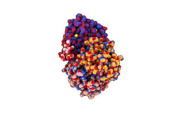
The Structure Of A Deoxygenated 400 Kda Hemoglobin Provides A More Accurate Description Of The Cooperative Mechanism Of Giant Hemoglobins: Oxygenated Form
Organism: Lamellibrachia satsuma
Method: X-RAY DIFFRACTION Resolution:2.40 Å Release Date: 2014-06-04 Classification: OXYGEN TRANSPORT Ligands: HEM, OXY, CA |
Organism: Lamellibrachia satsuma
Method: X-RAY DIFFRACTION
Release Date: 2014-06-04
Ligands: HEM, OXY, CA
 |
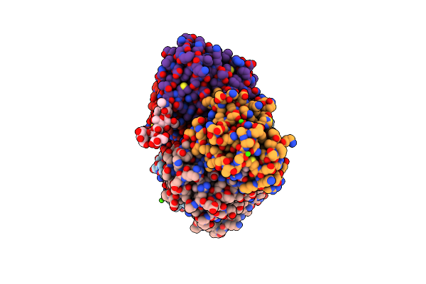
The Structure Of A Deoxygenated 400 Kda Hemoglobin Provides A More Accurate Description Of The Cooperative Mechanism Of Giant Hemoglobins: Deoxygenated Form
Organism: Lamellibrachia satsuma
Method: X-RAY DIFFRACTION Resolution:2.90 Å Release Date: 2014-06-04 Classification: OXYGEN TRANSPORT Ligands: HEM, CA |
Organism: Lamellibrachia satsuma
Method: X-RAY DIFFRACTION
Release Date: 2014-06-04
Ligands: HEM, CA
 |
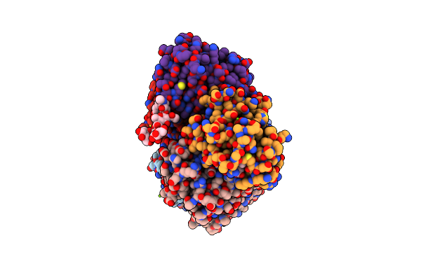
The Structure Of A Deoxygenated 400 Kda Hemoglobin Provides A More Accurate Description Of The Cooperative Mechanism Of Giant Hemoglobins: Ca Bound Form
Organism: Lamellibrachia satsuma
Method: X-RAY DIFFRACTION Resolution:2.60 Å Release Date: 2014-06-04 Classification: OXYGEN TRANSPORT Ligands: HEM, OXY, CA |
Organism: Lamellibrachia satsuma
Method: X-RAY DIFFRACTION
Release Date: 2014-06-04
Ligands: HEM, OXY, CA
 |
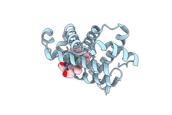
The Structure Of A Deoxygenated 400 Kda Hemoglobin Provides A More Accurate Description Of The Cooperative Mechanism Of Giant Hemoglobins: Mg Bound Form
Organism: Lamellibrachia satsuma
Method: X-RAY DIFFRACTION Resolution:2.50 Å Release Date: 2014-06-04 Classification: OXYGEN TRANSPORT Ligands: HEM, OXY, MG |
Organism: Lamellibrachia satsuma
Method: X-RAY DIFFRACTION
Release Date: 2014-06-04
Ligands: HEM, OXY, MG
 |
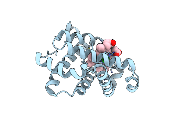
Cl- Binding Hemoglobin Component V Form Propsilocerus Akamusi Under 500 Mm Nacl At Ph 4.6
Organism: Tokunagayusurika akamusi
Method: X-RAY DIFFRACTION Resolution:1.81 Å Release Date: 2011-04-27 Classification: OXYGEN TRANSPORT Ligands: HEM, CL |
Organism: Tokunagayusurika akamusi
Method: X-RAY DIFFRACTION
Release Date: 2011-04-27
Ligands: HEM, CL
 |
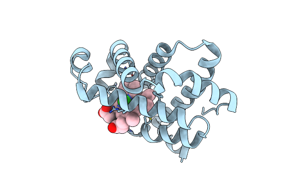
Cl- Binding Hemoglobin Component V Form Propsilocerus Akamusi Under 1 M Nacl At Ph 4.6
Organism: Tokunagayusurika akamusi
Method: X-RAY DIFFRACTION Resolution:1.81 Å Release Date: 2011-04-27 Classification: OXYGEN TRANSPORT Ligands: HEM, CL |
Organism: Tokunagayusurika akamusi
Method: X-RAY DIFFRACTION
Release Date: 2011-04-27
Ligands: HEM, CL
 |
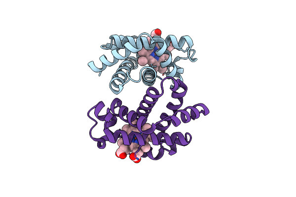
Cl- Binding Hemoglobin Component V Form Propsilocerus Akamusi Under 500 Mm Nacl At Ph 5.5
Organism: Tokunagayusurika akamusi
Method: X-RAY DIFFRACTION Resolution:1.81 Å Release Date: 2011-04-27 Classification: OXYGEN TRANSPORT Ligands: HEM, CL |
Organism: Tokunagayusurika akamusi
Method: X-RAY DIFFRACTION
Release Date: 2011-04-27
Ligands: HEM, CL
 |
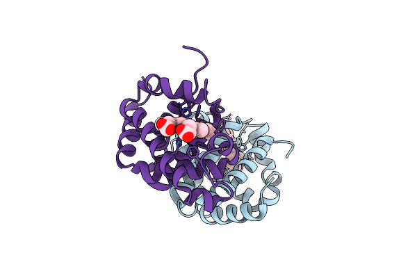
Side-Necked Turtle (Pleurodira, Chelonia, Reptilia) Hemoglobin: Cdna-Derived Primary Structures And X-Ray Crystal Structures Of Hb A
Organism: Podocnemis unifilis
Method: X-RAY DIFFRACTION Resolution:2.20 Å Release Date: 2011-04-20 Classification: OXYGEN TRANSPORT Ligands: HEM |
Organism: Podocnemis unifilis
Method: X-RAY DIFFRACTION
Release Date: 2011-04-20
Ligands: HEM
 |
Side-Necked Turtle (Pleurodira, Chelonia, Reptilia) Hemoglobin: Cdna-Derived Primary Structures And X-Ray Crystal Structures Of Hb A
Organism: Podocnemis unifilis
Method: X-RAY DIFFRACTION Resolution:2.35 Å Release Date: 2011-04-20 Classification: OXYGEN TRANSPORT Ligands: HEM |
Organism: Podocnemis unifilis
Method: X-RAY DIFFRACTION
Release Date: 2011-04-20
Ligands: HEM