

Search Count: 79
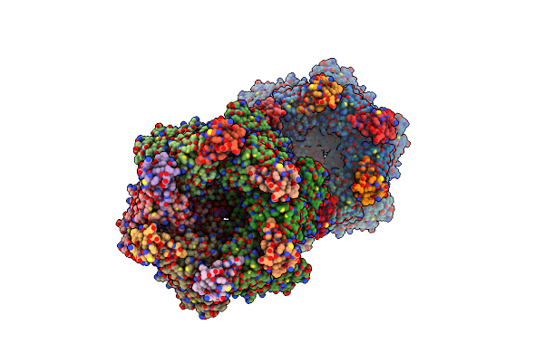 |
Structure Of A Rho-Nusg Kow Domain Complex
Organism: Escherichia coli m718, Escherichia coli m605, Synthetic construct
Method: X-RAY DIFFRACTION Resolution:3.70 Å Release Date: 2018-09-05 Classification: TRANSCRIPTION/RNA Ligands: ADP, MG, BEF |
Organism: Escherichia coli m718, Escherichia coli m605, Synthetic construct
Method: X-RAY DIFFRACTION
Release Date: 2018-09-05
Ligands: ADP, MG, BEF
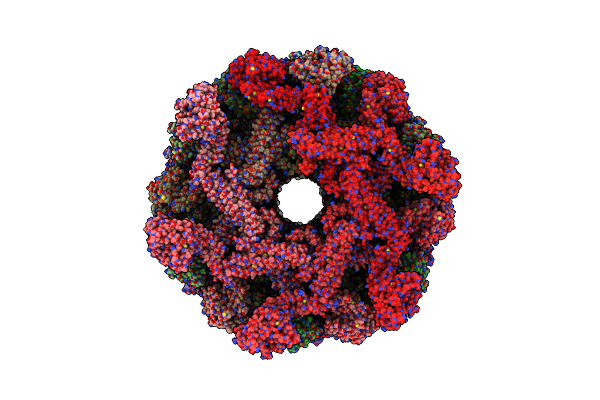 |
Cryoem Structure Of The Helical Assembly Of Full Length Mxb
Organism: Homo sapiens
Method: ELECTRON MICROSCOPY Release Date: 2018-02-21 Classification: ANTIVIRAL PROTEIN |
Organism: Homo sapiens
Method: ELECTRON MICROSCOPY
Release Date: 2018-02-21
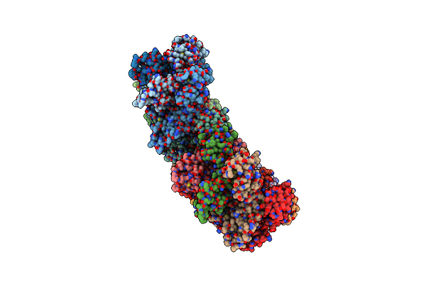 |
Structure Of The Hiv-1 Capsid Protein And Spacer Peptide 1 By Cryo-Em
Organism: Human immunodeficiency virus 1
Method: ELECTRON MICROSCOPY Release Date: 2017-12-06 Classification: VIRUS |
Organism: Human immunodeficiency virus 1
Method: ELECTRON MICROSCOPY
Release Date: 2017-12-06
 |
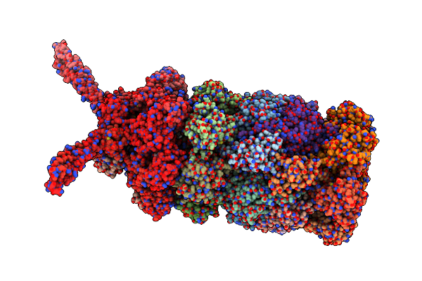
26S Proteasome In Presence Of Atp (S1)
Organism: Saccharomyces cerevisiae (strain atcc 204508 / s288c)
Method: ELECTRON MICROSCOPY Release Date: 2017-03-08 Classification: HYDROLASE Ligands: ATP, MG, ADP |
Organism: Saccharomyces cerevisiae (strain atcc 204508 / s288c)
Method: ELECTRON MICROSCOPY
Release Date: 2017-03-08
Ligands: ATP, MG, ADP
 |
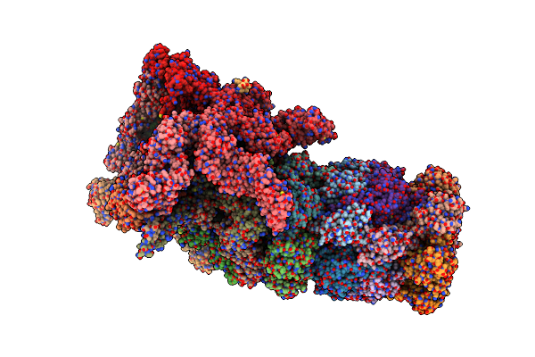
26S Proteasome In Presence Of Atp (S2)
Organism: Saccharomyces cerevisiae (strain atcc 204508 / s288c)
Method: ELECTRON MICROSCOPY Release Date: 2017-03-08 Classification: HYDROLASE Ligands: MG, ATP, ADP |
Organism: Saccharomyces cerevisiae (strain atcc 204508 / s288c)
Method: ELECTRON MICROSCOPY
Release Date: 2017-03-08
Ligands: MG, ATP, ADP
 |
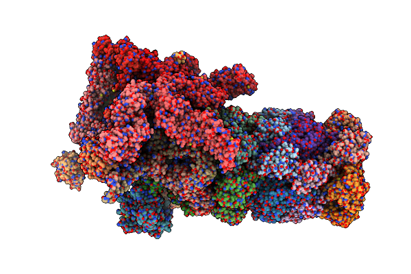
26S Proteasome In Presence Of Amp-Pnp (S3)
Organism: Saccharomyces cerevisiae (strain atcc 204508 / s288c)
Method: ELECTRON MICROSCOPY Release Date: 2017-03-08 Classification: HYDROLASE Ligands: ANP, MG, ADP |
Organism: Saccharomyces cerevisiae (strain atcc 204508 / s288c)
Method: ELECTRON MICROSCOPY
Release Date: 2017-03-08
Ligands: ANP, MG, ADP
 |
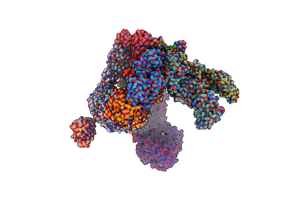
26S Proteasome In Presence Of Befx (S4)
Organism: Saccharomyces cerevisiae (strain atcc 204508 / s288c)
Method: ELECTRON MICROSCOPY Release Date: 2017-03-08 Classification: HYDROLASE Ligands: ATP, MG, ADP |
Organism: Saccharomyces cerevisiae (strain atcc 204508 / s288c)
Method: ELECTRON MICROSCOPY
Release Date: 2017-03-08
Ligands: ATP, MG, ADP
 |
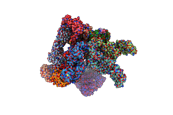
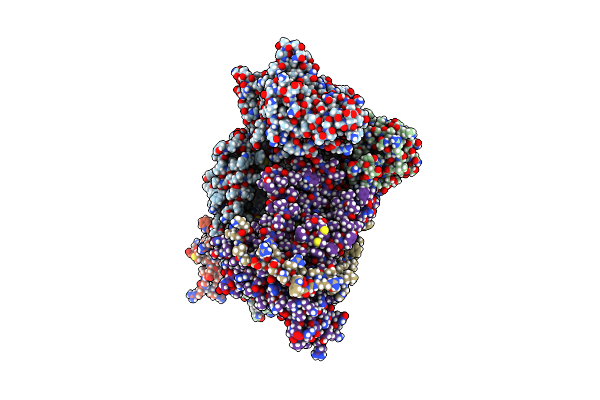
26S Proteasome In Presence Of Atp (S1)
Organism: Saccharomyces cerevisiae (strain atcc 204508 / s288c)
Method: ELECTRON MICROSCOPY Release Date: 2017-03-08 Classification: HYDROLASE |
Organism: Saccharomyces cerevisiae (strain atcc 204508 / s288c)
Method: ELECTRON MICROSCOPY
Release Date: 2017-03-08
 |
26S Proteasome In Presence Of Atp (S2)
Organism: Saccharomyces cerevisiae (strain atcc 204508 / s288c)
Method: ELECTRON MICROSCOPY Release Date: 2017-03-08 Classification: HYDROLASE |
Organism: Saccharomyces cerevisiae (strain atcc 204508 / s288c)
Method: ELECTRON MICROSCOPY
Release Date: 2017-03-08
 |
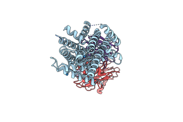
Em Fitted Model Of Bacterial Holo-Translocon
Organism: Escherichia coli
Method: ELECTRON MICROSCOPY Release Date: 2016-12-28 Classification: CHAPERONE |
Organism: Escherichia coli
Method: ELECTRON MICROSCOPY
Release Date: 2016-12-28
 |
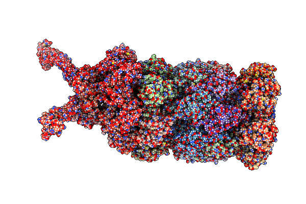
Crystal Structure Of Deinococcus Radiodurans Mnth, An Nramp-Family Transition Metal Transporter
Organism: Deinococcus radiodurans, Mus musculus
Method: X-RAY DIFFRACTION Resolution:3.94 Å Release Date: 2016-11-23 Classification: TRANSPORT PROTEIN/IMMUNE SYSTEM Ligands: OS |
Organism: Deinococcus radiodurans, Mus musculus
Method: X-RAY DIFFRACTION
Release Date: 2016-11-23
Ligands: OS
 |
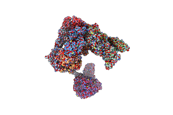
The Human 26S Proteasome At 3.9 A
Organism: Homo sapiens
Method: ELECTRON MICROSCOPY Resolution:3.90 Å Release Date: 2016-09-07 Classification: HYDROLASE Ligands: ATP, MG, ADP |
Organism: Homo sapiens
Method: ELECTRON MICROSCOPY
Release Date: 2016-09-07
Ligands: ATP, MG, ADP
 |
The Human 26S Proteasome Lid
Organism: Homo sapiens
Method: ELECTRON MICROSCOPY Resolution:3.90 Å Release Date: 2016-09-07 Classification: STRUCTURAL PROTEIN |
Organism: Homo sapiens
Method: ELECTRON MICROSCOPY
Release Date: 2016-09-07
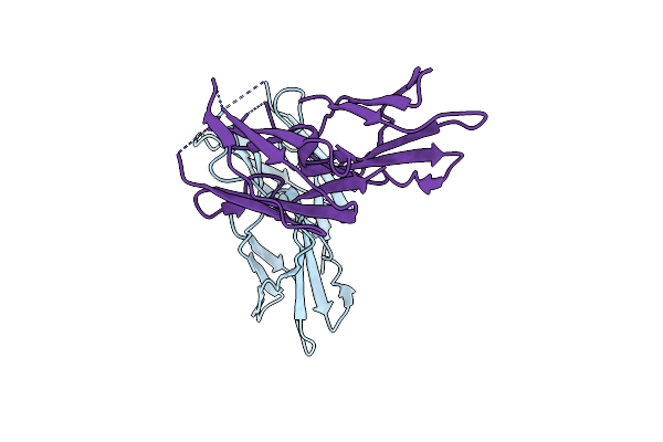 |
Crystal Structure Of The Human Filamin B Ig-Like Domains 16-17
Organism: Homo sapiens
Method: X-RAY DIFFRACTION Resolution:2.49 Å Release Date: 2016-08-24 Classification: STRUCTURAL PROTEIN |
Organism: Homo sapiens
Method: X-RAY DIFFRACTION
Release Date: 2016-08-24
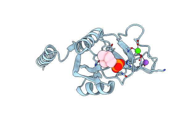 |
Crystal Structure Of Surfactant Protein-A Complexed With Phosphocholine
Organism: Rattus norvegicus
Method: X-RAY DIFFRACTION Resolution:2.20 Å Release Date: 2016-07-06 Classification: SUGAR BINDING PROTEIN Ligands: CA, PC, NA |
Organism: Rattus norvegicus
Method: X-RAY DIFFRACTION
Release Date: 2016-07-06
Ligands: CA, PC, NA
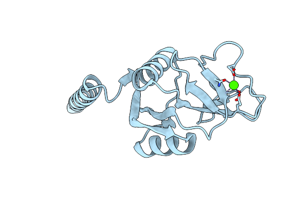 |
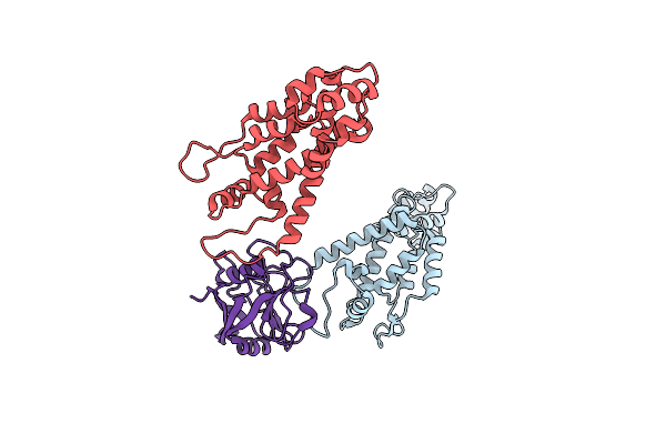
Crystal Structure Of Surfactant Protein-A Y164A Mutant
Organism: Rattus norvegicus
Method: X-RAY DIFFRACTION Resolution:1.80 Å Release Date: 2016-07-06 Classification: SUGAR BINDING PROTEIN Ligands: CA |
Organism: Rattus norvegicus
Method: X-RAY DIFFRACTION
Release Date: 2016-07-06
Ligands: CA
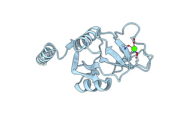 |
Crystal Structure Of Surfactant Protein-A Y221A Mutant
Organism: Rattus norvegicus
Method: X-RAY DIFFRACTION Resolution:2.20 Å Release Date: 2016-07-06 Classification: SUGAR BINDING PROTEIN Ligands: CA |
Organism: Rattus norvegicus
Method: X-RAY DIFFRACTION
Release Date: 2016-07-06
Ligands: CA
 |
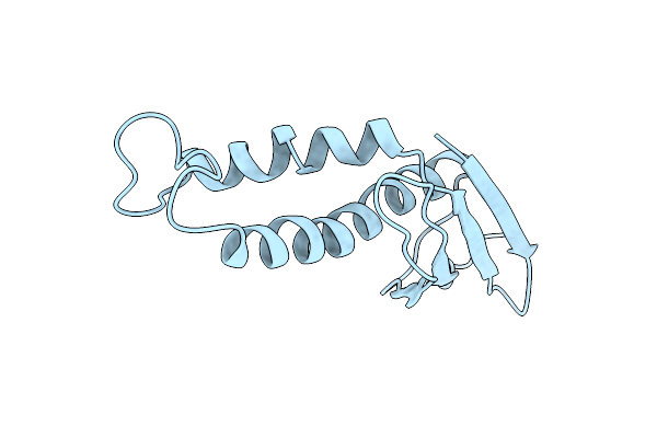
Cyclophilin A Stabilize Hiv-1 Capsid Through A Novel Non- Canonical Binding Site
Organism: Human immunodeficiency virus 1, Homo sapiens
Method: ELECTRON MICROSCOPY Resolution:9.00 Å Release Date: 2016-03-16 Classification: ISOMERASE |
Organism: Human immunodeficiency virus 1, Homo sapiens
Method: ELECTRON MICROSCOPY
Release Date: 2016-03-16
 |
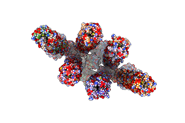
Cryo-Em Map Of Trigger Factor Bound To A Translating Ribosome
Organism: Escherichia coli
Method: ELECTRON MICROSCOPY Resolution:7.70 Å Release Date: 2016-01-13 Classification: ISOMERASE |
Organism: Escherichia coli
Method: ELECTRON MICROSCOPY
Release Date: 2016-01-13
 |
Cryo-Electron Tomography And All-Atom Molecular Dynamics Simulations Reveal A Novel Kinase Conformational Switch In Bacterial Chemotaxis Signaling
Organism: Escherichia coli
Method: ELECTRON MICROSCOPY Release Date: 2015-12-09 Classification: SIGNALING PROTEIN |
Organism: Escherichia coli
Method: ELECTRON MICROSCOPY
Release Date: 2015-12-09