

Search Count: 17
 |
Crystal Structure Of Mouse Galectin-3 In Complex With Small Molecule Inhibitor
Organism: Homo sapiens
Method: X-RAY DIFFRACTION Resolution:2.00 Å Release Date: 2025-03-05 Classification: SUGAR BINDING PROTEIN Ligands: A1L0Q, CL |
Organism: Homo sapiens
Method: X-RAY DIFFRACTION
Release Date: 2025-03-05
Ligands: A1L0Q, CL
 |
Crystal Structure Of Mouse Galectin-3 In Complex With Small Molecule Inhibitor
Organism: Homo sapiens
Method: X-RAY DIFFRACTION Resolution:2.00 Å Release Date: 2025-03-05 Classification: SUGAR BINDING PROTEIN Ligands: A1L0Q, CL |
Organism: Homo sapiens
Method: X-RAY DIFFRACTION
Release Date: 2025-03-05
Ligands: A1L0Q, CL
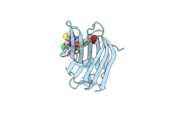 |
Crystal Structure Of Mouse Galectin-3 In Complex With Small Molecule Inhibitor
Organism: Homo sapiens
Method: X-RAY DIFFRACTION Resolution:1.72 Å Release Date: 2025-03-05 Classification: SUGAR BINDING PROTEIN Ligands: MG, A1L0R |
Organism: Homo sapiens
Method: X-RAY DIFFRACTION
Release Date: 2025-03-05
Ligands: MG, A1L0R
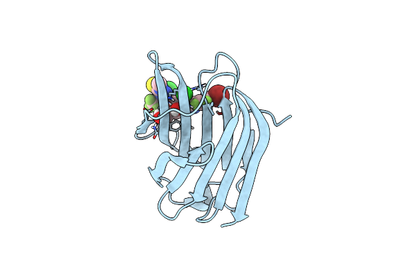 |
Crystal Structure Of Mouse Galectin-3 In Complex With Small Molecule Inhibitor
Organism: Homo sapiens
Method: X-RAY DIFFRACTION Resolution:1.45 Å Release Date: 2025-03-05 Classification: SUGAR BINDING PROTEIN Ligands: A1L0R, CL |
Organism: Homo sapiens
Method: X-RAY DIFFRACTION
Release Date: 2025-03-05
Ligands: A1L0R, CL
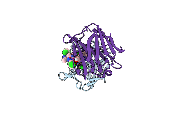 |
Crystal Structure Of Human Galectin-3 In Complex With Small Molecule Inhibitor
Organism: Homo sapiens
Method: X-RAY DIFFRACTION Resolution:1.12 Å Release Date: 2024-03-27 Classification: SUGAR BINDING PROTEIN/INHIBITOR Ligands: CL, SXP, PZE |
Organism: Homo sapiens
Method: X-RAY DIFFRACTION
Release Date: 2024-03-27
Ligands: CL, SXP, PZE
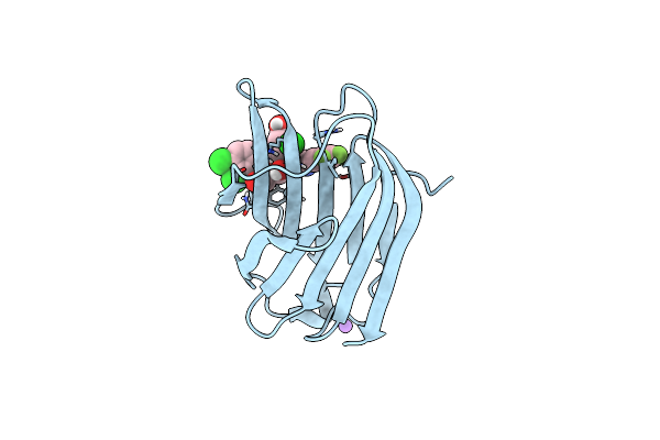 |
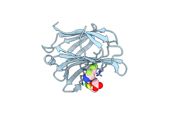
Crystal Structure Of Human Galectin-3 In Complex With Small Molecule Inhibitor
Organism: Homo sapiens
Method: X-RAY DIFFRACTION Resolution:1.22 Å Release Date: 2024-03-27 Classification: SUGAR BINDING PROTEIN Ligands: CL, QB2, NA |
Organism: Homo sapiens
Method: X-RAY DIFFRACTION
Release Date: 2024-03-27
Ligands: CL, QB2, NA
 |
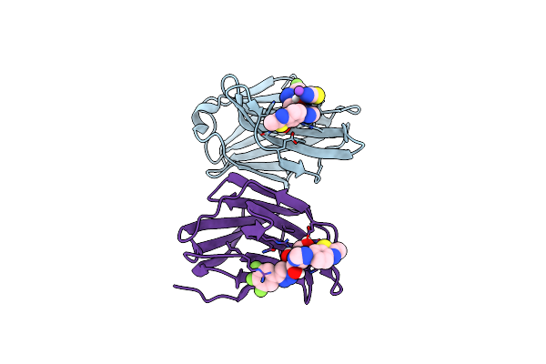
Crystal Structure Of Mouse Galectin-3 In Complex With Small Molecule Inhibitor
Organism: Homo sapiens
Method: X-RAY DIFFRACTION Resolution:1.97 Å Release Date: 2024-03-27 Classification: SUGAR BINDING PROTEIN Ligands: QB2, MG |
Organism: Homo sapiens
Method: X-RAY DIFFRACTION
Release Date: 2024-03-27
Ligands: QB2, MG
 |
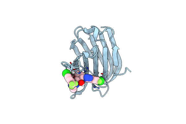
Crystal Structure Of Mouse Galectin-3 In Complex With Small Molecule Inhibitor
Organism: Mus musculus
Method: X-RAY DIFFRACTION Resolution:1.80 Å Release Date: 2024-03-06 Classification: SUGAR BINDING PROTEIN Ligands: Q1L, SCN, NA |
Organism: Mus musculus
Method: X-RAY DIFFRACTION
Release Date: 2024-03-06
Ligands: Q1L, SCN, NA
 |
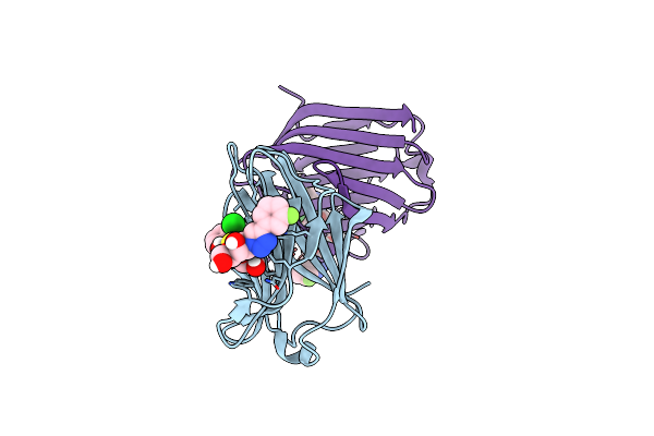
Structure Of Human Galectin-3 Crd In Complex With Monosaccharide Inhibitor
Organism: Homo sapiens
Method: X-RAY DIFFRACTION Resolution:0.98 Å Release Date: 2022-10-12 Classification: Galactoside-binding lectin Ligands: D9J |
Organism: Homo sapiens
Method: X-RAY DIFFRACTION
Release Date: 2022-10-12
Ligands: D9J
 |
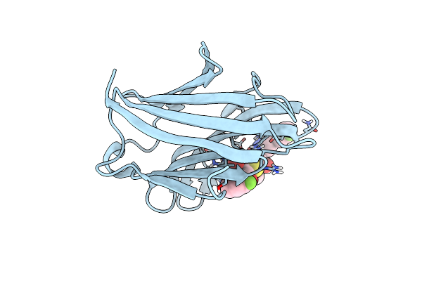
Structure Of Human Galectin-3 Crd In Complex With Td-139 Belonging To P31 Space Group.
Organism: Homo sapiens
Method: X-RAY DIFFRACTION Resolution:1.97 Å Release Date: 2021-09-01 Classification: SUGAR BINDING PROTEIN Ligands: CL, TD2 |
Organism: Homo sapiens
Method: X-RAY DIFFRACTION
Release Date: 2021-09-01
Ligands: CL, TD2
 |
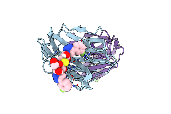
Structure Of Mouse Galectin-3 Crd In Complex With Td-139 Belonging To P6522 Space Group.
Organism: Mus musculus
Method: X-RAY DIFFRACTION Resolution:1.46 Å Release Date: 2021-09-01 Classification: SUGAR BINDING PROTEIN Ligands: TD2, CL |
Organism: Mus musculus
Method: X-RAY DIFFRACTION
Release Date: 2021-09-01
Ligands: TD2, CL
 |
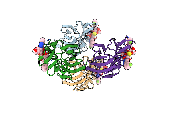
Structure Of Mouse Galectin-3 Crd Point Mutant (V160A) In Complex With Td-139 Belonging To P121 Space Group.
Organism: Mus musculus
Method: X-RAY DIFFRACTION Resolution:1.40 Å Release Date: 2021-09-01 Classification: SUGAR BINDING PROTEIN Ligands: TD2 |
Organism: Mus musculus
Method: X-RAY DIFFRACTION
Release Date: 2021-09-01
Ligands: TD2
 |
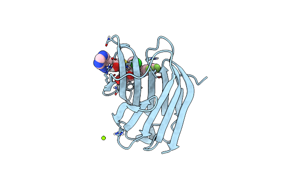
Xray Structure Of Rat Galectin-3 Crd In Complex With Td-139 Belonging To P121 Space Group
Organism: Rattus norvegicus
Method: X-RAY DIFFRACTION Resolution:1.71 Å Release Date: 2021-09-01 Classification: SUGAR BINDING PROTEIN Ligands: BR, TD2 |
Organism: Rattus norvegicus
Method: X-RAY DIFFRACTION
Release Date: 2021-09-01
Ligands: BR, TD2
 |
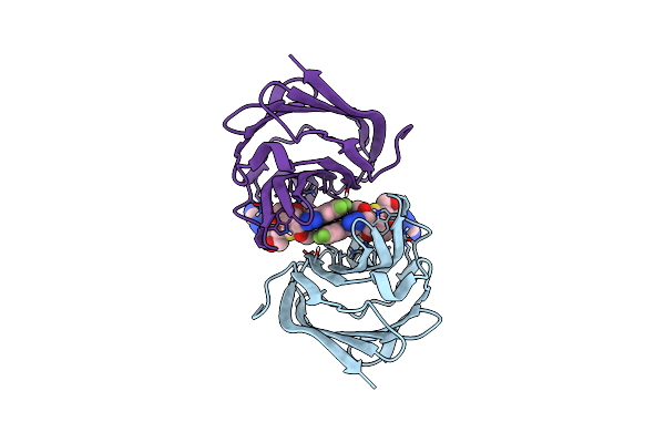
Human Galectin-3 Crd In Complex With Novel Tetrahydropyran-Based Thiodisaccharide Mimic Inhibitor
Organism: Homo sapiens
Method: X-RAY DIFFRACTION Resolution:1.08 Å Release Date: 2021-06-23 Classification: CARBOHYDRATE Ligands: MG, CL, H5O |
Organism: Homo sapiens
Method: X-RAY DIFFRACTION
Release Date: 2021-06-23
Ligands: MG, CL, H5O
 |
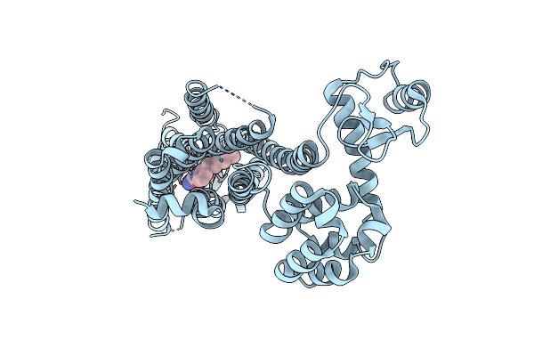
Mouse Galectin-3 Crd In Complex With Novel Tetrahydropyran-Based Thiodisaccharide Mimic Inhibitor
Organism: Mus musculus
Method: X-RAY DIFFRACTION Resolution:1.80 Å Release Date: 2021-06-23 Classification: CARBOHYDRATE Ligands: H5O |
Organism: Mus musculus
Method: X-RAY DIFFRACTION
Release Date: 2021-06-23
Ligands: H5O
 |
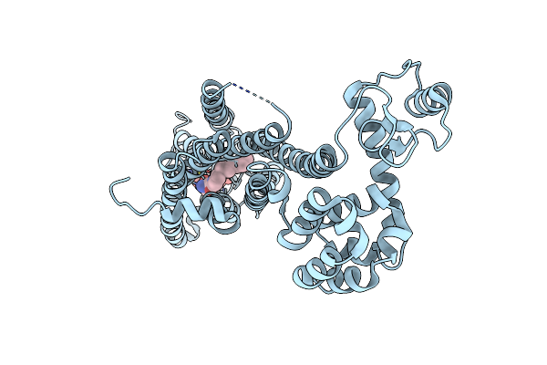
Crystal Structure Of A Lipid G Protein-Coupled Receptor At 3.35A
Organism: Homo sapiens, Enterobacteria phage t4
Method: X-RAY DIFFRACTION Resolution:3.35 Å Release Date: 2012-02-15 Classification: HYDROLASE Ligands: ML5, NAG |
Organism: Homo sapiens, Enterobacteria phage t4
Method: X-RAY DIFFRACTION
Release Date: 2012-02-15
Ligands: ML5, NAG
 |
Crystal Structure Of A Lipid G Protein-Coupled Receptor At 2.80A
Organism: Homo sapiens, Enterobacteria phage t4
Method: X-RAY DIFFRACTION Resolution:2.80 Å Release Date: 2012-02-15 Classification: HYDROLASE Ligands: ML5, NAG |
Organism: Homo sapiens, Enterobacteria phage t4
Method: X-RAY DIFFRACTION
Release Date: 2012-02-15
Ligands: ML5, NAG