

Search Count: 34
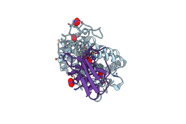 |
Crystal Structure Of Tyrosinase From Streptomyces Castaneoglobisporus In Complex With The Caddie Protein Obtained By Soaking In The Solution Containing Cu(Ii) And Hydroxylamine For 24 H
Organism: Streptomyces castaneoglobisporus
Method: X-RAY DIFFRACTION Resolution:1.50 Å Release Date: 2021-06-16 Classification: METAL BINDING PROTEIN Ligands: PEO, CU, NO3 |
Organism: Streptomyces castaneoglobisporus
Method: X-RAY DIFFRACTION
Release Date: 2021-06-16
Ligands: PEO, CU, NO3
 |
Crystal Structure Of N191G-Mutated Tyrosinase From Streptomyces Castaneoglobisporus In Complex With The Caddie Protein Obtained By Soaking In The Solution Containing Cu(Ii) And Hydroxylamine For 24 H
Organism: Streptomyces castaneoglobisporus
Method: X-RAY DIFFRACTION Resolution:1.47 Å Release Date: 2021-06-16 Classification: METAL BINDING PROTEIN Ligands: PEO, CU, NO3 |
Organism: Streptomyces castaneoglobisporus
Method: X-RAY DIFFRACTION
Release Date: 2021-06-16
Ligands: PEO, CU, NO3
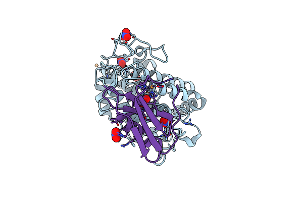 |
1.16 A-Resolution Crystal Structure Of The Deoxy-Form Tyrosinase From Streptomyces Castaneoglobisporus In Complex With The Caddie Protein
Organism: Streptomyces castaneoglobisporus
Method: X-RAY DIFFRACTION Resolution:1.16 Å Release Date: 2018-12-26 Classification: OXIDOREDUCTASE/METAL BINDING PROTEIN Ligands: CU, NO3 |
Organism: Streptomyces castaneoglobisporus
Method: X-RAY DIFFRACTION
Release Date: 2018-12-26
Ligands: CU, NO3
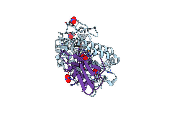 |
Crystal Structure Of Copper-Bound Tyrosinase From Streptomyces Castaneoglobisporus In Complex With The Y98F Mutant Of The Caddie Protein Obtained By Soaking In The Hydroxylamine-Containing Solution For 2 H At 298 K
Organism: Streptomyces castaneoglobisporus
Method: X-RAY DIFFRACTION Resolution:1.16 Å Release Date: 2018-12-26 Classification: OXIDOREDUCTASE/METAL BINDING PROTEIN Ligands: CU, PER, NO3 |
Organism: Streptomyces castaneoglobisporus
Method: X-RAY DIFFRACTION
Release Date: 2018-12-26
Ligands: CU, PER, NO3
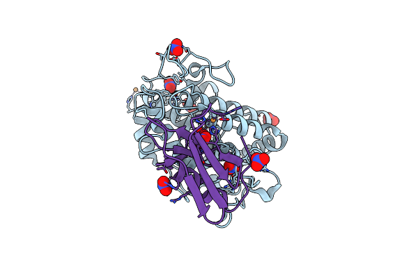 |
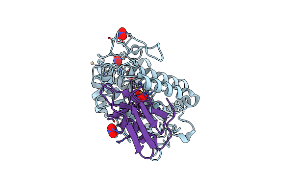
Crystal Structure Of Copper-Bound Tyrosinase From Streptomyces Castaneoglobisporus In Complex With The Caddie Protein Obtained By Soaking In The Hydroxylamine-Containing Solution For 10 Min At 298 K
Organism: Streptomyces castaneoglobisporus
Method: X-RAY DIFFRACTION Resolution:1.16 Å Release Date: 2018-12-26 Classification: OXIDOREDUCTASE/METAL BINDING PROTEIN Ligands: CU, PER, NO3 |
Organism: Streptomyces castaneoglobisporus
Method: X-RAY DIFFRACTION
Release Date: 2018-12-26
Ligands: CU, PER, NO3
 |
Crystal Structure Of Copper-Bound Tyrosinase From Streptomyces Castaneoglobisporus In Complex With The Caddie Protein Obtained By Soaking In The Hydroxylamine-Containing Solution For 20 Min At 298 K
Organism: Streptomyces castaneoglobisporus
Method: X-RAY DIFFRACTION Resolution:1.32 Å Release Date: 2018-12-26 Classification: OXIDOREDUCTASE/METAL BINDING PROTEIN Ligands: CU, PER, NO3 |
Organism: Streptomyces castaneoglobisporus
Method: X-RAY DIFFRACTION
Release Date: 2018-12-26
Ligands: CU, PER, NO3
 |
Crystal Structure Of Copper-Bound Tyrosinase From Streptomyces Castaneoglobisporus In Complex With The Caddie Protein Obtained By Soaking In The Hydroxylamine-Containing Solution For 2 H At 298 K
Organism: Streptomyces castaneoglobisporus
Method: X-RAY DIFFRACTION Resolution:1.18 Å Release Date: 2018-12-26 Classification: OXIDOREDUCTASE/METAL BINDING PROTEIN Ligands: CU, PER, NO3 |
Organism: Streptomyces castaneoglobisporus
Method: X-RAY DIFFRACTION
Release Date: 2018-12-26
Ligands: CU, PER, NO3
 |
Crystal Structure Of Copper-Bound Tyrosinase From Streptomyces Castaneoglobisporus In Complex With The Caddie Protein Obtained By Soaking In The Hydroxylamine-Containing Solution For 1 H At 277 K
Organism: Streptomyces castaneoglobisporus
Method: X-RAY DIFFRACTION Resolution:1.32 Å Release Date: 2018-12-26 Classification: OXIDOREDUCTASE/METAL BINDING PROTEIN Ligands: CU, PER, NO3 |
Organism: Streptomyces castaneoglobisporus
Method: X-RAY DIFFRACTION
Release Date: 2018-12-26
Ligands: CU, PER, NO3
 |
Crystal Structure Of Copper-Bound Tyrosinase From Streptomyces Castaneoglobisporus In Complex With The Caddie Protein Obtained By Soaking In The Hydroxylamine-Containing Solution For 2 H At 277 K
Organism: Streptomyces castaneoglobisporus
Method: X-RAY DIFFRACTION Resolution:1.35 Å Release Date: 2018-12-26 Classification: OXIDOREDUCTASE/METAL BINDING PROTEIN Ligands: CU, PER, NO3 |
Organism: Streptomyces castaneoglobisporus
Method: X-RAY DIFFRACTION
Release Date: 2018-12-26
Ligands: CU, PER, NO3
 |
Crystal Structure Of Copper-Bound Tyrosinase From Streptomyces Castaneoglobisporus In Complex With The Caddie Protein Obtained By Soaking In The Hydroxylamine-Containing Solution For 4 H At 277 K
Organism: Streptomyces castaneoglobisporus
Method: X-RAY DIFFRACTION Resolution:1.28 Å Release Date: 2018-12-26 Classification: OXIDOREDUCTASE/METAL BINDING PROTEIN Ligands: CU, PER, NO3 |
Organism: Streptomyces castaneoglobisporus
Method: X-RAY DIFFRACTION
Release Date: 2018-12-26
Ligands: CU, PER, NO3
 |
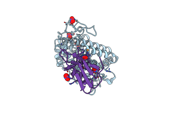
Crystal Structure Of Copper-Bound Tyrosinase From Streptomyces Castaneoglobisporus In Complex With The Caddie Protein Obtained By Soaking In The Hydroxylamine-Containing Solution For 9 H At 277 K
Organism: Streptomyces castaneoglobisporus
Method: X-RAY DIFFRACTION Resolution:1.17 Å Release Date: 2018-12-26 Classification: OXIDOREDUCTASE/METAL BINDING PROTEIN Ligands: CU, PER, NO3 |
Organism: Streptomyces castaneoglobisporus
Method: X-RAY DIFFRACTION
Release Date: 2018-12-26
Ligands: CU, PER, NO3
 |
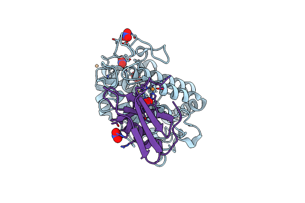
Crystal Structure Of Copper-Bound H63F-Mutated Tyrosinase From Streptomyces Castaneoglobisporus In Complex With The Caddie Protein Obtained By Soaking In The Hydroxylamine-Containing Solution For 12H At 298K
Organism: Streptomyces castaneoglobisporus
Method: X-RAY DIFFRACTION Resolution:1.70 Å Release Date: 2018-12-26 Classification: OXIDOREDUCTASE/METAL BINDING PROTEIN Ligands: CU, NO3 |
Organism: Streptomyces castaneoglobisporus
Method: X-RAY DIFFRACTION
Release Date: 2018-12-26
Ligands: CU, NO3
 |
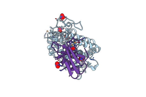
Crystal Structure Of Streptomyces Tyrosinase In A Complex With Caddie Soaked In A Cu(Ii)-Containing Solution For 20 Hr: Occupancy Of Cu(Ii) Is Low
Organism: Streptomyces castaneoglobisporus
Method: X-RAY DIFFRACTION Resolution:1.24 Å Release Date: 2011-06-29 Classification: OXIDOREDUCTASE/METAL TRANSPORT Ligands: CU, NO3 |
Organism: Streptomyces castaneoglobisporus
Method: X-RAY DIFFRACTION
Release Date: 2011-06-29
Ligands: CU, NO3
 |
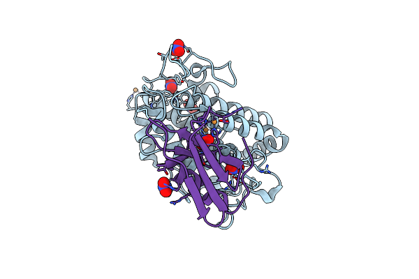
Crystal Structure Of Streptomyces Tyrosinase In A Complex With Caddie Soaked In A Cu(Ii)-Containing Solution For 20 Hr: Occupancy Of Cu(Ii) Is High
Organism: Streptomyces castaneoglobisporus
Method: X-RAY DIFFRACTION Resolution:1.35 Å Release Date: 2011-06-29 Classification: OXIDOREDUCTASE/METAL TRANSPORT Ligands: CU, NO3 |
Organism: Streptomyces castaneoglobisporus
Method: X-RAY DIFFRACTION
Release Date: 2011-06-29
Ligands: CU, NO3
 |
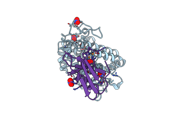
Crystal Structure Of Streptomyces Tyrosinase In A Complex With Caddie Soaked In A Cu(Ii)-Containing Solution For 40 H
Organism: Streptomyces castaneoglobisporus
Method: X-RAY DIFFRACTION Resolution:1.16 Å Release Date: 2011-06-29 Classification: OXIDOREDUCTASE/METAL TRANSPORT Ligands: CU, NO3 |
Organism: Streptomyces castaneoglobisporus
Method: X-RAY DIFFRACTION
Release Date: 2011-06-29
Ligands: CU, NO3
 |
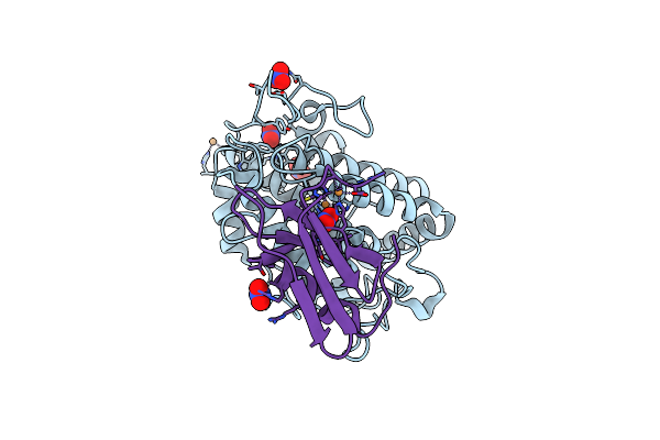
Crystal Structure Of Streptomyces Tyrosinase In A Complex With Caddie Soaked In A Cu(Ii)-Containing Solution For 80 Hr: Occupancy Of Cua Is Low
Organism: Streptomyces castaneoglobisporus
Method: X-RAY DIFFRACTION Resolution:1.40 Å Release Date: 2011-06-29 Classification: OXIDOREDUCTASE/METAL TRANSPORT Ligands: CU, NO3 |
Organism: Streptomyces castaneoglobisporus
Method: X-RAY DIFFRACTION
Release Date: 2011-06-29
Ligands: CU, NO3
 |
Crystal Structure Of Streptomyces Tyrosinase In A Complex With Caddie Soaked In A Cu(Ii)-Containing Solution For 80 Hr: Occupancy Of Cua Is High
Organism: Streptomyces castaneoglobisporus
Method: X-RAY DIFFRACTION Resolution:1.35 Å Release Date: 2011-06-29 Classification: OXIDOREDUCTASE/METAL TRANSPORT Ligands: CU, NO3 |
Organism: Streptomyces castaneoglobisporus
Method: X-RAY DIFFRACTION
Release Date: 2011-06-29
Ligands: CU, NO3
 |
Crystal Structure Of Streptomyces Tyrosinase In A Complex With Caddie H82Q Mutant Soaked In A Cu(Ii)-Containing Solution For 80 Hr
Organism: Streptomyces castaneoglobisporus
Method: X-RAY DIFFRACTION Resolution:1.25 Å Release Date: 2011-06-29 Classification: OXIDOREDUCTASE/METAL TRANSPORT Ligands: CU, NO3 |
Organism: Streptomyces castaneoglobisporus
Method: X-RAY DIFFRACTION
Release Date: 2011-06-29
Ligands: CU, NO3
 |
Crystal Structure Of Streptomyces Tyrosinase In A Complex With Caddie M84L Mutant Soaked In A Cu(Ii)-Containing Solution For 80 Hr
Organism: Streptomyces castaneoglobisporus
Method: X-RAY DIFFRACTION Resolution:1.58 Å Release Date: 2011-06-29 Classification: OXIDOREDUCTASE/METAL TRANSPORT Ligands: CU, NO3 |
Organism: Streptomyces castaneoglobisporus
Method: X-RAY DIFFRACTION
Release Date: 2011-06-29
Ligands: CU, NO3
 |
Crystal Structure Of Streptomyces Tyrosinase In A Complex With Caddie H97Q Mutant Soaked In A Cu(Ii)-Containing Solution For 80 Hr
Organism: Streptomyces castaneoglobisporus
Method: X-RAY DIFFRACTION Resolution:1.43 Å Release Date: 2011-06-29 Classification: OXIDOREDUCTASE/METAL TRANSPORT Ligands: CU, NO3 |
Organism: Streptomyces castaneoglobisporus
Method: X-RAY DIFFRACTION
Release Date: 2011-06-29
Ligands: CU, NO3