

Search Count: 208
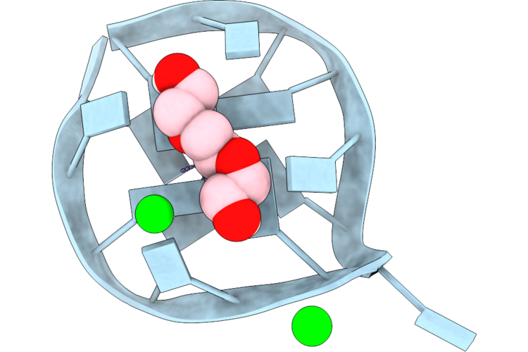 |
Jlm3 Left-Handed, Parallel G-Quadruplex In P65 2 2 Spacegroup
Organism: Synthetic construct
Method: X-RAY DIFFRACTION Resolution:1.54 Å Release Date: 2026-07-22 Classification: DNA Ligands: PGE, K, SR |
Organism: Synthetic construct
Method: X-RAY DIFFRACTION
Release Date: 2026-07-22
Ligands: PGE, K, SR
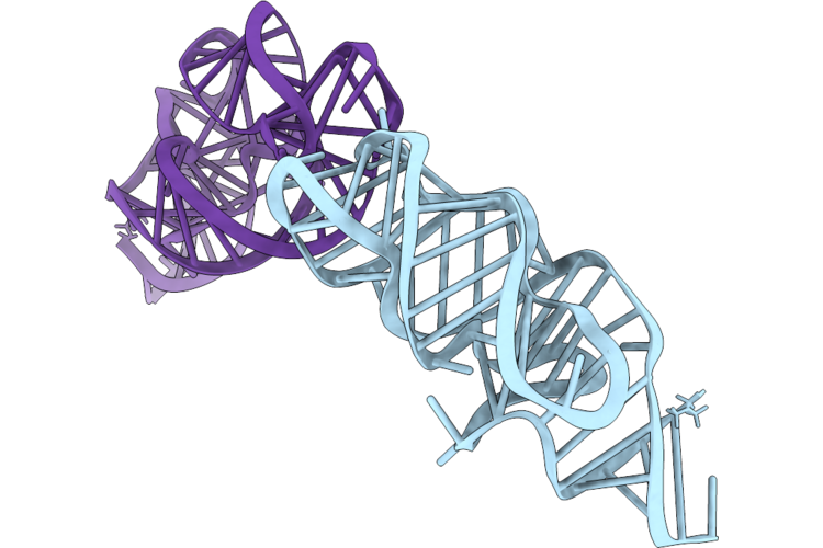 |
Apo Structure Of Dopamine-Binding Aptamer, Dgr-1B
Organism: Synthetic construct
Method: X-RAY DIFFRACTION Resolution:2.93 Å Release Date: 2026-05-13 Classification: RNA Ligands: SR |
Organism: Synthetic construct
Method: X-RAY DIFFRACTION
Release Date: 2026-05-13
Ligands: SR
 |
Probing Positions 3 And 8: Insights Into A 960 Nm Emissive Dna-Stabilized Silver Nanocluster
Organism: Synthetic construct
Method: X-RAY DIFFRACTION Resolution:1.90 Å Release Date: 2026-01-28 Classification: DNA Ligands: AG, CL, SR |
Organism: Synthetic construct
Method: X-RAY DIFFRACTION
Release Date: 2026-01-28
Ligands: AG, CL, SR
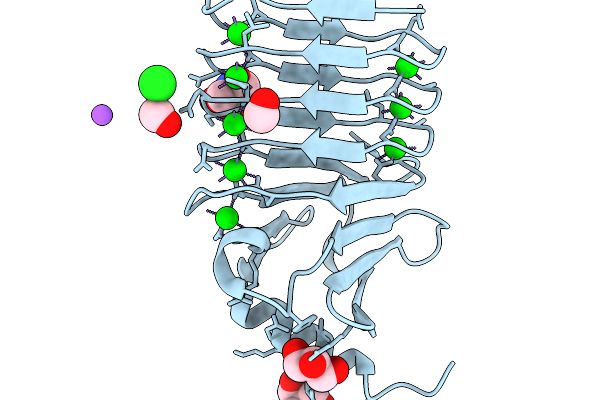 |
Crystal Structure Of Sr2+-Bound Rtx Domain Block V Of Adenylate Cyclase Toxin From Bordetella Pertussis
Organism: Bordetella pertussis
Method: X-RAY DIFFRACTION Resolution:1.50 Å Release Date: 2026-01-07 Classification: TOXIN Ligands: SR, CL, NA, FOR, GOL, TRS |
Organism: Bordetella pertussis
Method: X-RAY DIFFRACTION
Release Date: 2026-01-07
Ligands: SR, CL, NA, FOR, GOL, TRS
 |
Crystal Structure Of Ash1L Phd-Bah Domains
Organism: Homo sapiens
Method: X-RAY DIFFRACTION Resolution:2.40 Å Release Date: 2025-03-19 Classification: TRANSFERASE Ligands: ZN, SR |
Organism: Homo sapiens
Method: X-RAY DIFFRACTION
Release Date: 2025-03-19
Ligands: ZN, SR
 |
Dna Major Groove Binding By Lambda-[Ru(Phen)2(Phi)]2+
Organism: Synthetic construct
Method: X-RAY DIFFRACTION Resolution:2.21 Å Release Date: 2024-03-13 Classification: DNA Ligands: V7F, SR |
Organism: Synthetic construct
Method: X-RAY DIFFRACTION
Release Date: 2024-03-13
Ligands: V7F, SR
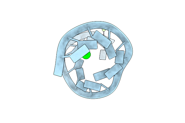 |
16Mer Self-Complementary Duplex Rna With Two Separated Native U:U Pairs
Organism: Synthetic construct
Method: X-RAY DIFFRACTION Resolution:1.52 Å Release Date: 2024-01-24 Classification: RNA Ligands: SR, MG |
Organism: Synthetic construct
Method: X-RAY DIFFRACTION
Release Date: 2024-01-24
Ligands: SR, MG
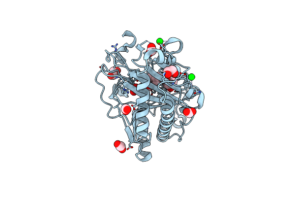 |
Structure Of Hyoscyamine 6-Beta Hydroxylase In Complex With Iron, 2-Oxoglutarate, And Hyoscyamine
Organism: Atropa belladonna
Method: X-RAY DIFFRACTION Resolution:1.53 Å Release Date: 2023-11-22 Classification: OXIDOREDUCTASE Ligands: SR, EDO, FMT, FE2, AKG, HYO |
Organism: Atropa belladonna
Method: X-RAY DIFFRACTION
Release Date: 2023-11-22
Ligands: SR, EDO, FMT, FE2, AKG, HYO
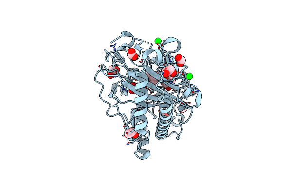 |
Structure Of Hyoscyamine 6-Beta Hydroxylase In Complex With Vanadyl, Succinate, And Hyoscyamine
Organism: Atropa belladonna
Method: X-RAY DIFFRACTION Resolution:1.79 Å Release Date: 2023-11-22 Classification: OXIDOREDUCTASE Ligands: EDO, HYO, SIN, FMT, V, O, SR |
Organism: Atropa belladonna
Method: X-RAY DIFFRACTION
Release Date: 2023-11-22
Ligands: EDO, HYO, SIN, FMT, V, O, SR
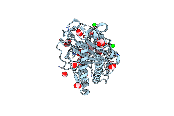 |
Structure Of Hyoscyamine 6-Beta Hydroxylase In Complex With Iron, Succinate, And 6-Oh-Hyoscyamine
Organism: Atropa belladonna
Method: X-RAY DIFFRACTION Resolution:1.58 Å Release Date: 2023-11-22 Classification: OXIDOREDUCTASE Ligands: FE2, EDO, FMT, OVR, SIN, SR |
Organism: Atropa belladonna
Method: X-RAY DIFFRACTION
Release Date: 2023-11-22
Ligands: FE2, EDO, FMT, OVR, SIN, SR
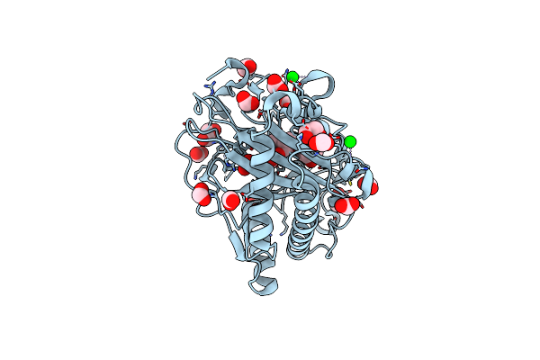 |
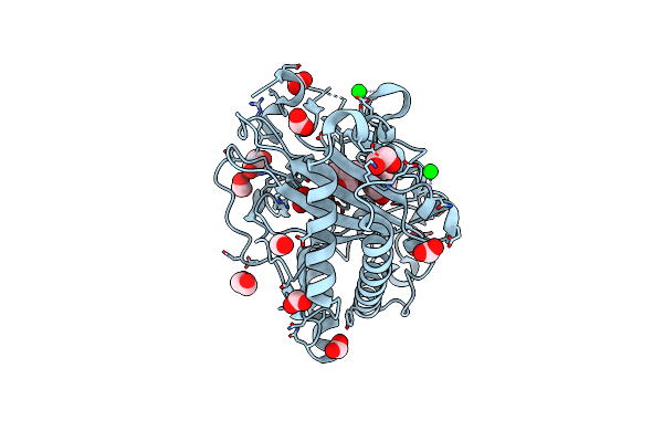
Structure Of Hyoscyamine 6-Beta Hydroxylase In Complex With Iron, 2-Oxoglutarate, And 6-Oh-Hyoscyamine
Organism: Atropa belladonna
Method: X-RAY DIFFRACTION Resolution:1.53 Å Release Date: 2023-11-22 Classification: OXIDOREDUCTASE Ligands: FE2, EDO, FMT, AKG, OVR, SR |
Organism: Atropa belladonna
Method: X-RAY DIFFRACTION
Release Date: 2023-11-22
Ligands: FE2, EDO, FMT, AKG, OVR, SR
 |
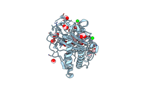
Structure Of Hyoscyamine 6-Beta Hydroxylase In Complex With Vanadyl, Succinate, And 6-Oh-Hyoscyamine
Organism: Atropa belladonna
Method: X-RAY DIFFRACTION Resolution:1.79 Å Release Date: 2023-11-22 Classification: OXIDOREDUCTASE Ligands: EDO, SIN, FMT, OVR, V, O, SR |
Organism: Atropa belladonna
Method: X-RAY DIFFRACTION
Release Date: 2023-11-22
Ligands: EDO, SIN, FMT, OVR, V, O, SR
 |
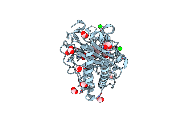
Structure Of Hyoscyamine 6-Beta Hydroxylase In Complex With Iron, Succinate, And Scopolamine
Organism: Atropa belladonna
Method: X-RAY DIFFRACTION Resolution:1.72 Å Release Date: 2023-11-22 Classification: OXIDOREDUCTASE Ligands: FE2, EDO, FMT, OW0, SIN, SR |
Organism: Atropa belladonna
Method: X-RAY DIFFRACTION
Release Date: 2023-11-22
Ligands: FE2, EDO, FMT, OW0, SIN, SR
 |
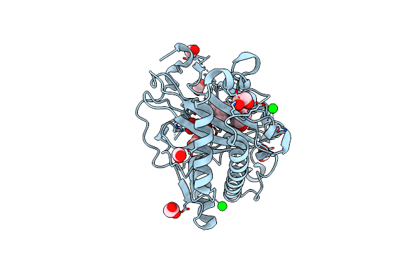
Structure Of L289F Hyoscyamine 6-Beta Hydroxylase In Complex With Iron, 2-Oxoglutarate, And Hyoscyamine
Organism: Atropa belladonna
Method: X-RAY DIFFRACTION Resolution:1.53 Å Release Date: 2023-11-22 Classification: OXIDOREDUCTASE Ligands: FE2, EDO, FMT, HYO, AKG, SR |
Organism: Atropa belladonna
Method: X-RAY DIFFRACTION
Release Date: 2023-11-22
Ligands: FE2, EDO, FMT, HYO, AKG, SR
 |
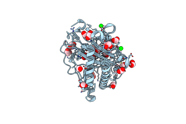
Structure Of L289F Hyoscyamine 6-Beta Hydroxylase In Complex With Vanadyl, Succinate, And Hyoscyamine
Organism: Atropa belladonna
Method: X-RAY DIFFRACTION Resolution:1.53 Å Release Date: 2023-11-22 Classification: OXIDOREDUCTASE Ligands: EDO, FMT, HYO, SIN, V, O, SR |
Organism: Atropa belladonna
Method: X-RAY DIFFRACTION
Release Date: 2023-11-22
Ligands: EDO, FMT, HYO, SIN, V, O, SR
 |
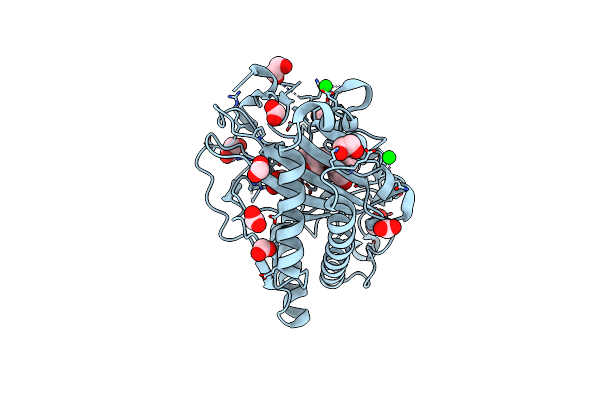
Structure Of L289F Hyoscyamine 6-Beta Hydroxylase In Complex With Iron, 2-Oxoglutarate, And 6-Oh-Hyoscyamine
Organism: Atropa belladonna
Method: X-RAY DIFFRACTION Resolution:1.56 Å Release Date: 2023-11-22 Classification: OXIDOREDUCTASE Ligands: FE2, EDO, FMT, AKG, OVR, SR |
Organism: Atropa belladonna
Method: X-RAY DIFFRACTION
Release Date: 2023-11-22
Ligands: FE2, EDO, FMT, AKG, OVR, SR
 |
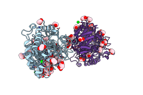
Structure Of L289F Hyoscyamine 6-Beta Hydroxylase In Complex With Vanadyl, Succinate, And 6-Oh-Hyoscyamine
Organism: Atropa belladonna
Method: X-RAY DIFFRACTION Resolution:2.03 Å Release Date: 2023-11-22 Classification: OXIDOREDUCTASE Ligands: EDO, FMT, SIN, OVR, V, SR |
Organism: Atropa belladonna
Method: X-RAY DIFFRACTION
Release Date: 2023-11-22
Ligands: EDO, FMT, SIN, OVR, V, SR
 |
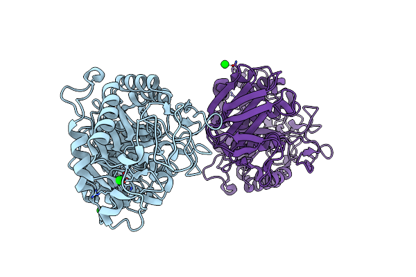
Crystal Structure Of Drosophila Melanogaster Alpha-Amylase In Complex With The Inhibitor Acarbose
Organism: Drosophila melanogaster
Method: X-RAY DIFFRACTION Resolution:2.00 Å Release Date: 2023-08-16 Classification: HYDROLASE Ligands: CL, EDO, SR |
Organism: Drosophila melanogaster
Method: X-RAY DIFFRACTION
Release Date: 2023-08-16
Ligands: CL, EDO, SR
 |
Crystal Structure Of Drosophila Melanogaster Alpha-Amylase
Organism: Drosophila melanogaster
Method: X-RAY DIFFRACTION Resolution:2.50 Å Release Date: 2023-08-09 Classification: HYDROLASE Ligands: CL, SR |
Organism: Drosophila melanogaster
Method: X-RAY DIFFRACTION
Release Date: 2023-08-09
Ligands: CL, SR
 |
Rep-Related Chom18 Variant With Double At Base Pairing
Organism: Cardiobacterium hominis
Method: X-RAY DIFFRACTION Resolution:2.70 Å Release Date: 2023-03-29 Classification: DNA Ligands: SR |
Organism: Cardiobacterium hominis
Method: X-RAY DIFFRACTION
Release Date: 2023-03-29
Ligands: SR