

Search Count: 16
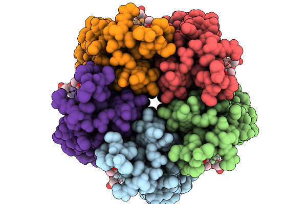 |
Structure Of The Honeybee Gabaa Rdl Receptor With Gaba And Abamectin
Organism: Apis mellifera
Method: ELECTRON MICROSCOPY Release Date: 2026-02-18 Classification: MEMBRANE PROTEIN Ligands: ABU, NAG, A1JNZ |
Organism: Apis mellifera
Method: ELECTRON MICROSCOPY
Release Date: 2026-02-18
Ligands: ABU, NAG, A1JNZ
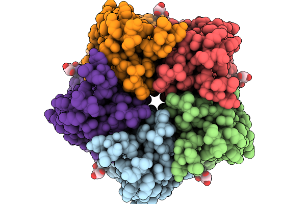 |
Structure Of The Honeybee Gabaa Rdl Receptor With Gaba
Organism: Apis mellifera
Method: ELECTRON MICROSCOPY Release Date: 2026-02-11 Classification: MEMBRANE PROTEIN Ligands: NAG, ABU |
Organism: Apis mellifera
Method: ELECTRON MICROSCOPY
Release Date: 2026-02-11
Ligands: NAG, ABU
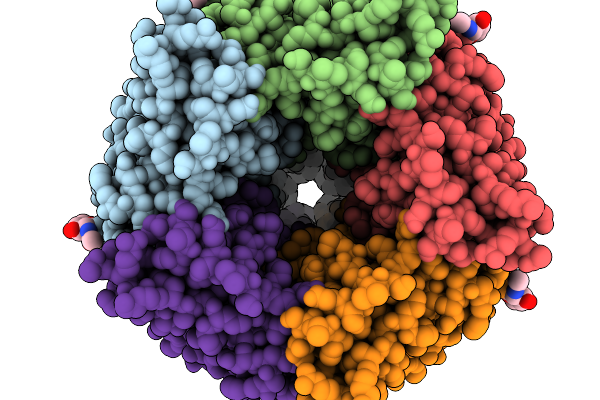 |
Structure Of The Honeybee Gabaa Rdl Receptor Apo State
Organism: Apis mellifera
Method: ELECTRON MICROSCOPY Release Date: 2026-02-11 Classification: MEMBRANE PROTEIN Ligands: NAG |
Organism: Apis mellifera
Method: ELECTRON MICROSCOPY
Release Date: 2026-02-11
Ligands: NAG
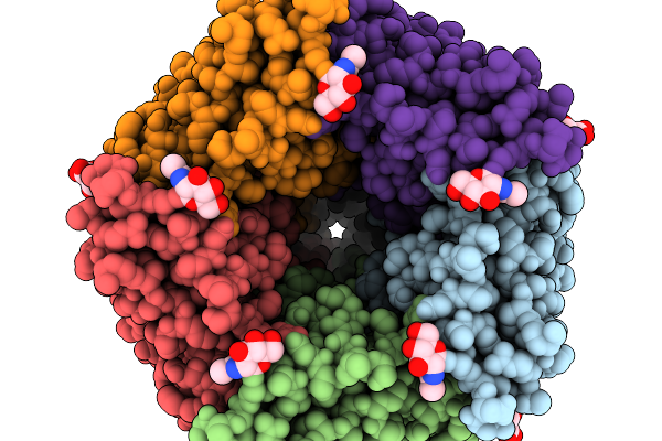 |
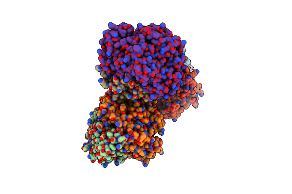
Structure Of The Honeybee Gabaa Rdl Receptor With Chrodrimanin B
Organism: Apis mellifera
Method: ELECTRON MICROSCOPY Release Date: 2026-02-11 Classification: MEMBRANE PROTEIN Ligands: NAG, PT5, A1JN7 |
Organism: Apis mellifera
Method: ELECTRON MICROSCOPY
Release Date: 2026-02-11
Ligands: NAG, PT5, A1JN7
 |
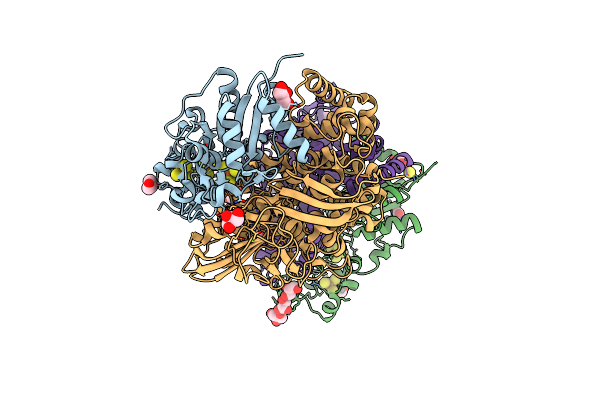
Structure Of The Unready Ni-A State Of The S499C Mutant Of D. Fructosovorans Nife-Hydrogenase
Organism: Desulfovibrio fructosovorans
Method: X-RAY DIFFRACTION Resolution:1.80 Å Release Date: 2014-10-29 Classification: OXIDOREDUCTASE Ligands: SF4, F3S, FCO, NI, CA, GOL, PO4, MES |
Organism: Desulfovibrio fructosovorans
Method: X-RAY DIFFRACTION
Release Date: 2014-10-29
Ligands: SF4, F3S, FCO, NI, CA, GOL, PO4, MES
 |
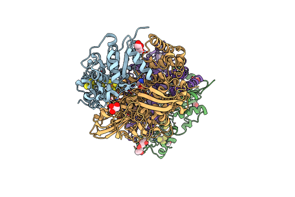
Low X-Ray Dose Structure Of A Ni-A Ni-Sox Mixture Of The D. Fructosovorans Nife-Hydrogenase L122A Mutant
Organism: Desulfovibrio fructosovorans
Method: X-RAY DIFFRACTION Resolution:1.52 Å Release Date: 2014-10-29 Classification: OXIDOREDUCTASE Ligands: SF4, F3S, GOL, GLY, SOT, H2S, FCO, NI, MG, CL |
Organism: Desulfovibrio fructosovorans
Method: X-RAY DIFFRACTION
Release Date: 2014-10-29
Ligands: SF4, F3S, GOL, GLY, SOT, H2S, FCO, NI, MG, CL
 |
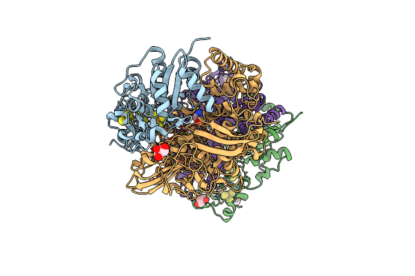
High-Resolution Structure Of A Ni-A Ni-Sox Mixture Of The D. Fructosovorans Nife-Hydrogenase L122A Mutant
Organism: Desulfovibrio fructosovorans
Method: X-RAY DIFFRACTION Resolution:1.22 Å Release Date: 2014-10-29 Classification: OXIDOREDUCTASE Ligands: SF4, F3S, GOL, GLY, SOT, H2S, FCO, NI, MG, CL |
Organism: Desulfovibrio fructosovorans
Method: X-RAY DIFFRACTION
Release Date: 2014-10-29
Ligands: SF4, F3S, GOL, GLY, SOT, H2S, FCO, NI, MG, CL
 |
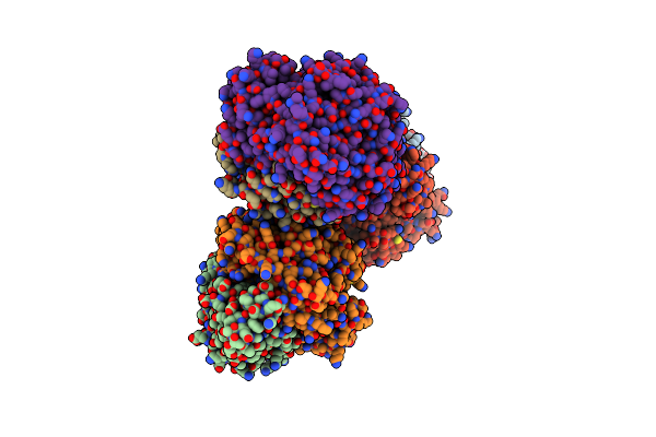
High-Resolution Structure Of The D. Fructosovorans Nife-Hydrogenase L122A Mutant After Exposure To Air
Organism: Desulfovibrio fructosovorans
Method: X-RAY DIFFRACTION Resolution:1.42 Å Release Date: 2014-10-29 Classification: OXIDOREDUCTASE Ligands: SF4, F3S, GOL, SOT, H2S, FCO, NI, MG, GLY |
Organism: Desulfovibrio fructosovorans
Method: X-RAY DIFFRACTION
Release Date: 2014-10-29
Ligands: SF4, F3S, GOL, SOT, H2S, FCO, NI, MG, GLY
 |
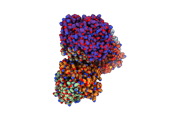
High-Resolution Structure Of Partially Oxidized D. Fructosovorans Nife-Hydrogenase
Organism: Desulfovibrio fructosovorans
Method: X-RAY DIFFRACTION Resolution:1.44 Å Release Date: 2014-10-29 Classification: OXIDOREDUCTASE Ligands: SF4, F3S, GOL, FCO, NI, MG |
Organism: Desulfovibrio fructosovorans
Method: X-RAY DIFFRACTION
Release Date: 2014-10-29
Ligands: SF4, F3S, GOL, FCO, NI, MG
 |
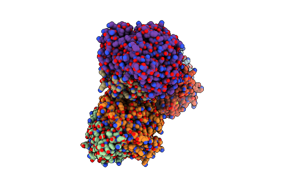
Structure Of The V74M Large Subunit Mutant Of Ni-Fe Hydrogenase In An Oxidized State
Organism: Desulfovibrio fructosovorans
Method: X-RAY DIFFRACTION Resolution:2.70 Å Release Date: 2009-07-21 Classification: OXIDOREDUCTASE Ligands: SF4, F3S, GOL, FCO, NI, MG |
Organism: Desulfovibrio fructosovorans
Method: X-RAY DIFFRACTION
Release Date: 2009-07-21
Ligands: SF4, F3S, GOL, FCO, NI, MG
 |
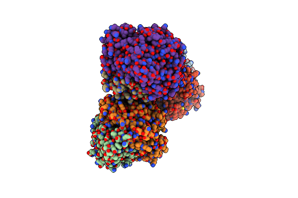
Structure Of A Double Methionine Mutant Of Ni-Fe Hydrogenase
Organism: Desulfovibrio fructosovorans
Method: X-RAY DIFFRACTION Resolution:2.40 Å Release Date: 2008-08-05 Classification: OXIDOREDUCTASE Ligands: SF4, F3S, NI, MG, FCO, PER, GOL |
Organism: Desulfovibrio fructosovorans
Method: X-RAY DIFFRACTION
Release Date: 2008-08-05
Ligands: SF4, F3S, NI, MG, FCO, PER, GOL
 |
Structure Of A Double Ile/Phe Mutant Of Ni-Fe Hydrogenase Refined At 2.2 Angstrom Resolution
Organism: Desulfovibrio fructosovorans
Method: X-RAY DIFFRACTION Resolution:2.20 Å Release Date: 2008-08-05 Classification: OXIDOREDUCTASE Ligands: SF4, F3S, GOL, NI, MG, FCO |
Organism: Desulfovibrio fructosovorans
Method: X-RAY DIFFRACTION
Release Date: 2008-08-05
Ligands: SF4, F3S, GOL, NI, MG, FCO
 |
Structure Of The Unready Oxidized Form Of [Nife] Hydrogenase
Organism: Desulfovibrio gigas
Method: X-RAY DIFFRACTION Resolution:2.35 Å Release Date: 2005-04-19 Classification: OXIDOREDUCTASE Ligands: SF4, F3S, H2S, GOL, NI, MG, FCO, PER |
Organism: Desulfovibrio gigas
Method: X-RAY DIFFRACTION
Release Date: 2005-04-19
Ligands: SF4, F3S, H2S, GOL, NI, MG, FCO, PER
 |
Structure Of The Oxidized Unready Form Of Ni-Fe Hydrogenase
Organism: Desulfovibrio fructosovorans
Method: X-RAY DIFFRACTION Resolution:1.83 Å Release Date: 2005-04-19 Classification: OXIDOREDUCTASE Ligands: SF4, F3S, GOL, NI, FE2, BCT, MG, FCO, PER |
Organism: Desulfovibrio fructosovorans
Method: X-RAY DIFFRACTION
Release Date: 2005-04-19
Ligands: SF4, F3S, GOL, NI, FE2, BCT, MG, FCO, PER
 |
Structure Of The Ready Oxidized Form Of [Nife]-Hydrogenase
Organism: Desulfovibrio fructosovorans
Method: X-RAY DIFFRACTION Resolution:2.10 Å Release Date: 2005-04-19 Classification: OXIDOREDUCTASE Ligands: SF4, F3S, NI, MG, FCO |
Organism: Desulfovibrio fructosovorans
Method: X-RAY DIFFRACTION
Release Date: 2005-04-19
Ligands: SF4, F3S, NI, MG, FCO
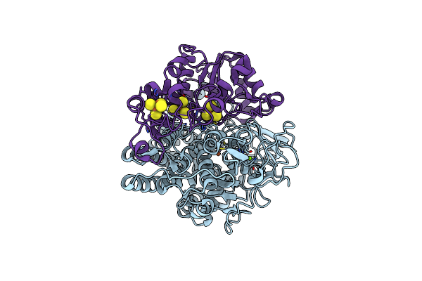 |
Crystal Structure Of The Ni-Fe Hydrogenase From Desulfovibrio Fructosovorans
Organism: Desulfovibrio fructosovorans
Method: X-RAY DIFFRACTION Resolution:2.70 Å Release Date: 1998-07-29 Classification: OXIDOREDUCTASE Ligands: SF4, F3S, FE, NI, MG |
Organism: Desulfovibrio fructosovorans
Method: X-RAY DIFFRACTION
Release Date: 1998-07-29
Ligands: SF4, F3S, FE, NI, MG