

Search Count: 11
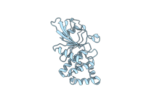 |
Human Ptpru D1 Domain, Reduced Form
Organism: Homo sapiens
Method: X-RAY DIFFRACTION Resolution:1.72 Å Release Date: 2020-07-08 Classification: HYDROLASE Ligands: CL |
Organism: Homo sapiens
Method: X-RAY DIFFRACTION
Release Date: 2020-07-08
Ligands: CL
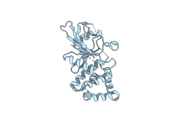 |
Human Ptpru D1 Domain, Oxidised Form
Organism: Homo sapiens
Method: X-RAY DIFFRACTION Resolution:1.97 Å Release Date: 2020-07-08 Classification: HYDROLASE |
Organism: Homo sapiens
Method: X-RAY DIFFRACTION
Release Date: 2020-07-08
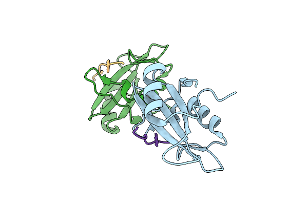 |
Structure Of The N-Sh2 Domain Of The Human Tyrosine-Protein Phosphatase Non-Receptor Type 11 In Complex With The Phosphorylated Immune Receptor Tyrosine-Based Inhibitory Motif
Organism: Homo sapiens, Synthetic construct
Method: X-RAY DIFFRACTION Resolution:2.10 Å Release Date: 2020-02-19 Classification: CELL CYCLE |
Organism: Homo sapiens, Synthetic construct
Method: X-RAY DIFFRACTION
Release Date: 2020-02-19
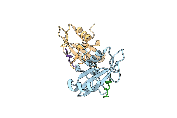 |
Structure Of The N-Sh2 Domain Of The Human Tyrosine-Protein Phosphatase Non-Receptor Type 11 In Complex With The Phosphorylated Immune Receptor Tyrosine-Based Switch Motif
Organism: Homo sapiens, Synthetic construct
Method: X-RAY DIFFRACTION Resolution:2.89 Å Release Date: 2020-02-19 Classification: CELL CYCLE |
Organism: Homo sapiens, Synthetic construct
Method: X-RAY DIFFRACTION
Release Date: 2020-02-19
 |
C-Sh2 Domain Of Shp-2 In Complex With Phospho-Itsm Of Pd-1
Organism: Homo sapiens
Method: SOLUTION NMR Release Date: 2020-02-05 Classification: PEPTIDE BINDING PROTEIN |
Organism: Homo sapiens
Method: SOLUTION NMR
Release Date: 2020-02-05
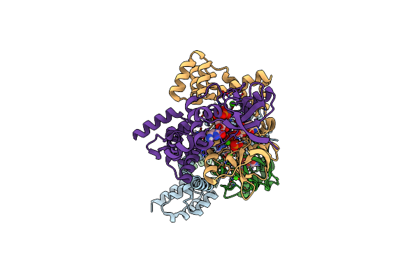 |
Crystal Structure Of Bacterial Parb Dimer Bound To Cdp
Organism: Bacillus subtilis (strain 168)
Method: X-RAY DIFFRACTION Resolution:1.81 Å Release Date: 2019-10-23 Classification: DNA BINDING PROTEIN Ligands: CDP, CA |
Organism: Bacillus subtilis (strain 168)
Method: X-RAY DIFFRACTION
Release Date: 2019-10-23
Ligands: CDP, CA
 |
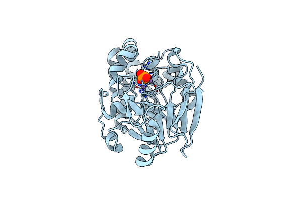
Active Fe-Pp1
Organism: Homo sapiens
Method: X-RAY DIFFRACTION Resolution:2.00 Å Release Date: 2018-11-21 Classification: HYDROLASE Ligands: PO4, FE, MN |
Organism: Homo sapiens
Method: X-RAY DIFFRACTION
Release Date: 2018-11-21
Ligands: PO4, FE, MN
 |
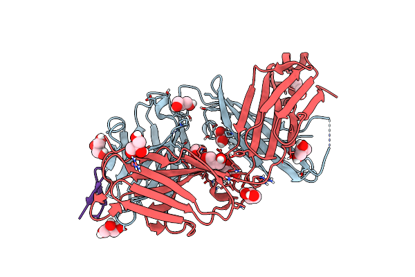
Inactive Fe-Pp1
Organism: Homo sapiens
Method: X-RAY DIFFRACTION Resolution:2.10 Å Release Date: 2018-11-21 Classification: HYDROLASE Ligands: FE, MN, PO4 |
Organism: Homo sapiens
Method: X-RAY DIFFRACTION
Release Date: 2018-11-21
Ligands: FE, MN, PO4
 |
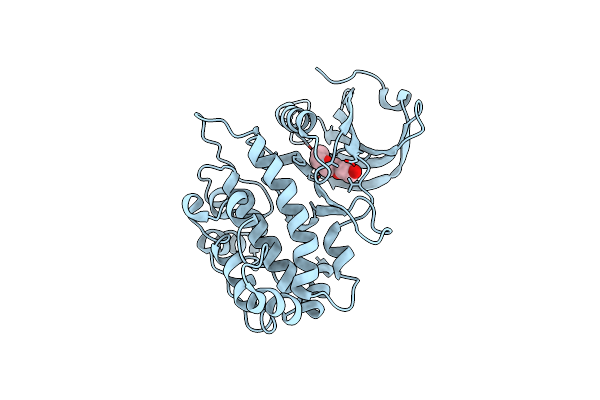
The Antigen-Binding Fragment Of Mab24 In Complex With A Peptide From Hepatitis C Virus E2 Epitope I (412-423)
Organism: Hepatitis c virus, Mus musculus
Method: X-RAY DIFFRACTION Resolution:1.40 Å Release Date: 2018-02-21 Classification: IMMUNE SYSTEM Ligands: GOL |
Organism: Hepatitis c virus, Mus musculus
Method: X-RAY DIFFRACTION
Release Date: 2018-02-21
Ligands: GOL
 |
Structure Of A Semisynthetic Phosphorylated Dapk
Organism: Homo sapiens
Method: X-RAY DIFFRACTION Resolution:2.49 Å Release Date: 2015-08-12 Classification: TRANSFERASE Ligands: PGE |
Organism: Homo sapiens
Method: X-RAY DIFFRACTION
Release Date: 2015-08-12
Ligands: PGE
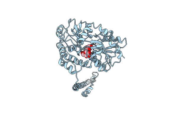 |
Htlv-1 Gp21 Ectodomain/Maltose-Binding Protein Chimera
Organism: Human t-lymphotropic virus 1
Method: X-RAY DIFFRACTION Resolution:2.50 Å Release Date: 1999-04-06 Classification: VIRAL PROTEIN Ligands: CL |
Organism: Human t-lymphotropic virus 1
Method: X-RAY DIFFRACTION
Release Date: 1999-04-06
Ligands: CL