

Search Count: 25
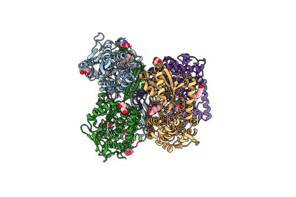 |
1.65 Angstrom Resolution Crystal Structure Of Betaine Aldehyde Dehydrogenase (Betb) From Staphylococcus Aureus With Bme-Modified Cys289 And Peg Molecule In Active Site
Organism: Staphylococcus aureus subsp. aureus col
Method: X-RAY DIFFRACTION Resolution:1.65 Å Release Date: 2014-07-16 Classification: OXIDOREDUCTASE |
Organism: Staphylococcus aureus subsp. aureus col
Method: X-RAY DIFFRACTION
Release Date: 2014-07-16
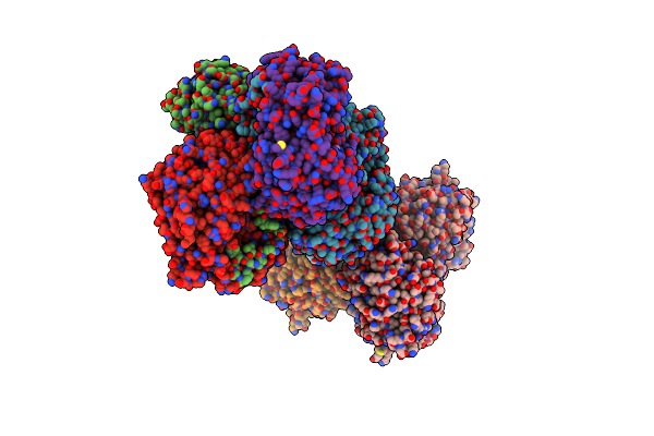 |
2.6 Angstrom Resolution Crystal Structure Of Betaine Aldehyde Dehydrogenase (Betb) G234S Mutant From Staphylococcus Aureus (Idp00699) In Complex With Nad+ And Bme-Free Cys289
Organism: Staphylococcus aureus subsp. aureus
Method: X-RAY DIFFRACTION Resolution:2.60 Å Release Date: 2014-07-02 Classification: OXIDOREDUCTASE Ligands: NAD, ACT |
Organism: Staphylococcus aureus subsp. aureus
Method: X-RAY DIFFRACTION
Release Date: 2014-07-02
Ligands: NAD, ACT
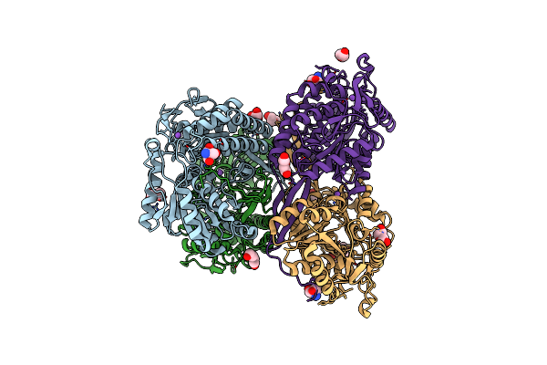 |
1.85 Angstrom Resolution Crystal Structure Of Apo Betaine Aldehyde Dehydrogenase (Betb) G234S Mutant From Staphylococcus Aureus (Idp00699) With Bme-Free Sulfinic Acid Form Of Cys289
Organism: Staphylococcus aureus subsp. aureus
Method: X-RAY DIFFRACTION Resolution:1.85 Å Release Date: 2014-06-18 Classification: OXIDOREDUCTASE |
Organism: Staphylococcus aureus subsp. aureus
Method: X-RAY DIFFRACTION
Release Date: 2014-06-18
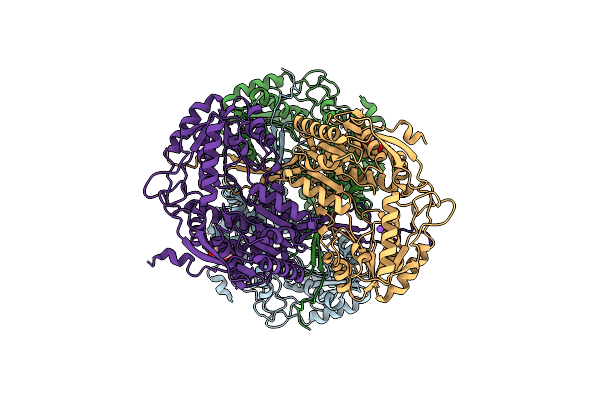 |
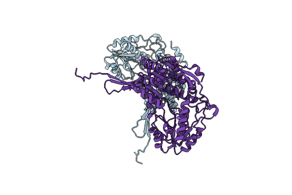
1.90 Angstrom Resolution Crystal Structure Of Apo Betaine Aldehyde Dehydrogenase (Betb) G234S Mutant From Staphylococcus Aureus (Idp00699) With Bme-Modified Cys289
Organism: Staphylococcus aureus subsp. aureus
Method: X-RAY DIFFRACTION Resolution:1.90 Å Release Date: 2014-05-07 Classification: OXIDOREDUCTASE |
Organism: Staphylococcus aureus subsp. aureus
Method: X-RAY DIFFRACTION
Release Date: 2014-05-07
 |
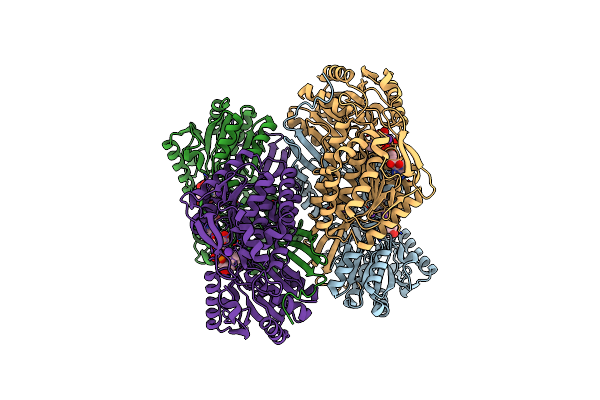
2.30 Angstrom Resolution Crystal Structure Of Betaine Aldehyde Dehydrogenase (Betb) From Staphylococcus Aureus With Bme-Free Cys289
Organism: Staphylococcus aureus subsp. aureus
Method: X-RAY DIFFRACTION Resolution:2.30 Å Release Date: 2013-12-11 Classification: OXIDOREDUCTASE |
Organism: Staphylococcus aureus subsp. aureus
Method: X-RAY DIFFRACTION
Release Date: 2013-12-11
 |
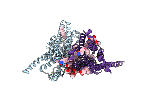
1.90 Angstrom Resolution Crystal Structure Of Betaine Aldehyde Dehydrogenase (Betb) From Staphylococcus Aureus In Complex With Nad+ And Bme-Free Cys289
Organism: Staphylococcus aureus subsp. aureus
Method: X-RAY DIFFRACTION Resolution:1.90 Å Release Date: 2013-11-06 Classification: OXIDOREDUCTASE Ligands: NAD, K |
Organism: Staphylococcus aureus subsp. aureus
Method: X-RAY DIFFRACTION
Release Date: 2013-11-06
Ligands: NAD, K
 |
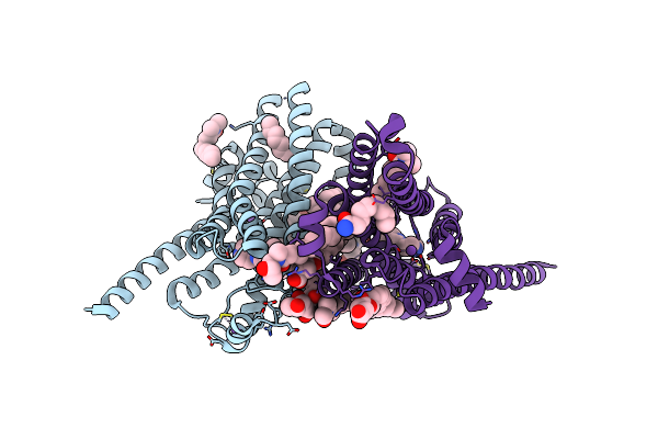
Thermostabilised Turkey Beta1 Adrenergic Receptor With 4-(Piperazin-1- Yl)-1H-Indole Bound (Compound 19)
Organism: Meleagris gallopavo
Method: X-RAY DIFFRACTION Resolution:2.80 Å Release Date: 2013-04-03 Classification: MEMBRANE PROTEIN Ligands: Y01, 2CV, XF5, NA |
Organism: Meleagris gallopavo
Method: X-RAY DIFFRACTION
Release Date: 2013-04-03
Ligands: Y01, 2CV, XF5, NA
 |
Thermostabilised Turkey Beta1 Adrenergic Receptor With 4-Methyl-2-(Piperazin-1-Yl) Quinoline Bound
Organism: Meleagris gallopavo
Method: X-RAY DIFFRACTION Resolution:2.70 Å Release Date: 2013-04-03 Classification: MEMBRANE PROTEIN |
Organism: Meleagris gallopavo
Method: X-RAY DIFFRACTION
Release Date: 2013-04-03
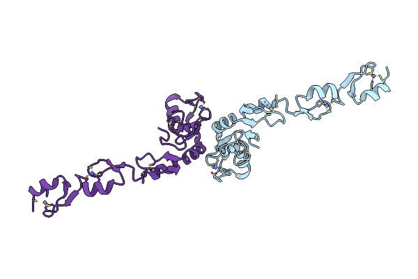 |
Crystal Structure Of The N-Terminal Domain Of Traf6
Organism: Homo sapiens
Method: X-RAY DIFFRACTION Resolution:2.20 Å Release Date: 2009-05-26 Classification: SIGNALING PROTEIN Ligands: ZN |
Organism: Homo sapiens
Method: X-RAY DIFFRACTION
Release Date: 2009-05-26
Ligands: ZN
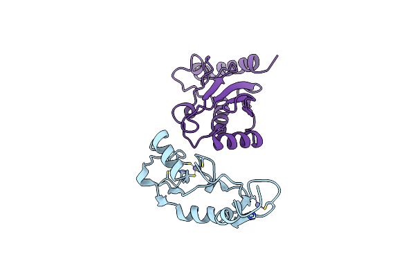 |
Crystal Structure Of Traf6 In Complex With Ubc13 In The P1 Space Group
Organism: Homo sapiens
Method: X-RAY DIFFRACTION Resolution:2.10 Å Release Date: 2009-05-26 Classification: SIGNALING PROTEIN/LIGASE Ligands: ZN |
Organism: Homo sapiens
Method: X-RAY DIFFRACTION
Release Date: 2009-05-26
Ligands: ZN
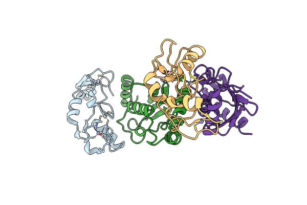 |
Crystal Structure Of Traf6 In Complex With Ubc13 In The C2 Space Group
Organism: Homo sapiens
Method: X-RAY DIFFRACTION Resolution:2.60 Å Release Date: 2009-05-26 Classification: SIGNALING PROTEIN/LIGASE Ligands: ZN |
Organism: Homo sapiens
Method: X-RAY DIFFRACTION
Release Date: 2009-05-26
Ligands: ZN
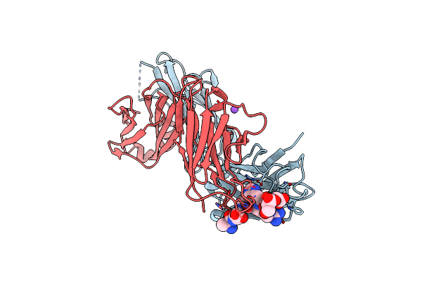 |
Pfa2 Fab Complexed With Abeta1-8
Organism: Mus musculus
Method: X-RAY DIFFRACTION Resolution:2.50 Å Release Date: 2007-10-16 Classification: IMMUNE SYSTEM |
Organism: Mus musculus
Method: X-RAY DIFFRACTION
Release Date: 2007-10-16
 |
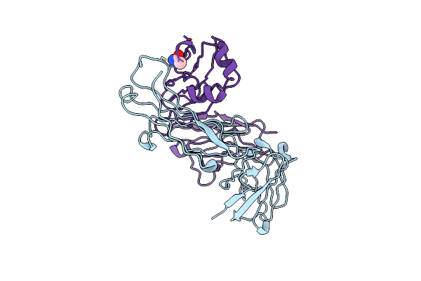
Pfa1 Fab Complexed With Gripi Peptide Fragment
Organism: Mus musculus
Method: X-RAY DIFFRACTION Resolution:2.10 Å Release Date: 2007-10-16 Classification: IMMUNE SYSTEM Ligands: GOL |
Organism: Mus musculus
Method: X-RAY DIFFRACTION
Release Date: 2007-10-16
Ligands: GOL
 |
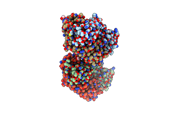
Pfa1 Fab Fragment
Organism: Mus musculus
Method: X-RAY DIFFRACTION Resolution:2.00 Å Release Date: 2007-10-09 Classification: IMMUNE SYSTEM Ligands: ACM |
Organism: Mus musculus
Method: X-RAY DIFFRACTION
Release Date: 2007-10-09
Ligands: ACM
 |
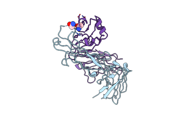
Pfa1 Fab Fragment Complexed With Abeta 1-8 Peptide
Organism: Mus musculus
Method: X-RAY DIFFRACTION Resolution:1.65 Å Release Date: 2007-10-09 Classification: IMMUNE SYSTEM Ligands: ACM, GOL |
Organism: Mus musculus
Method: X-RAY DIFFRACTION
Release Date: 2007-10-09
Ligands: ACM, GOL
 |
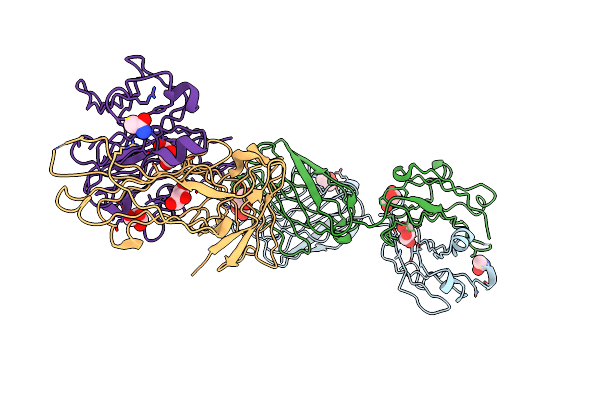
Pfa2 Fab Fragment, Triclinic Apo Form
Organism: Mus musculus
Method: X-RAY DIFFRACTION Resolution:2.30 Å Release Date: 2007-10-09 Classification: IMMUNE SYSTEM Ligands: ACM |
Organism: Mus musculus
Method: X-RAY DIFFRACTION
Release Date: 2007-10-09
Ligands: ACM
 |
Pfa2 Fab Fragment, Monoclinic Apo Form
Organism: Mus musculus
Method: X-RAY DIFFRACTION Resolution:2.00 Å Release Date: 2007-10-09 Classification: IMMUNE SYSTEM Ligands: ACM, GOL, CL |
Organism: Mus musculus
Method: X-RAY DIFFRACTION
Release Date: 2007-10-09
Ligands: ACM, GOL, CL
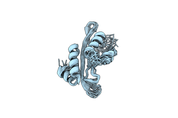 |
Structure Of The Tsg101 Uev Domain
|
Organism: Homo sapiens
Method: SOLUTION NMR
Release Date: 2002-05-25
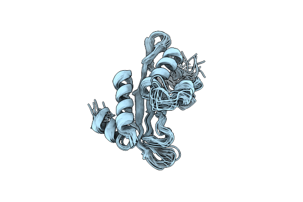 |
Structure Of The Tsg101 Uev Domain
|
Organism: Homo sapiens
Method: SOLUTION NMR
Release Date: 2002-05-24
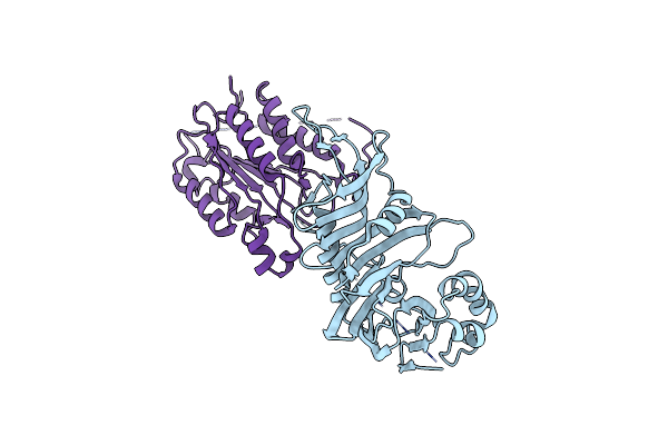 |
Crystal Structure Of The Caspase-8/P35 Complex
Organism: Autographa californica nucleopolyhedrovirus, Homo sapiens
Method: X-RAY DIFFRACTION Resolution:3.00 Å Release Date: 2001-03-28 Classification: APOPTOSIS/HYDROLASE |
Organism: Autographa californica nucleopolyhedrovirus, Homo sapiens
Method: X-RAY DIFFRACTION
Release Date: 2001-03-28