

Search Count: 9
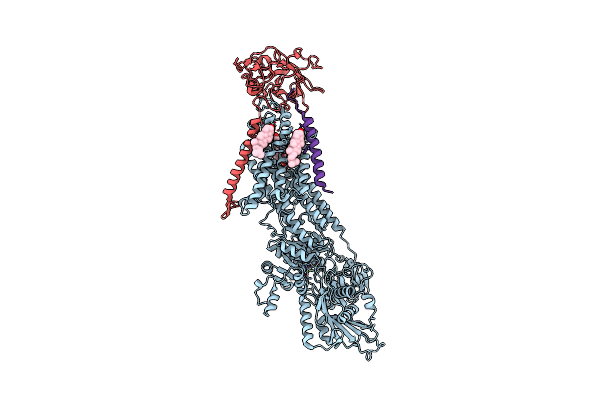 |
Crystal Structure Of The Na,K-Atpase From Bovine
Organism: Bos taurus
Method: X-RAY DIFFRACTION Resolution:3.90 Å Release Date: 2016-03-09 Classification: HYDROLASE Ligands: MG, OBN, CLR |
Organism: Bos taurus
Method: X-RAY DIFFRACTION
Release Date: 2016-03-09
Ligands: MG, OBN, CLR
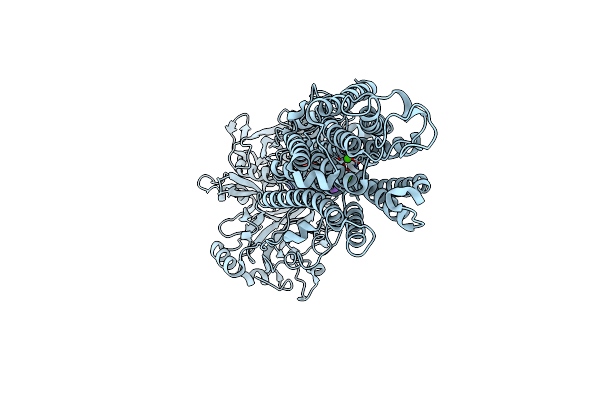 |
Crystal Structure Of The Sr Ca2+-Atpase In The Ca2-E1-Mgamppcp Form Determined By Serial Femtosecond Crystallography Using An X-Ray Free-Electron Laser.
Organism: Oryctolagus cuniculus
Method: X-RAY DIFFRACTION Resolution:2.80 Å Release Date: 2015-06-10 Classification: HYDROLASE Ligands: CA, K, ACP |
Organism: Oryctolagus cuniculus
Method: X-RAY DIFFRACTION
Release Date: 2015-06-10
Ligands: CA, K, ACP
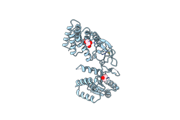 |
Structure Of The Nuclease Subunit Of Human Mitochondrial Rnase P (Mrpp3) At 1.8A
Organism: Homo sapiens
Method: X-RAY DIFFRACTION Resolution:1.80 Å Release Date: 2015-05-13 Classification: HYDROLASE Ligands: ZN, GOL |
Organism: Homo sapiens
Method: X-RAY DIFFRACTION
Release Date: 2015-05-13
Ligands: ZN, GOL
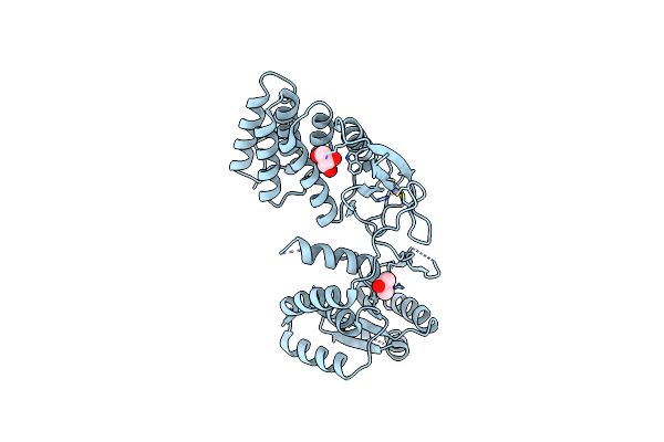 |
Structure Of The Nuclease Subunit Of Human Mitochondrial Rnase P (Mrpp3) At 1.98A
Organism: Homo sapiens
Method: X-RAY DIFFRACTION Resolution:1.98 Å Release Date: 2015-05-13 Classification: HYDROLASE Ligands: ZN, GOL |
Organism: Homo sapiens
Method: X-RAY DIFFRACTION
Release Date: 2015-05-13
Ligands: ZN, GOL
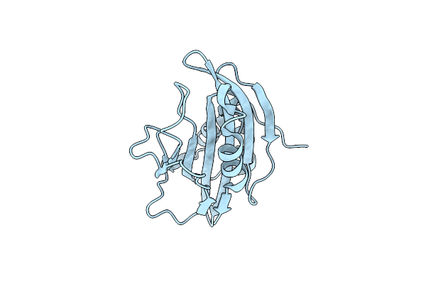 |
Crystal Structure Of Secreted Proline Rich Antigen Mtc28 (Rv0040C) At 2.15 A With Bound Chloride From Mycobacterium Tuberculosis
Organism: Mycobacterium tuberculosis
Method: X-RAY DIFFRACTION Resolution:2.15 Å Release Date: 2015-03-25 Classification: IMMUNE SYSTEM Ligands: CL |
Organism: Mycobacterium tuberculosis
Method: X-RAY DIFFRACTION
Release Date: 2015-03-25
Ligands: CL
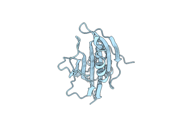 |
Crystal Structure Of Secreted Proline Rich Antigen Mtc28 (Rv0040C) From Mycobacterium Tuberculosis
Organism: Mycobacterium tuberculosis
Method: X-RAY DIFFRACTION Resolution:2.80 Å Release Date: 2015-01-28 Classification: LIPID BINDING PROTEIN |
Organism: Mycobacterium tuberculosis
Method: X-RAY DIFFRACTION
Release Date: 2015-01-28
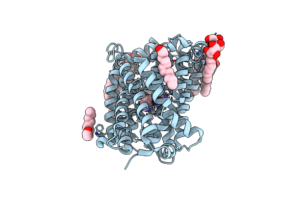 |
Crystal Structure Of The Bacterial Nss Member Mhst In An Occluded Inward-Facing State
Organism: Bacillus halodurans
Method: X-RAY DIFFRACTION Resolution:2.10 Å Release Date: 2014-09-24 Classification: TRANSPORT PROTEIN Ligands: TRP, NA, BNG, LMU |
Organism: Bacillus halodurans
Method: X-RAY DIFFRACTION
Release Date: 2014-09-24
Ligands: TRP, NA, BNG, LMU
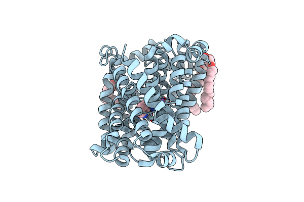 |
Crystal Structure Of The Bacterial Nss Member Mhst In An Occluded Inward-Facing State (Lipidic Cubic Phase Form)
Organism: Bacillus halodurans
Method: X-RAY DIFFRACTION Resolution:2.60 Å Release Date: 2014-09-24 Classification: TRANSPORT PROTEIN Ligands: TRP, NA, 78M, 78N |
Organism: Bacillus halodurans
Method: X-RAY DIFFRACTION
Release Date: 2014-09-24
Ligands: TRP, NA, 78M, 78N
 |
Crystal Structure Of Na+,K+-Atpase In The Na+-Bound State
Organism: Sus scrofa
Method: X-RAY DIFFRACTION Resolution:4.30 Å Release Date: 2013-10-02 Classification: HYDROLASE/TRANSPORT PROTEIN Ligands: ADP, ALF, MG, NA, CLR |
Organism: Sus scrofa
Method: X-RAY DIFFRACTION
Release Date: 2013-10-02
Ligands: ADP, ALF, MG, NA, CLR