

Search Count: 11
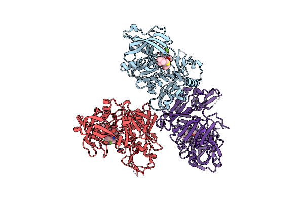 |
Spr And Structural Analysis Yield Insight Towards Mechanism Of Inhibition Of Bace Inhibitors
Organism: Homo sapiens
Method: X-RAY DIFFRACTION Resolution:2.20 Å Release Date: 2013-11-20 Classification: hydrolase/hydrolase inhibitor Ligands: 957 |
Organism: Homo sapiens
Method: X-RAY DIFFRACTION
Release Date: 2013-11-20
Ligands: 957
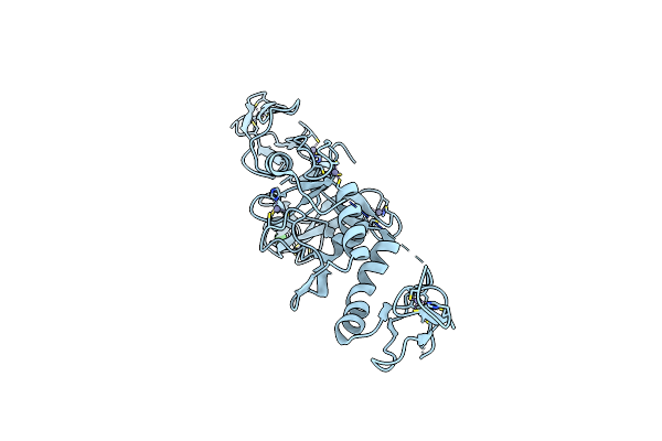 |
Structure Of Parkin-S223P E3 Ligase
Organism: Homo sapiens
Method: X-RAY DIFFRACTION Resolution:1.58 Å Release Date: 2013-06-19 Classification: LIGASE Ligands: ZN, BA |
Organism: Homo sapiens
Method: X-RAY DIFFRACTION
Release Date: 2013-06-19
Ligands: ZN, BA
 |
Structure Of Parkin E3 Ligase
Organism: Homo sapiens
Method: X-RAY DIFFRACTION Resolution:2.00 Å Release Date: 2013-06-19 Classification: LIGASE Ligands: ZN |
Organism: Homo sapiens
Method: X-RAY DIFFRACTION
Release Date: 2013-06-19
Ligands: ZN
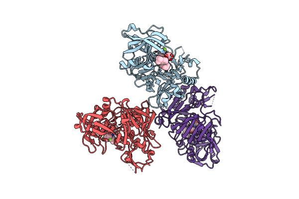 |
Spr And Structural Analysis Yield Insight Towards Mechanism Of Inhibition Of Bace Inhibitors.
Organism: Homo sapiens
Method: X-RAY DIFFRACTION Resolution:2.20 Å Release Date: 2013-03-06 Classification: hydrolase/hydrolase inhibitor Ligands: 1BL |
Organism: Homo sapiens
Method: X-RAY DIFFRACTION
Release Date: 2013-03-06
Ligands: 1BL
 |
Spr And Structural Analysis Yield Insight Towards Mechanism Of Inhibition Of Bace Inhibitors
Organism: Homo sapiens
Method: X-RAY DIFFRACTION Resolution:1.99 Å Release Date: 2013-03-06 Classification: hydrolase/hydrolase inhibitor Ligands: ZN, 842 |
Organism: Homo sapiens
Method: X-RAY DIFFRACTION
Release Date: 2013-03-06
Ligands: ZN, 842
 |
Ribonucleotide Reductase R1 Protein Mutant Y730F With A Reduced Active Site From Escherichia Coli
Organism: Escherichia coli
Method: X-RAY DIFFRACTION Resolution:2.90 Å Release Date: 1998-01-28 Classification: OXIDOREDUCTASE |
Organism: Escherichia coli
Method: X-RAY DIFFRACTION
Release Date: 1998-01-28
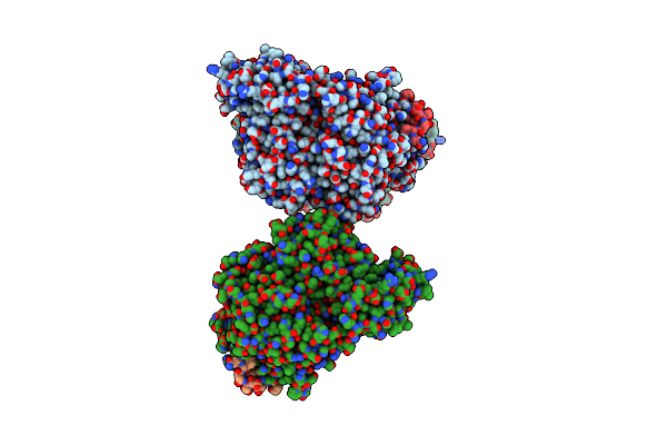 |
Ribonucleotide Reductase R1 Protein With Dttp Occupying The Specificity Site From Escherichia Coli
Organism: Escherichia coli
Method: X-RAY DIFFRACTION Resolution:3.00 Å Release Date: 1998-01-28 Classification: COMPLEX (OXIDOREDUCTASE/PEPTIDE) Ligands: TTP |
Organism: Escherichia coli
Method: X-RAY DIFFRACTION
Release Date: 1998-01-28
Ligands: TTP
 |
Ribonucleotide Reductase R1 Protein With Amppnp Occupying The Activity Site From Escherichia Coli
Organism: Escherichia coli
Method: X-RAY DIFFRACTION Resolution:3.00 Å Release Date: 1998-01-28 Classification: COMPLEX (OXIDOREDUCTASE/PEPTIDE) Ligands: ATP |
Organism: Escherichia coli
Method: X-RAY DIFFRACTION
Release Date: 1998-01-28
Ligands: ATP
 |
Ribonucleotide Reductase R1 Protein With Substrate, Gdp And Effector Dttp From Escherichia Coli
Organism: Escherichia coli
Method: X-RAY DIFFRACTION Resolution:3.20 Å Release Date: 1998-01-28 Classification: COMPLEX (OXIDOREDUCTASE/PEPTIDE) Ligands: TTP, GDP |
Organism: Escherichia coli
Method: X-RAY DIFFRACTION
Release Date: 1998-01-28
Ligands: TTP, GDP
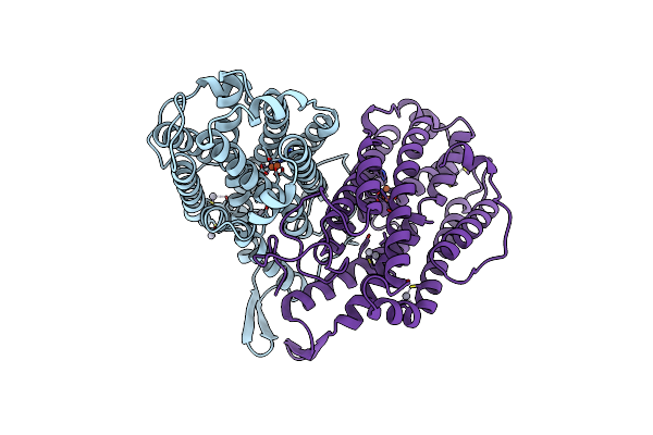 |
Ribonucleoside-Diphosphate Reductase 1 Beta Chain
Organism: Escherichia coli
Method: X-RAY DIFFRACTION Resolution:2.20 Å Release Date: 1997-03-12 Classification: REDUCTASE Ligands: FE, HG |
Organism: Escherichia coli
Method: X-RAY DIFFRACTION
Release Date: 1997-03-12
Ligands: FE, HG
 |
Ribonucleoside-Diphosphate Reductase 1 Beta Chain
Organism: Escherichia coli
Method: X-RAY DIFFRACTION Resolution:1.70 Å Release Date: 1997-03-12 Classification: OXIDOREDUCTASE Ligands: FE2, HG |
Organism: Escherichia coli
Method: X-RAY DIFFRACTION
Release Date: 1997-03-12
Ligands: FE2, HG