

Search Count: 300
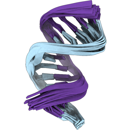 |
Nmr Structure And Free-Energy Simulations For Rational Design Of Myotonic Dystrophy Rna Inhibitors
|
Organism: Homo sapiens
Method: SOLUTION NMR
Release Date: 2026-10-07
 |
Nmr Structure And Free-Energy Simulations For Rational Design Of Myotonic Dystrophy Rna Inhibitors
Organism: Homo sapiens
Method: SOLUTION NMR Release Date: 2026-10-07 Classification: RNA Ligands: A1DA4 |
Organism: Homo sapiens
Method: SOLUTION NMR
Release Date: 2026-10-07
Ligands: A1DA4
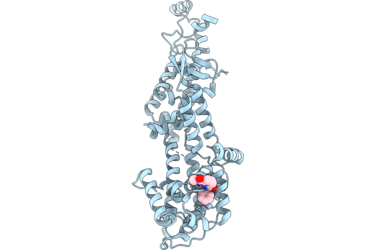 |
Sos1 In Complex With Compound 3
Organism: Homo sapiens
Method: X-RAY DIFFRACTION Resolution:1.92 Å Release Date: 2026-06-24 Classification: SIGNALING PROTEIN Ligands: A1JWK |
Organism: Homo sapiens
Method: X-RAY DIFFRACTION
Release Date: 2026-06-24
Ligands: A1JWK
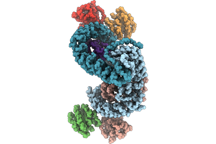 |
Mammalian Ap3 Complex On Tubular Membranes (Ap3 Centered)
Organism: Pan troglodytes, Homo sapiens, Rattus norvegicus
Method: ELECTRON MICROSCOPY Release Date: 2026-05-27 Classification: ENDOCYTOSIS |
Organism: Pan troglodytes, Homo sapiens, Rattus norvegicus
Method: ELECTRON MICROSCOPY
Release Date: 2026-05-27
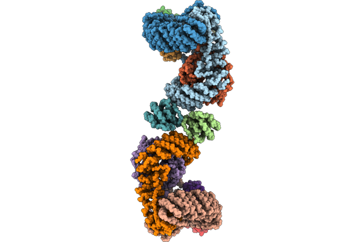 |
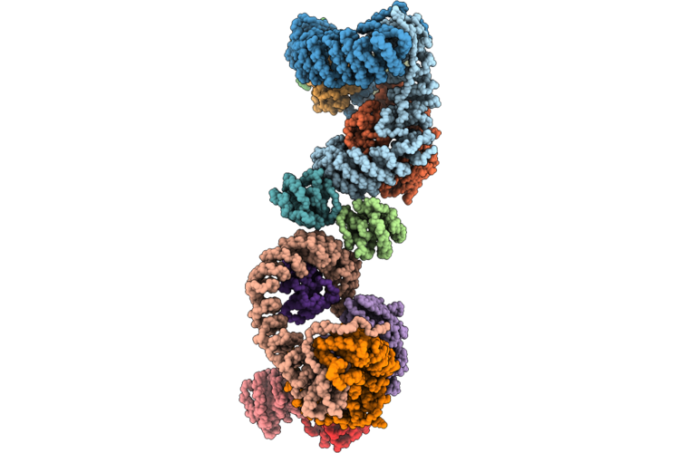
Mammalian Ap3 Complex On Tubular Membranes (Arf1 Centered Beta3-Arf1 Dimer-Beta3 Interface)
Organism: Pan troglodytes, Homo sapiens, Rattus norvegicus
Method: ELECTRON MICROSCOPY Release Date: 2026-05-27 Classification: ENDOCYTOSIS |
Organism: Pan troglodytes, Homo sapiens, Rattus norvegicus
Method: ELECTRON MICROSCOPY
Release Date: 2026-05-27
 |
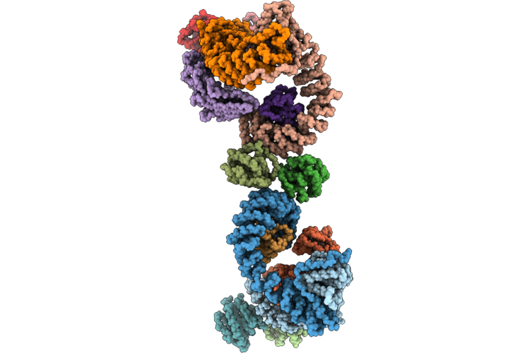
Mammalian Ap3 Complex On Tubular Membranes (Arf1 Centered Beta3-Arf1 Dimer-Delta Interface)
Organism: Pan troglodytes, Homo sapiens, Rattus norvegicus
Method: ELECTRON MICROSCOPY Release Date: 2026-05-27 Classification: ENDOCYTOSIS |
Organism: Pan troglodytes, Homo sapiens, Rattus norvegicus
Method: ELECTRON MICROSCOPY
Release Date: 2026-05-27
 |
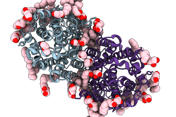
Mammalian Ap3 Complex On Tubular Membranes (Arf1 Centered Delta-Arf1 Dimer-Delta3 Interface)
Organism: Pan troglodytes, Homo sapiens, Rattus norvegicus
Method: ELECTRON MICROSCOPY Release Date: 2026-05-27 Classification: ENDOCYTOSIS |
Organism: Pan troglodytes, Homo sapiens, Rattus norvegicus
Method: ELECTRON MICROSCOPY
Release Date: 2026-05-27
 |
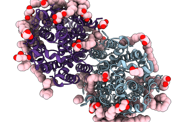
X-Ray Structure Of The Bacteroides Fragilis Nramp/Mnth Divalent Transition Metal Transporter Wt In An Inward-Open, State
Organism: Bacteroides fragilis
Method: X-RAY DIFFRACTION Resolution:2.48 Å Release Date: 2026-03-04 Classification: METAL TRANSPORT Ligands: OLC, PEG |
Organism: Bacteroides fragilis
Method: X-RAY DIFFRACTION
Release Date: 2026-03-04
Ligands: OLC, PEG
 |
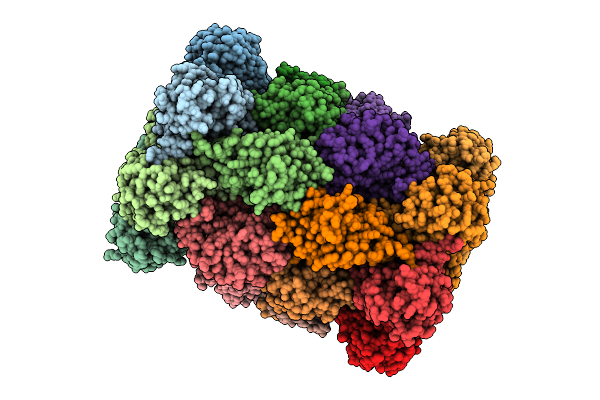
X-Ray Structure Of The Bacteroides Fragilis Nramp/Mnth Divalent Transition Metal Transporter Wt In An Inward-Open, Manganese-Bound State
Organism: Bacteroides fragilis
Method: X-RAY DIFFRACTION Resolution:2.40 Å Release Date: 2026-03-04 Classification: METAL TRANSPORT Ligands: MN, OLC, PEG |
Organism: Bacteroides fragilis
Method: X-RAY DIFFRACTION
Release Date: 2026-03-04
Ligands: MN, OLC, PEG
 |
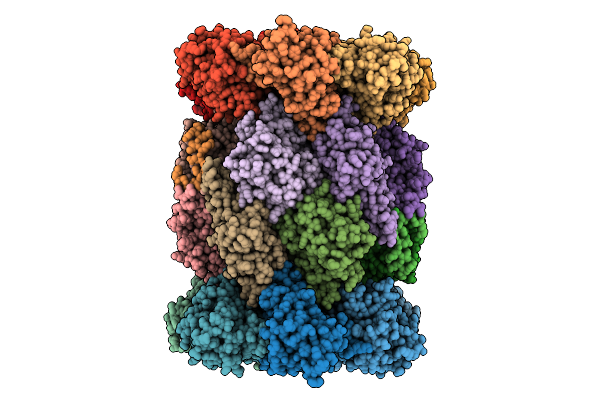
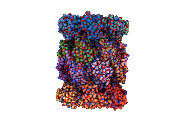
Plasmodium Falciparum 20S Proteasome Bound To Inhibitor 159
Organism: Plasmodium falciparum 3d7
Method: ELECTRON MICROSCOPY Resolution:2.79 Å Release Date: 2025-11-12 Classification: CYTOSOLIC PROTEIN Ligands: A1B74 |
Organism: Plasmodium falciparum 3d7
Method: ELECTRON MICROSCOPY
Release Date: 2025-11-12
Ligands: A1B74
 |
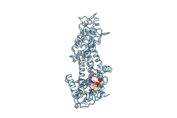
Plasmodium Falciparum 20S Proteasome Bound To Inhibitor 296
Organism: Plasmodium falciparum 3d7
Method: ELECTRON MICROSCOPY Resolution:2.28 Å Release Date: 2025-11-12 Classification: CYTOSOLIC PROTEIN Ligands: A1B73 |
Organism: Plasmodium falciparum 3d7
Method: ELECTRON MICROSCOPY
Release Date: 2025-11-12
Ligands: A1B73
 |
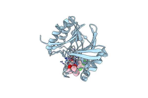
Structure Of Sos1 In Complex With Compound 3
Organism: Homo sapiens
Method: X-RAY DIFFRACTION Resolution:1.88 Å Release Date: 2025-09-17 Classification: CYTOSOLIC PROTEIN Ligands: A1I6H |
Organism: Homo sapiens
Method: X-RAY DIFFRACTION
Release Date: 2025-09-17
Ligands: A1I6H
 |
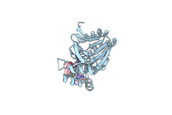
Crystal Structure Of Escherichia Coli Lpxh In Complex With Ebl-2805
Organism: Escherichia coli
Method: X-RAY DIFFRACTION Resolution:1.60 Å Release Date: 2024-09-18 Classification: HYDROLASE Ligands: MN, A1H5P |
Organism: Escherichia coli
Method: X-RAY DIFFRACTION
Release Date: 2024-09-18
Ligands: MN, A1H5P
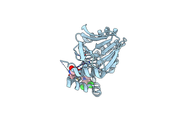 |
Structure Of K.Pneumoniae Lpxh In Complex With Ebl-3218
Organism: Klebsiella pneumoniae
Method: X-RAY DIFFRACTION Resolution:2.20 Å Release Date: 2024-09-18 Classification: HYDROLASE Ligands: MN, A1H55 |
Organism: Klebsiella pneumoniae
Method: X-RAY DIFFRACTION
Release Date: 2024-09-18
Ligands: MN, A1H55
 |
Plasmodium Falciparum 20S Proteasome Bound To An Inhibitor
Organism: Plasmodium falciparum 3d7
Method: ELECTRON MICROSCOPY Release Date: 2024-07-31 Classification: CYTOSOLIC PROTEIN/INHIBITOR Ligands: A1AE6 |
Organism: Plasmodium falciparum 3d7
Method: ELECTRON MICROSCOPY
Release Date: 2024-07-31
Ligands: A1AE6
 |
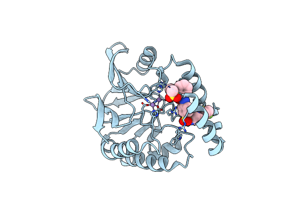
Crystal Structure Of E. Coli Lpxh In Complex With Lipid X
Organism: Escherichia coli
Method: X-RAY DIFFRACTION Resolution:1.35 Å Release Date: 2024-04-17 Classification: HYDROLASE Ligands: MN, LP5 |
Organism: Escherichia coli
Method: X-RAY DIFFRACTION
Release Date: 2024-04-17
Ligands: MN, LP5
 |
Structure Of K.Pneumoniae Lpxh In Complex With Ebl-3339
Organism: Klebsiella pneumoniae
Method: X-RAY DIFFRACTION Resolution:1.95 Å Release Date: 2024-04-17 Classification: HYDROLASE Ligands: MN, VTF |
Organism: Klebsiella pneumoniae
Method: X-RAY DIFFRACTION
Release Date: 2024-04-17
Ligands: MN, VTF
 |
Structure Of K. Pneumoniae Lpxh In Complex With Ebl-3647
Organism: Klebsiella pneumoniae
Method: X-RAY DIFFRACTION Resolution:1.95 Å Release Date: 2024-04-17 Classification: HYDROLASE Ligands: MN, VN6 |
Organism: Klebsiella pneumoniae
Method: X-RAY DIFFRACTION
Release Date: 2024-04-17
Ligands: MN, VN6
 |
Structure Of E. Coli Lpxh In Complex With Jedi-1444
Organism: Escherichia coli
Method: X-RAY DIFFRACTION Resolution:1.90 Å Release Date: 2024-04-17 Classification: HYDROLASE Ligands: MN, VQ9 |
Organism: Escherichia coli
Method: X-RAY DIFFRACTION
Release Date: 2024-04-17
Ligands: MN, VQ9
 |
Structure Of K. Pneumoniae Lpxh In Complex With Jedi-852
Organism: Klebsiella pneumoniae
Method: X-RAY DIFFRACTION Resolution:2.00 Å Release Date: 2024-04-17 Classification: HYDROLASE Ligands: MN, VTK |
Organism: Klebsiella pneumoniae
Method: X-RAY DIFFRACTION
Release Date: 2024-04-17
Ligands: MN, VTK