

Search Count: 24
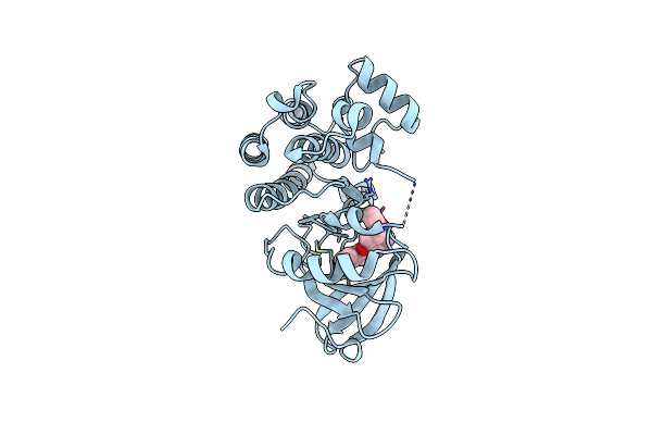 |
Bruton'S Tyrosine Kinase In Complex With Covalent Inhibitor Compound 10
Organism: Homo sapiens
Method: X-RAY DIFFRACTION Resolution:1.70 Å Release Date: 2024-06-26 Classification: TRANSFERASE/INHIBITOR Ligands: UEO |
Organism: Homo sapiens
Method: X-RAY DIFFRACTION
Release Date: 2024-06-26
Ligands: UEO
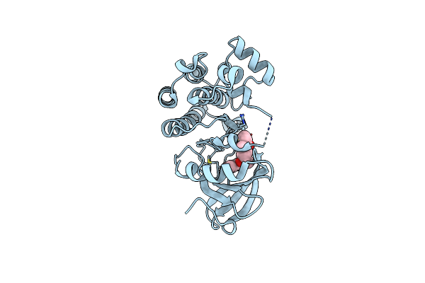 |
Bruton'S Tyrosine Kinase In Complex With Covalent Inhibitor Compound 25
Organism: Homo sapiens
Method: X-RAY DIFFRACTION Resolution:1.60 Å Release Date: 2024-06-26 Classification: TRANSFERASE/INHIBITOR Ligands: V72 |
Organism: Homo sapiens
Method: X-RAY DIFFRACTION
Release Date: 2024-06-26
Ligands: V72
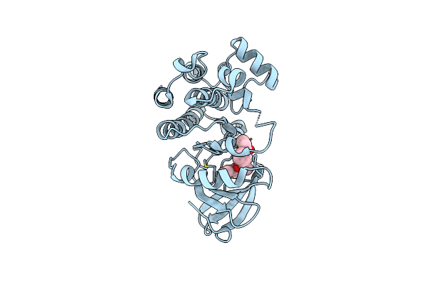 |
Bruton'S Tyrosine Kinase In Complex With Covalent Inhibitor Compound 27
Organism: Homo sapiens
Method: X-RAY DIFFRACTION Resolution:2.10 Å Release Date: 2024-06-26 Classification: TRANSFERASE/INHIBITOR Ligands: V7I |
Organism: Homo sapiens
Method: X-RAY DIFFRACTION
Release Date: 2024-06-26
Ligands: V7I
 |
Bio-2895 (Brd0705) Bound Gsk3Alpha-Axin Complex
Organism: Homo sapiens
Method: X-RAY DIFFRACTION Resolution:1.94 Å Release Date: 2023-06-14 Classification: HYDROLASE Ligands: 6VL, CA |
Organism: Homo sapiens
Method: X-RAY DIFFRACTION
Release Date: 2023-06-14
Ligands: 6VL, CA
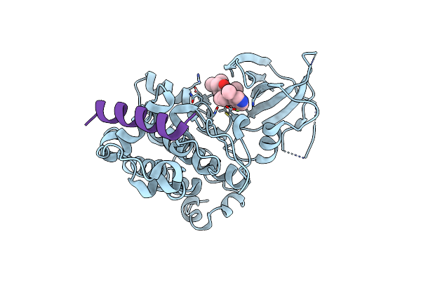 |
Bio-8546 Bound Gsk3Alpha-Axin Complex
Organism: Homo sapiens
Method: X-RAY DIFFRACTION Resolution:2.40 Å Release Date: 2023-06-14 Classification: HYDROLASE Ligands: D1E |
Organism: Homo sapiens
Method: X-RAY DIFFRACTION
Release Date: 2023-06-14
Ligands: D1E
 |
Bio-8546 Bound Gsk3Beta-Axin Complex
Organism: Homo sapiens
Method: X-RAY DIFFRACTION Resolution:2.09 Å Release Date: 2023-06-14 Classification: HYDROLASE Ligands: D1E |
Organism: Homo sapiens
Method: X-RAY DIFFRACTION
Release Date: 2023-06-14
Ligands: D1E
 |
Bio-2895 (Brd0705) Bound Gsk3Beta-Axin Complex
Organism: Homo sapiens
Method: X-RAY DIFFRACTION Resolution:1.85 Å Release Date: 2023-06-14 Classification: HYDROLASE Ligands: 6VL |
Organism: Homo sapiens
Method: X-RAY DIFFRACTION
Release Date: 2023-06-14
Ligands: 6VL
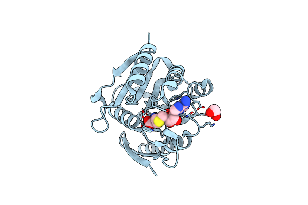 |
Structural Basis For Targeted P97 Remodeling By Aspl As Prerequisite For P97 Trimethylation By Mettl21D
Organism: Arabidopsis thaliana
Method: X-RAY DIFFRACTION Resolution:1.82 Å Release Date: 2022-08-03 Classification: TRANSFERASE Ligands: SAH, EDO |
Organism: Arabidopsis thaliana
Method: X-RAY DIFFRACTION
Release Date: 2022-08-03
Ligands: SAH, EDO
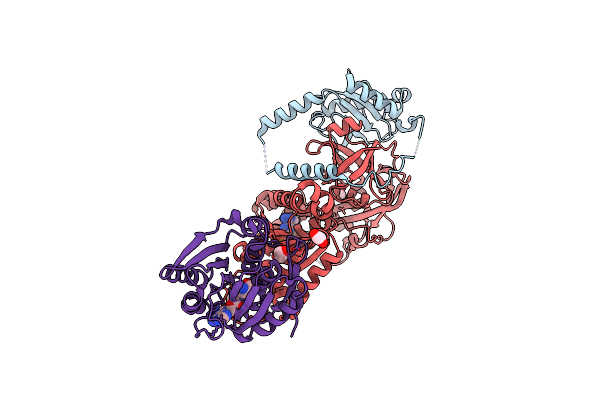 |
Structural Basis For Targeted P97 Remodelling By Aspl As Prerequisite For P97 Trimethylation By Mettl21D
Organism: Homo sapiens
Method: X-RAY DIFFRACTION Resolution:3.00 Å Release Date: 2022-08-03 Classification: TRANSFERASE Ligands: EDO, ATP, MG, SAH |
Organism: Homo sapiens
Method: X-RAY DIFFRACTION
Release Date: 2022-08-03
Ligands: EDO, ATP, MG, SAH
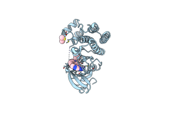 |
Bruton'S Tyrosine Kinase In Complex With Compound 23
Organism: Homo sapiens
Method: X-RAY DIFFRACTION Resolution:1.69 Å Release Date: 2022-01-12 Classification: TRANSFERASE/TRANSFERASE INHIBITOR Ligands: YD7, DMS |
Organism: Homo sapiens
Method: X-RAY DIFFRACTION
Release Date: 2022-01-12
Ligands: YD7, DMS
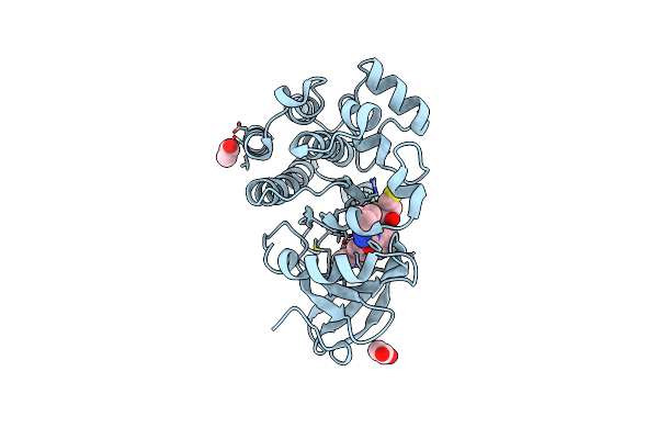 |
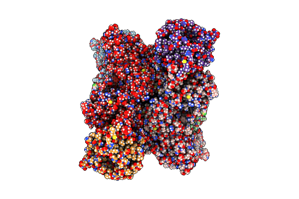
Bruton'S Tyrosine Kinase In Complex With Compound 51
Organism: Homo sapiens
Method: X-RAY DIFFRACTION Resolution:1.53 Å Release Date: 2022-01-12 Classification: TRANSFERASE/TRANSFERASE INHIBITOR Ligands: YDA, DTT, ACT |
Organism: Homo sapiens
Method: X-RAY DIFFRACTION
Release Date: 2022-01-12
Ligands: YDA, DTT, ACT
 |
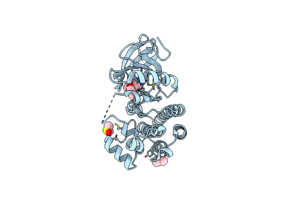
Cryoem Structure Of Prmt5 Bound To Covalent Pbm-Site Inhibitor Brd-6988
Organism: Homo sapiens
Method: ELECTRON MICROSCOPY Release Date: 2021-03-17 Classification: TRANSFERASE/INHIBITOR Ligands: YJG |
Organism: Homo sapiens
Method: ELECTRON MICROSCOPY
Release Date: 2021-03-17
Ligands: YJG
 |
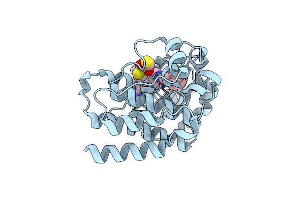
Bruton'S Tyrosine Kinase In Complex With Compound 5
Organism: Homo sapiens
Method: X-RAY DIFFRACTION Resolution:1.40 Å Release Date: 2020-12-16 Classification: TRANSFERASE/TRANSFERASE INHIBITOR Ligands: DMS, RQS |
Organism: Homo sapiens
Method: X-RAY DIFFRACTION
Release Date: 2020-12-16
Ligands: DMS, RQS
 |
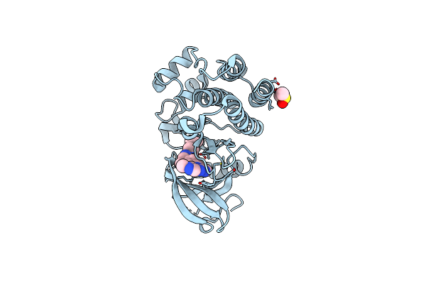
Bruton'S Tyrosine Kinase In Complex With Compound 6
Organism: Homo sapiens
Method: X-RAY DIFFRACTION Resolution:1.55 Å Release Date: 2020-12-16 Classification: TRANSFERASE/TRANSFERASE INHIBITOR Ligands: DMS, DTT, S5M |
Organism: Homo sapiens
Method: X-RAY DIFFRACTION
Release Date: 2020-12-16
Ligands: DMS, DTT, S5M
 |
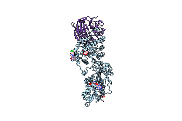
Bruton'S Tyrosine Kinase In Complex With Compound 1
Organism: Homo sapiens
Method: X-RAY DIFFRACTION Resolution:1.51 Å Release Date: 2020-12-16 Classification: TRANSFERASE/TRANSFERASE INHIBITOR Ligands: S9A, DMS |
Organism: Homo sapiens
Method: X-RAY DIFFRACTION
Release Date: 2020-12-16
Ligands: S9A, DMS
 |
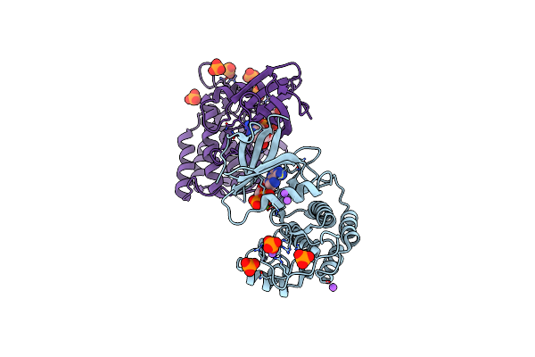
Prmt5 Complex Bound To Covalent Pbm Inhibitor Brd6711
Organism: Homo sapiens
Method: X-RAY DIFFRACTION Resolution:1.88 Å Release Date: 2020-11-25 Classification: SPLICING/TRANSFERASE Ligands: QN4, CL, SFG, GOL |
Organism: Homo sapiens
Method: X-RAY DIFFRACTION
Release Date: 2020-11-25
Ligands: QN4, CL, SFG, GOL
 |
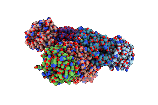
Adp Bound Ttbk2 Kinase Domain
Organism: Homo sapiens
Method: X-RAY DIFFRACTION Resolution:1.50 Å Release Date: 2020-06-03 Classification: TRANSFERASE Ligands: ADP, PO4, NA, MG, GOL |
Organism: Homo sapiens
Method: X-RAY DIFFRACTION
Release Date: 2020-06-03
Ligands: ADP, PO4, NA, MG, GOL
 |
Crystal Structure Of Tryptophan Synthase From M. Tuberculosis - Ligand-Free Form
Organism: Mycobacterium tuberculosis (strain atcc 25618 / h37rv)
Method: X-RAY DIFFRACTION Resolution:2.46 Å Release Date: 2017-05-31 Classification: LYASE Ligands: MLI, FMT, K |
Organism: Mycobacterium tuberculosis (strain atcc 25618 / h37rv)
Method: X-RAY DIFFRACTION
Release Date: 2017-05-31
Ligands: MLI, FMT, K
 |
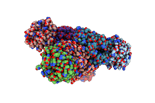
Crystal Structure Of Tryptophan Synthase From M. Tuberculosis - Aminoacrylate-Bound Form
Organism: Mycobacterium tuberculosis (strain atcc 25618 / h37rv)
Method: X-RAY DIFFRACTION Resolution:2.40 Å Release Date: 2017-05-31 Classification: LYASE Ligands: MLI, FMT, P1T, CS |
Organism: Mycobacterium tuberculosis (strain atcc 25618 / h37rv)
Method: X-RAY DIFFRACTION
Release Date: 2017-05-31
Ligands: MLI, FMT, P1T, CS
 |
Crystal Structure Of Tryptophan Synthase From M. Tuberculosis - Ligand-Free Form, Trpa-G66V Mutant
Organism: Mycobacterium tuberculosis (strain atcc 25618 / h37rv)
Method: X-RAY DIFFRACTION Resolution:2.35 Å Release Date: 2017-05-31 Classification: LYASE Ligands: MLI, FMT |
Organism: Mycobacterium tuberculosis (strain atcc 25618 / h37rv)
Method: X-RAY DIFFRACTION
Release Date: 2017-05-31
Ligands: MLI, FMT